Chronic osteomyelitis of the femur in a 5-year-old girl
Received: 06-May-2021 / Accepted Date: 09-May-2021 / Published Date: 16-May-2021 DOI: 10.4172/2167-7964.1000326
Abstract
Chronic osteomyelitis is a well-known disease which represents a progressive inflammatory process caused by pathogens, resulting in bone destruction and sequestrum formation which is a challenging condition to diagnose and to treat. We report a case of a 5-year-old girl who suffered from a right femur chronic osteomyelitis with sequestrum and fistula demonstrated at CT scan.
Keywords: Osteomyelitis, Chronic, CT, Sequestrum, Fistulas
Text
Chronic osteomyelitis is defined as a long-standing infection of bone and marrow, characterized by persistence of microorganisms, presence of sequestrum, low-grade inflammation and fistulae [1]. Usually associated with open fractures or after a long period of infection through blood transmission It may present as a recurrent or intermittent disease. The symptoms and their duration may vary considerably, whereas periods of quiescence can also be of variable duration [2].
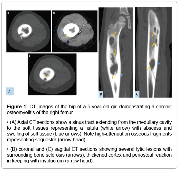
The imaging diagnostic of osteomyelitis can require the combination of diverse imaging techniques for an accurate diagnosis. CT demonstrates abnormal thickening of the affected cortical bone, with sclerotic changes, encroachment of the medullary cavity, and chronic draining sinus Figure 1. Although CT may show these changes earlier than do plain radiographs, CT is less desirable than MRI because of decreased soft tissue contrast as well as exposure to ionizing radiation [3].
Figure 1: CT images of the hip of a 5-year-old girl demonstrating a chronic osteomyelitis of the
right femur
• (A) Axial CT sections show a sinus tract extending from the medullary cavity to the soft tissues
representing a fistula (white arrow) with abscess and swelling of soft tissue (blue arrows). Note high-attenuation osseous
fragments representing sequestra (arrow head).
• (B) coronal and (C) sagittal CT sections showing several lytic lesions with surrounding bone sclerosis
(arrows), thickened cortex and periosteal reaction in keeping with involucrum (arrow head).
The « gold standard » for the diagnosis of chronic osteomyelitis is the presence of positive bone cultures and histopathology examination of the bone [2].
The incidence of relapse remains high, making its management challenging for the treating physician [4]. Assumed ‘remission’ should only be claimed after at least 12 months of follow-up, while ‘cure’ of the disease cannot be safely declared [2].
References
- Mouzopoulos G, Kanakaris NK, Kontakis G, Obakponovwe O,nTownsend R (2011) Management of bone infectionsnin adults: the surgeon's and microbiologist's perspectives. Injurie 42:18-23
- Panteli M, Giannoudis PV (2016)  Chronic osteomyelitis what the surgeon needs tonknow. EFORT Open Rev 1(5):128-135.
- Pineda C, Espinosa R, Pena A (2009)  Radiographic imaging in osteomyelitis  the rolenof plain radiography, computed tomography, ultrasonography, magnetic resonance imaging, and scintigraphy. SeminnPlast Surg 23(2):80-89.
- Spellberg B, Lipsky BA (2012) antibiotic therapy for chronic osteomyelitis innadults. Clin Infect Dis 54:394-407
Citation: Lrhorfi N, Moslih H, El haddad S, Allali N, Chat L (2021) Chronic osteomyelitis of the femur in a 5-year-old girl. OMICS J Radiol 10: 326. DOI: 10.4172/2167-7964.1000326
Copyright: © 2021 Lrhorfi N, et al. This is an open-access article distributed under the terms of the Creative Commons Attribution License, which permits unrestricted use, distribution, and reproduction in any medium, provided the original author and source are credited.
Select your language of interest to view the total content in your interested language
Share This Article
Open Access Journals
Article Tools
Article Usage
- Total views: 3329
- [From(publication date): 0-2021 - Nov 27, 2025]
- Breakdown by view type
- HTML page views: 2493
- PDF downloads: 836

