Review Article Open Access
Changes in the Anatomy and Physiology of the Distal Esophagus and Stomach after Sleeve Gastrectomy
| Attila Csendes* and Italo Braghetto | |
| Department of Surgery, University Hospital, University of Chile, Santiago, Chileia | |
| Corresponding Author : | Attila Csendes, MD Department of Surgery Hospital J.J. Aguirre Santos Dumont 999 Santiago, Chile Tel: 56 2-2978-8000 E-mail: acsendes@ hcuch.cl |
| Received January 12, 2016; Accepted January 21, 2016; Published January 24, 2016 | |
| Citation: Csendes A, Braghetto I (2016) Changes in the Anatomy and Physiology of the Distal Esophagus and Stomach after Sleeve Gastrectomy. J Obes Weight Loss Ther 6:297. doi:10.4172/2165-7904.1000297 | |
| Copyright: © 2016 Csendes A, et al. This is an open-access article distributed under the terms of the Creative Commons Attribution License, which permits unrestricted use, distribution, and reproduction in any medium, provided the original author and source are credited. | |
Visit for more related articles at Journal of Obesity & Weight Loss Therapy
Abstract
Aim: Sleeve gastrectomy is one of the most popular surgical procedures for patients with obesity. Its performance produces several pathophysiological changes at the esophago-gastric junction, gastric acid secretion, emptying and motility. Purpose: To review all pathophysiological changes of the distal esophagus and stomach after the resection of 80% of the stomach during sleeve gastrectomy. Material and Methods: Review of all publications concerning the measurements of lower esophageal sphincter after sleeve gastrectomy, as well as acid reflux, gastric motility and gastric emptying. Results: The section of some portion of the sling fibers produces dilatation of the cardia and development of pathologic acid reflux into the distal esophagus. The great majority of reports dealing with 24 h pH measurements or impedanciometry report severe acid and non-acid reflux. Gastric acid secretion is greatly diminished after sleeve gastrectomy in about 80% but the residual acid secretion is at least 20 times greater than after gastric bypass. Gastric motility and electric activity is also compromised due to resection of most of the fundus and the gastric pacemaker located at the greater curvature. As a consequence, gastric emptying of liquids and solids are greatly enhanced. Then a new swallow of food impacts against this elevated pressure which may overcome the hypotensive lower esophageal sphincter and pathologic reflux may occur into the esophagus. Conclusion: Sleeve gastrectomy is an operation which may produce severe pathologic reflux of acid, as
| Keywords |
| Distal esophagus; Sleeve gastrectomy |
| Introduction |
| The development of bariatric surgery in the last 2 decades has increased exponentially all over the world. Multiple surgical procedures have been designed over the digestive tract, trying to find the best technique in terms of simplicity and safety, but at the same time effective in achieving a permanent loss of weight and improvement of co-morbidities [1-3]. The early and late clinical follow up, as well as the impact on the nutritional state and metabolism, have revealed profound pathophysiological changes over the distal esophagus, stomach and small intestine, which were partially known before bariatric surgery, during the era of peptic ulcer surgery [4]. At present, two surgical procedures are widely employed due to their safeness and good clinical results: gastric bypass and sleeve gastrectomy [5-7]. This last procedure has increased significantly in United States, Europe and Latin America [7,8]. The long term results of this operation have shown an acceptable excess weight loss, however weight gain and “de novo” gastroesophageal reflux symptoms may appear [9,10]. In the literature there are very few mentions to the pathophysiologic changes that occur after sleeve gastrectomy contrasting with the enormous amount of information concerning the clinical changes. |
| The purpose of the present study is to review the different pathophysiological alterations after sleeve gastrectomy, which includes the lower esophageal sphincter, gastric acid secretion, serum gastrin changes, gastric emptying and gastric motility. No mention is included in reference to the alterations at the level of small intestine. Besides, the review will not deal with clinical symptoms or complications, but only to physiological functions and their changes after this operation determined by objective measurements. |
| Lower esophageal sphincter |
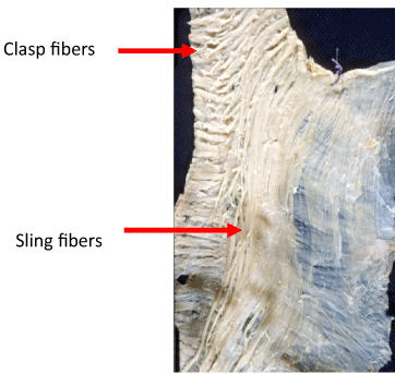
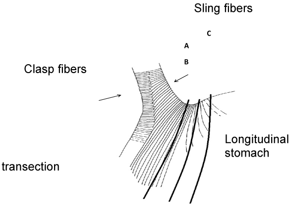
| It is well known that obesity increases the incidence of gastroesophageal reflux [11,12]. The etiology is not completely understood, but probably includes a mixture of hereditary, ambiental and functional factors, which mainly deal with the behavior of the lower esophageal sphincter (LES) [13-15].The anatomy of this sphincter has been carefully evaluated by Korn et al. [16,17] showing the presence of 2 different fibers: “clasps” and “sling” (Figure 1). The clasp fibers are located at the right portion of the sphincter and are the final prolongation of semicircular fibers. The sling fibers are located at the left side of the sphincter and are anchored at the antrum. According to the technique of sleeve gastrectomy, if the section is done near to the esophagus (A), an increase in the production of leaks may occur. In order to avoid or to diminish this complication as suggested by many authors gastric section should start 1-2 cms from the esophagogastric junction. If this is the situation (B), few sling fibers are divided. On the contrary, if this section is done 3 or more cms from the EG junction, probably most sling fibers are divided [18] (Figure 2), which may affect LES pressure and pathologic acid reflux many appear. However, it is well known that also other factors such as ischemia, staple mismatch, non-completion of the staple line, etc, may be related to this event. |
| The main function of the LES is to prevent the retrograde movement of gastric content into the esophagus. However if the normal function of this sphincter is altered, such as prolonged lower esophageal sphincter relaxation [19,20], a hypotensive lower esophageal sphincter [21,22] or anatomic disruption of the gastroesophageal junction as a hiatal hernia [23,24], gastroesophageal reflux of different severity can occur. As described before, the disruption of the angle of His causes a partial section of the sling fibers [18], according to the distance from the EG junction to the staple line. The transection of these fibers may produce an hypotensive sphincter and therefore, pathologic acid reflux. |
| Table 1 shows the lower esophageal sphincter pressure before and several months after sleeve gastrectomy in 7 reported studies. The first was published by Braghetto et al. showing a significant decrease in resting pressure [18]. Two other authors [25,26], also reported the same findings. On the contrary, 2 authors [27,28] published an increase in resting pressure. One group [29] found no significant alteration. Finally, the most recent study [30] divides patients in 2 groups before surgery: with pathologic or normal acid reflux. In both groups there was no change in sphincter pressure. However it is important to consider in these results the variations of the surgical technique performed by different authors, which might explain some of the contradictory results from different studies. |
| The total length and abdominal length of the lower esophageal sphincter before and after sleeve gastrectomy is shown in Table 2. Only one author [28] evaluated both parameters, showing a significant increase. Other 2 reports [29,30] published no change in total length. None of the published papers evaluated the presence of transient sphincter relaxation. Table 3 demonstrates the only two papers reporting measurement of the amplitude of distal esophageal contractions. In both groups there was no significant change after surgery [27,28]. |
| More important is the objective evaluation of acid and non-acid reflux after sleeve gastrectomy: The results are shown in Table 4. Two authors reported a significant increase of acid reflux measured by 24- hrs monitoring [25,26]. In one publication [29], although an increase of acid reflux after surgery was reported, it was not significant. Finally Rebecchi et al. [30] demonstrated a significant decrease of acid reflux after surgery in patients who showed pathologic acid esophageal reflux before surgery, but no change among patients who were asymptomatic before operation. Another way to evaluate acid and non-acid reflux is by impedanciometry (Table 5). Three reports deal with this topic. Two of them showed a significant increase after surgery measuring acid and non-acid reflux [25,29]. It is curious that one author, who demonstrated a significant increase of acid reflux by 24-hrs pHmetry [26], showed no increase in acid or non-acid reflux after surgery measured by impedanciometry. However this same author published that the episode of reflux which last more than 5 minutes increased from 0.8% to 5.7% after surgery (p < 0.01), while the longest single period of reflux was 6.4 minutes before surgery and increased to 25.4 minutes after sleeve gastrectomy. |
| Finally, there is a very recent study [31] measuring gastroesophageal reflux by scintigraphic method by Tc-99 labeled sulfur colloid, before and after sleeve (Table 6). Before operation, only 6.2% showed gastroesophageal reflux while 6 months after surgery 78% demonstrated reflux into the esophagus (p < 0.001). Besides, it has been shown that a decrease in plasma grelin may cause gastrointestinal dismotility in animal model and this effect is also seen after sleeve gastrectomy [32,33]. Therefore this alteration could predispose to reflux. |
| In conclusion, data concerning the effect of sleeve gastrectomy on the dynamics of lower esophageal sphincter and production of GERD are controversial [30]. During surgery there are 2 crucial points: |
| a-Avoid creating a mid-stomach stenosis that could increase reflux. |
| b- Careful dissection of the angle of His with a safe distance from the EG junction, in order to avoid the inclusion of esophageal tissue [28]. This “safe distance” has not been clearly determined. As seen in (Figure 2), the preservation of sling fibers integrity is almost impossible, due to their extension to the angle of His and gastric fundus. As seen in Table 1, multiple studies show different outcomes in regard to the effect of sleeve gastrectomy on LES pressure (in 3 studies LES pressure decreases in 1 increases and in 3 remains similar), reflecting the confusion that actually exists in the data. Isolated manometric increase in resting pressure is not enough to postulate that GERD is cured by sleeve as shown by Rebecchi et al. [30]. De-novo reflux could be induced by an adverse effect of sleeve on the function of the antireflux barrier and therefore pathologic acid exposure to the distal esophagus is mainly determined by lower esophageal sphincter insufficiency rather than loss of weight and increased intragastric pressure [34]. The normal LES has a mean pressure of 18 mmHg, a total length of 30 to 40 mm and an abdominal length of 15 mm [34,35]. Sleeve gastrectomy produces a reduction of gastric compliance due to resection of 75% to 85% of the oxintic area [28,34]. When the reduced stomach is full with meal, intraluminal pressure increases according to Laplace´s low [36]. Then a new swallow of food impacts against this elevated pressure which may overcome the hypotensive LES pressure and produces reflux into the esophagus. |
| The importance of several other factors that may have a role on producing or avoiding gastric content reflux into the esophagus will be discussed in the following points. |
| Gastric acid secretion |
| Anatomically the stomach is formed by 3 topographic areas: fundus, body and antrum [37] but functionally there are 2 glandular regions: oxyntic and antral mucosa. The oxintic area comprises 80% of the proximal stomach (fundus and body) while the antral mucosa , with G cells which produce gastrin, corresponds to 20% of all gastric surface. The normal human stomach has 1 x 10⁹ parietal cells and 9x10⁶ G cells [38]. |
| Oxintic cells are responsible for the production of hydrochloric acid and intrinsic factor, but other cells are also located in this region secreting pepsinogen and leptin [39]. Besides there are also enterochromafin cells which produce serotonin, adenomedulin and natriuretic peptide [40,41] and enterochomaffin-like cells which contain histamine, D cells (somatostatine) and Gr cells which produce ghrelin and obestatin [42- 47]. These last cells comprise 30% of the total neuroendocrine cells. |
| However in the present review we do not intend to describe the multiple complex mechanisms for acid secretion and production of different neuroendocrine peptides. The reader interested in these topic can read excellent updates in several recent articles [37,48-52]. |
| Gastric acid secretion is a complex process that involves neuronal, hormonal and endocrine pathways [48]. All of these pathways have one common target: the parietal cell, where the functional “factory” for acid secretion sits. These cells concentrate hydrochloric acid into the lumen, due to the fact that they have a proton pump (H+, K+, adenosive triphosphatase) which must travel to the surface of the cells, where it transports the protons necessary to acidify the gastric content at the expense of adenosine triphosphate: This high demand of energy is possible because the mitochondria represents 40% of the entire parietal cells volume. |
| This physiologic process may produce damage if an unprotected tissue is exposed to acid or there is a pathologic hypersecretion of acid. This may occur in some diseases such as gastroesophageal reflux or peptic ulcer. The actual therapy for these diseases is based on the reduction of the amount of acid secreted by the parietal cells by different mechanism. |
| There are several methods of measuring gastric acid secretion [49]. For us the classical methods are the best and more practical. They are based on the suction of gastric content through a nasogastric tube positioned at the most dependent part of the stomach, which can be confirmed either by radiological visualization or more easily by the water recovery test. This method measures both basal acid output (B.A.O.) and the maximal stimulated acid output (M.A.O.), either by histamine or penta-gastrin. It is very important to remember that the maximal acid secretion is closely related to the parietal cell mass and therefore different magnitude of gastrectomies produce a reduction of acid secretion parallel to the reduction of the parietal cell mass. We have performed 2 studies of gastric acid secretion in controls, as shown in Table 7 [53,54]. Basal acid secretion is near to 3 mMl/h and maximal acid output is near to 16 mMl/h. If we assume that sleeve gastrectomy produces a resection of 80% of parietal cells, BAO decreases to 0.6mMl/h. and MAO to 3.2 mMl/h. This value is in contrast to what has been published after gastric bypass in 2 papers [55,56] in which both basal and stimulated acid secretion are negligible and virtually absent after gastric bypass. It can be seen that after sleeve gastrectomy, there is still an acid output at least 20 times more than after gastric bypass. Therefore the damage to distal esophageal mucosa due to reflux is important and if also duodenal reflux occurs together to acid reflux, the damage may be severe [57,58]. |
| Serum gastrin after sleeve |
| Gastrin is the main regulator of gastric acid secretion during food intake. It is produced in G cells which are mainly located in the antral portion of the stomach, but also in the proximal duodenum: [37,38,51] It stimulates acid secretion by releasing histamine from enterochomaffin cells like [50]. Today it is included in the “family of gastrins” because the hormone cholecytokinin shares the same tetrapeptide at the terminal segment of the biologically active molecule. After several chemical complex steps, the final process is the amidation of the termical COOH, and the formation of the final G17 (most frequent) and G34 that are present at the antrum. |
| Proteins and peptides, as well as vagal stimulus release gastrin, but gastric acid inhibits gastrin release up to pH 3-4 (negative feedback). Gastrin besides stimulating acid secretion, has an important positive effect on the oxyntic mucosa. Serum gastrin increases in humans if there is a gastrin producing tumor (gastrinoma) either in the pancreas or duodenum [51]. It also increases in the presence of an important decrease of acid secretion such as an atrophic gastritis at the oxintic area or by the chronic use of inhibitors of acid secretion [50]. Finally if antral mucosa is excluded by an operation from the gastrointestinal tract, the retained antrum syndrome may occur: Up to now we have found only 2 reports concerning serum gastrin levels after sleeve gastrectomy. [59,60]. One experimental study (Table 8) was performed in Goto-Kakizaki rats who are diabetic in early life, but leans and not fatty, determining serum gastrin before and 36 weeks after surgery in fast condition and after a mixed meal. Sleeve was constructed from the middle point between the proximal limit of the antrum and the pylorus. They showed markedly elevated serum gastrin late after surgery. The same finding was published in one study in humans, demonstrating a significant increase after sleeve. This hypergastrinemia is probably due to the fact that acid secretion is greatly diminished (80%) in sleeve patients and therefore there is no acid inhibition of gastrin release, because sleeve gastrectomy leaves antral function intact. |
| Gastric motility |
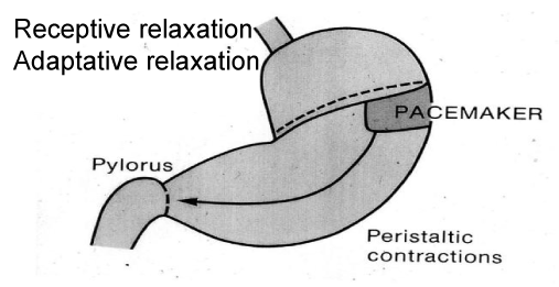
| The anatomic structures of the stomach are the fundus, body and antrum. Similar to what happens with gastric acid secretion, gastric motility has also 2 different functional portions. The upper third serves as reservoir of food, and includes the fundus and the proximal one third of the corpus (Figure 3). The distal portion which extends from the 2 distal thirds of the body including the antrum has electric and motor activity, which results in the mixture of foods with the digestive secretions and facilitates gastric emptying. The pylorus regulates the outflow of food to the duodenum [61,62]. |
| Gastric motility is controlled by several stimulus such as myogenic, neural and hormonal influences. However the intrinsic myogenic activity is the most important factor for gastric motility. It is initiated by an electric activity of a pacemaker located in the midportion of the corpus and it is, composed by the interstitial cells of Cajal. This pacemaker origins a depolarization of smooth muscle membrane at a rate of 3 cycles per minute [63]. This depolarization can be nodulated either by vagal stimulus or hormonal influences. Only when an action potential results by a complete depolarization, contractile muscle activity appears, at a maximum rate of 3/minute. |
| Fasting gastric motility is composed by the migrating motor complex which has a special function: to clear gastric and intestine lumen from indigested debris. It is composed of 4 different phases with a total duration of 90 to 120 minutes per cycle, being phase III the most important, with a short duration of 10 minutes, but due to the high amplitude contractions, it cleans gastric lumen [64]. |
| Function of the proximal stomach |
| This portion during the post prandial period exhibits a tonic contraction due to slow sustained contractions. However, when food is ingested, it has 2 physiological properties: |
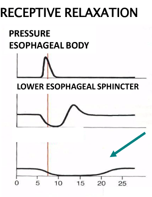
| a.- Receptive relaxation of the muscle parallel to lower esophageal sphincter relaxation, in order to receive the ingested food without pressure against swallowing [65,66] (Figure 4). |
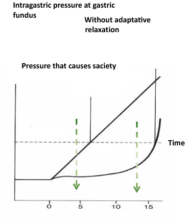
| b.- Accommodation to distension, that is, expansion of proximal gastric lumen without increase of intraluminal pressure, in which intrinsic and vasovagal reflexes are involved [67] (Figure 5). |
| These 2 mechanisms allow the entrance of great amount of food ingested without an increase of intragastric pressure until a certain point where increase in pressure occurs (Figure 5). In this way the meal is mixed with HCl and pepsin in order to start the digestion of protein. |
| Funcion of the distal stomach |
| Peristaltic activity starts at the middle third portion of corpus due to the presence of gastric pacemaker. Due to complete depolarization of smooth muscle membrane by creation of an action potential, a muscular contractions occurs [61,63]. Besides vagal stimulus, these are several hormones and neurotransmitters which modulate this activity. Motor contractions of the antrum mix and crush food into a small particulate facilitating the emptying to the duodenum. The larger particles are forced retrograde in order to reduce them in smaller particles. |
| The terminal antral contraction (TAC) produces a contraction of the pylorus, therefore allowing the passage of small particles less than 5 mm and the rest is expelled retrogdaly. |
| After sleeve gastrectomy, there is a profound change in gastric motor physiology. Due to resection of almost all gastric fundus, receptive relaxation is abolished and gastric accommodations or receptive adaptation is greatly altered (Figure 5). In this scenario, small amount of ingested food can create high intragastric pressure. |
| Anatomic and histologic landmark of the antrum |
| There are very few reports concerning the anatomic features of the stomach in humans. The stomach measures 18 cms by the lesser curvature and 36 to 40 cms by the greater curvature [68]. Its weight is near to 200 gr and the volume capacity to an acute distension is near to 1200 ml either in normal subjects or patients with different degrees of obesity [69]. |
| The precise histologic landmarks of the oxyntic area and antral are few. Lopetegui [70] studied the stomachs of humans who recently died by accident before 12 hrs of death and measured the extension of the antrum by lesser curvature, corresponding to 50% of its length (9 cms from pylorus). In the greater curvature it corresponded to 25% of the length (9 cms from pylorus). In another study determining the extension of oxintic area in patients with duodenal ulcer, [71], we found that the proximal branch of Latarjet´s crow-foot corresponded to oxintic-antral limit, and this distance was 8.5+/-1 cms from the pylorus, similar to Lopetegui´s findings. |
| A recent paper evaluating the width of the different part of stomach wall concluded that the antrum measured twice the width of the fundus [72]. This finding has a tremendous importance when performing a sleeve, in order to use the proper staples. |
| Intragastric pressure |
| This topic is also very controversial, but crucial in the understanding of the different mechanisms which may create a pathologic reflux. Yehoshua et al. [36] measured gastric pressure and distensibility in the remnant sleeve, under special condition during the operation, by insufflating the abdomen for laparoscopic surgery and distending the stomach with saline infusion (Table 9). The high resting intraluminal gastric pressure (remember that the mean abdominal pressure during inflation with carbon dioxide due to laparoscopic surgery is 15 mmHg) increased rapidly and significantly with a small volume of saline (100-150 ml), which demonstrates a low distensibility and to effect of Laplace´s law. This is due to the resection of the fundus where accommodation to the swallowed meal occurs (Figure 5). |
| Burgerhart et al. [26] reported a non-significant decrease of resting intragastric pressure 3 months after surgery, parallel to the important loss of weight. They postulate that acid reflux is largely determined by LES insufficiency independent of weight loss and intragastric pressure. Hayat et al. [34] also measured intragastric pressure 3 months after surgery. They found a significant increase in intragastric pressure, parallel to the increase in gastroesophageal reflux by 31% compared to preoperative values. However it is important to clarify that both measurements 3 months after surgery where performed on basal situation without the presence of swallowed food, which clearly can increase intragastric pressure. This diminished gastic compliance with increased intra-luminal pressure not only can overcome the basal sphincter pressure, but also may stimulate an increase in the number of transient lower esophageal sphincter relaxation [34]. |
| As it can be seen, there are no direct measurements of gastric motility after surgery. Antral function can remain stable if sleeve is short and preserves the antrum (9 cms form pylorus). Howerver, we and others start the performance of sleeve 3 to 4 cms from the pylorus, creating a true “complete sleeve”. In this situation, terminal antral contractions do not occur and pylorus remains open without contractions during the digestive period. There is only one study up to now to evaluate gastric motility after sleeve gastrectomy [73]. Dynamic steady-state free procession sequences were employed after the ingestion of 500 ml of water and intragastric fluid volume was determined before and 6 months after surgery. They employed a 32-F bougie to create the sleeve starting 6 cms from the pylorus. Five patients completed the protocol. Six months after surgery antral motility was preserved, but the sleeve did not show any motility. The speed of antral contractions increased from 2.7 mm/s to 4.4 mm/s which was significative (Table 10). This was accompanied by a decrease of intragastric volume to 118 ml 6 months after surgery, with a significantly faster gastric emptying compared to preoperative values (p < 0.04). Therefore, gastric emptying is closely related to antral motor activity, although in patients leaving 2-3 cms of antrum, they also exhibit faster emptying [74]. |
| Gastric emptying |
| This normal physiological process is closely related to a normal gastric motility with passage of gastric content containing small particles which are easier to digest. |
| The regulation of gastric emptying starts at the upper portion of the stomach with 2 processes: |
| a.- Receptive relaxation of the fundus. |
| b.- Receptive accommodation of the fundus. Both mechanisms allow the entrance of great quantity of food without increase in pressure. If one of these is altered, an increase in intragastric pressure occurs. This expansion of the fundus allows a controlled flow to the distal stomach, where the powerful antral peristalsis mixes and grinds the food. A small constant portion is delivered to the duodenum through the pylorus, while the majority is expelled backward in order to a better mixture and digestion. |
| This emptying is normally dependent on several factors: composition of the food (faster emptying with carbohydrates, then proteins and fat slowly), state of the food (liquid faster than solids), caloric content (1 to 4 kilocalories per minute to proximal small intestine), psicological influences (anxiety, depression, etc) temperature (cold food slower). |
| It is important to remember that the duodenum has another pacing system with 12 cycles/minute. Therefore gastric motility must synchronize with duodenal motility in order to assure clearance of duodenal lumen [75]. |
| Sleeve gastrectomy creates a profound change in gastric motility and emptying. As shown before, sleeve gastrectomy eliminates 80% of the oxintic area specially towards greater curvature, with resection of the fundus and gastric peacemaker. According to the surgical technique employed and the anatomic landmarks of the antrum, always some portion of the antrum is resected if the section starts less than 9 cms from the pylorus. |
| Hormonal influences on gastric motility and emptying. |
| There are several peptides which have a modulatory effect on gastric motility and emptying [75]. They can be divided into 2 categories: stimulants and inhibitors. |
| 1.- Stimulants: 3 hormones are included mainly in this category: |
| a.- gastrin which is secreted in the antrum and duodenum. |
| b.- motilin secreted by H cells located in the stomach and duodenum. It is a powerful stimulant of gastric motility by inducing phase III of migratory motor complex. |
| c.- ghrelin secreted at the upper portion of the stomach. |
| 2.- Inhibitors. Although several peptides can be included in this category, mainly 3 are the most important: |
| a.- cholecystokin secreted at duodenum and jejunum. |
| b.- glucagon secreted by the alfa cells of the pancreas. |
| c.- somatostatin secreted by gastric antrum. |
| After sleeve gastrectomy, gastrin is elevated and ghrelin is greatly diminished. The only inhibitory hormone that may decrease significantly is somatostatin by partial resection of the antrum. |
| Gastric emptying of liquids after sleeve gastrectomy has been evaluated in 2 clinical studies [73,74] as seen in Table 11. Both authors describe a significant faster emptying of liquids 3 or 6 months after surgery. The amount of antrum resected during the sleeve procedure was different, but final results were similar. Gastric emptying of solids has been measured by 7 different authors in 9 reports [74,76- 83] as shown in Table 12. All studies were performed between 3 months and 24 months after surgery. Several authors leave the great majority of the antrum (6 to 7 cm from the pylorus is the beginning of gastric resection) while we [74] start 3 cm from the pylorus. In only 2 publications the measurement of gastric emptying was similar before and after surgery. [78,79]. However in 7 reports gastric emptying of solids was significantly faster after sleeve gastrectomy [74,76,77,80-83]. |
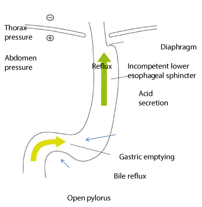
| In conclusion, sleeve gastrectomy has a profound pathophysiological effect on esophageal and gastric function, such as lower esophageal sphincter, gastric acid secretion, gastric motility and gastric emptying. |
| The most important clinical effect is the development of gastroesophageal reflux after sleeve. As has been discussed before, there are several pathological alterations which can explain this adverse effect and can predispose to reflux: |
| a.- disruption of the angle of His. |
| b.- Partial resection of sling fibers and development of an hypotensive lower esophageal sphincter. |
| c.- reduced gastric compliance due to loss of receptive relaxation and receptive accommodation to food. |
| d.- higher intragastric pressure |
| e.- although gastric acid secretion is diminished, there is still some amount of acid produced, much larger than after gastric bypass. |
| f.- neo-fundus and mid-stomach stenosis. |
| On the other hand, there are some authors who postulate that sleeve gastrectomy decreased the incidence of reflux esophagits due to: |
| a.- accelerated gastric emptying |
| b.- loss of weight |
| c.- decrease in acid secretion (but still there is some) |
| d.- increase in lower esophageal sphincter pressure |
| We postulate that sleeve gastrectomy is “pro-reflux” operation (Figure 6). The different physiopathological alterations demonstrate that sleeve gastrectomy may induce an adverse effect on antireflux barrier, and acid exposure of the distal esophagus is mainly determined by LES insufficiency rather than loss of weight. Surgeons dedicated to this operation should be aware of all these pathophysiological changes and of the clinical consequences that may occur. |
References
- Buchwald H, Avidor Y, Braunwald E, Jensen MD, Pories W, et al. (2004) Bariatric surgery: a systematic review and meta-analysis JAMA 292: 1724-1737.
- Fobi MA, Lee H, Holness R, Cabinda D (1998) Gastric bypass operation for obesity. J Surg 22: 925-935.
- Lee SM, Pryor AD (2011) Future directions in bariatric surgery. Surg Clin North Am 91: 1373-1395
- Csendes A, Lembach H (2010) Pathophysiology of bariatric surgery gastric bypass and sleeve gastrectomy. Rev Chil Cir 62:525-531.
- Peterli R, Borbely Y, Kean B, Gass H, Peter J et al.(2013)Early results of the Swiss Multicentre bypass or sleeve gastrectomy: a prospective randomized trial comparing sleeve gastrectomy and Roux-en-Y gastric bypass. Ann Surg 58:690-694.
- Crookes PF (2006) Surgical treatment of morbid obesity. Rev Med 57: 243-264.
- Buchwald H, Oien DM (2013) Metabolic/bariatric surgery worldwide 2011. Obes Surg 23: 427-436.
- Buchwald H, Oien DM (2009) Metabolic/bariatric surgery worldwide 2008. Obes Surg 19: 1605-1611.
- Himpens J, Dobbeleir J, Peeters G (2010) Long-term results of laparoscopic sleeve gastrectomy for obesity. Ann Surg 252: 319-324.
- Braghetto I, Csendes A, Lanzarini E, Papapietro K, Cárcamo C, et al. (2012) Is laparoscopic sleeve gastrectomy an acceptable primary bariatric procedure in obese patients? Early and 5-year postoperative results. Surg LaparoscEndoscPercutan Tech 22: 479-486.
- Csendes A, Burdiles P, Rojas J, Burgos A, Henríquez A (2001) [Pathological gastroesophageal reflux in patients with severe, morbid and hyper obesity]. Rev Med Chil 129: 1038-1043.
- Hampel H, Abraham NS, El-Serag HB (2005) Meta-analysis: obesity and the risk for gastroesophageal reflux disease and its complications. Ann Intern Med 143: 199-211.
- O'Brien TF Jr (1980) Lower esophageal sphincter pressure (LESP) and esophageal function in obese humans. J Clin Gastroenterol 2: 145-148.
- Orlando RC (2001) Overview of the mechanism of gastroesophageal reflux. Ann J Med 111: 1745-1775
- Mion F, Dargent J (2014) Gastro-oesophageal reflux disease and obesity: pathogenesis and response to treatment.Best Pract Res Clin Gastroenterol 28: 611-622.
- Korn O, Stein HJ, Richter TH, Liebermann-Meffert D (1997) Gastroesophageal sphincter: a model. Dis Esophagus 10: 105-109.
- Korn O, Csendes A, Burdiles P, Braghetto I, Stein HJ (2000) Anatomic dilatation of the cardia and competence of the lower esophageal sphincter: a clinical and experimental study. J Gastrointest Surg 4: 398-406.
- Braghetto I, Lanzarini E, Korn O, Valladares H, Molina JC, et al. (2010) Manometric changes of the lower esophageal sphincter after sleeve gastrectomy in obese patients. See comment in PubMed Commons below Obes Surg 20: 357-362.
- Dodds WJ, Dent J, Hogan WJ, Helm JF, Hauser R, et al. (1982) Mechanisms of gastroesophageal reflux in patients with reflux esophagitis. N Engl J Med 307: 1547-1552.
- Mitta RK, Holloway RH, Penagini R, Blackshaw LA, Dent J (1995) Transient lower esophageal sphincter relaxation. Gastroenterology 109: 601-610.
- Dent J, Dodds WJ, Hogan WJ, Toouli J (1988) Factors that influence induction of gastroesophageal reflux in normal human subjects. Dig Dis Sci 33: 270-275.
- Csendes A, Maluenda F, Braghetto I, Csendes P, Henriquez A, et al. (1993) Location of the lower oesophageal sphincter and the squamous columnar mucosal junction in 109 healthy controls and 778 patients with different degrees of endoscopic oesophagitis. Gut 34: 21-27.
- Pandolfino JE, Kim H, Ghosh SK, Clarke JO, Zhang Q, et al. (2007) High-resolution manometry of the EGJ: an analysis of crural diaphragm function in GERD. Am J Gastroenterol 102: 1056-1063.
- Sloan S, Rademaker AW, Kahrilas PJ (1992) Determinants of gastroesophageal junction incompetence: hiatal hernia, lower esophageal sphincter, or both? Ann Intern Med 117: 977-982.
- Hayat JO, Manson S, Wan A, Poulis AP, Yazaki E et al. (2014) Effects of sleeve gastrectomy on gastro-oesophageal reflux and oesophago-gastric motility? Expert Rev GastroenterolHepatol8: 445-52
- Berger E (2013) Electronic health records, promises promises: experts advise patience for return on investment. Ann Emerg Med 62: A19-21.
- Burgerhart JS, Schotborgh CA, Schoon EJ, Smulders JF, van de Meeberg PC, et al. (2014) Effect of sleeve gastrectomy on gastroesophageal reflux. Obes Surg 24: 1436-1441.
- Petersen WV, Meile T, Kuper MA, Zdichoesky KM, Korigsrainer A, Schneider JH (2012) Functional importance of laparoscopic sleeve gastrectomy for the lower esophageal sphincter in patients with morbid obesity. Obes Surg 23:360-366
- Kleidi E, Theodorou D, Albanopoulos K, Menenakos E, Karvelis MA, et al. (2013) The effect of laparoscopic sleeve gastrectomy on the antireflux mechanism: can it be minimized? Surg Endosc 27: 4625-4630.
- Del Genio G, Tolone S, Limongelli P, Brusciano L, D'Alessandro A, et al. (2014) Sleeve gastrectomy and development of "de novo" gastroesophageal reflux. Obes Surg 24: 71-77.
- Rebecchi F, Allaix ME, Giaccone C, Ugliono E, Scozzari G, et al. (2014) Gastroesophageal reflux disease and laparoscopic sleeve gastrectomy: a physiopathologic evaluation. Ann Surg 260: 909-914.
- Sharma A, Aggarwal S, Ahuja V, Bal C (2014) Evaluation of gastroesophageal reflux before and after sleeve gastrectomy using symptom scoring, scintigraphy, and endoscopy. ObesRelat Dis 10: 600-605.
- Nahata M, Muto S, Oridate N, Ohnishi S, Nakagawa K, et al. (2012) Impaired ghrelin signaling is associated with gastrointestinal dysmotility in rats with gastroesophageal reflux disease. Am J PhysiolGastrointest Liver Physiol 303: G42-53.
- Karamanakos SN, Vagenas K, Kalfarentzos F, Alexandrides TK. (2008) Weight loss, appetite suppression, and changes in fasting and postprandial ghrelin and peptide-YY levels after Roux-en-Y gastric bypass and sleeve gastrectomy: a prospective, double blind study. Ann Surg 247:401-7.
- Hayat JO, Wan A (2014) The effects of sleeve gastectomy on gastro-esophageal reflux and gastro-esophageal motility. Expert Rev GastroenterolHepatol 8: 445-452.
- Laffin M, Chau J, Gill RS, Birch DW, Karmali S (2013) Sleeve gastrectomy and gastroesophageal reflux disease. J Obes 2013: 741097.
- Yehoshua RT, Eidelman LA, Stein M, Fichman S, Mazor A, et al. (2008) Laparoscopic sleeve gastrectomy--volume and pressure assessment. Obes Surg 18: 1083-1088.
- Best Pract Res Clin Gastroenterol 28: 953-965.
- Joseph IM, Zavros Y, Merchant JL, Kirschner D (2003) A model for integrative study of human gastric acid secretion. J ApplPhysiol (1985) 94: 1602-1618.
- Mix H, Widjaja A, Jandl O, Cornberg M, Kaul A, et al. (2000) Expression of leptin and leptin receptor isoforms in the human stomach. Gut 47: 481-486.
- Hirsch AB, McCuen RW, Arimura A, Schubert ML (2003) Adrenomedullin stimulates somatostatin and thus inhibits histamine and acid secretion in the fundus of the stomach. RegulPept 110: 189-195.
- Gower WR Jr, Premaratne S, McCuen RW, Arimura A, McAfee Q, et al. (2003) Gastric atrial natriuretic peptide regulates endocrine secretion in antrum and fundus of human and rat stomach. Am J PhysiolGastrointest Liver Physiol 284: G638-645.
- Prinz C, Kajimura M, Scott DR, Mercier F, Helander HF, Sachs G(1993) Histamine secretion from rat enterochromaffin like cells. Gastroenterology105:449-461.
- Chen D, Zhao CM, Lindström E, Håkanson R (1999) Rat stomach ECL cells up-date of biology and physiology. Gen Pharmacol 32: 413-422.
- Zaki M, Koduru S, McCuen R, Vuyyuru L, Schubert ML (2002) Amylin, released from the gastric fundus, stimulates somatostatin and thus inhibits histamine and acid secretion in mice. Gastroenterology 123: 247-255.
- Larsson LI, Goltermann N, de Magistris L, Rehfeld JF, Schwartz TW (1979) Somatostatin cell processes as pathways for paracrine secretion. Science 205: 1393-1395.
- Asakawa A, Inui A, Kaga T, Yuzuriha H, Nagata T, et al. (2001) Ghrelin is an appetite-stimulatory signal from stomach with structural resemblance to motilin. Gastroenterology 120: 337-345.
- Zhao CM, Furnes MW, Stenström B, Kulseng B, Chen D (2008) Characterization of obestatin- and ghrelin-producing cells in the gastrointestinal tract and pancreas of rats: an immunohistochemical and electron-microscopic study. Cell Tissue Res 331: 575-587.
- Kopic S, Geibel JP (2010) Update on the mechanisms of gastric acid secretion. CurrGastroenterol Rep 12: 458-464.
- Ghosh T, Lewis DI, Axon AT, Everett SM (2011) Review article: methods of measuring gastric acid secretion. Aliment PharmacolTher 33: 768-781.
- Walderm HL, House O, Fossmark R (2014) the regulation of gastric acid secretion clinical prospective. ActaPhysiol210:239-256
- Schubert ML, Shamburek R (1990) Control of acid secretion. Gastroenterol Clin. North Am 19:1-25.
- Schubert ML, Edwards NF, Arimura A, Makhlouf GM (1987) Paracrine regulation of gastric acid secretion by fundicsomatostatin. Am J Physiol 252: G485-490.
- Csendes A, Orrego H, Heitmann P (1968) [Characteristics of gastric secretion in patients with duodenal ulcer]. Rev Med Chil 96: 788-794.
- Csendes A, Larrain A, Uribe P (1974) Gastric acid secretion in patients with a symptomatic gastroesophageal reflux and patients with esophageal strictures. Ann Surg 179: 119-122.
- Smith CD, Herkes SB, Behrns KE, Fairbanks VF, Kelly KA, et al. (1993) Gastric acid secretion and vitamin B12 absorption after vertical Roux-en-Y gastric bypass for morbid obesity. Ann Surg 218: 91-96.
- Behrns KE, Smith CD, Sarr MG (1994) Prospective evaluation of gastric acid secretion and cobalamin absorption following gastric bypass for clinically severe obesity. Dig Dis Sci 39: 315-320.
- Attwood SE, Smyrk TC, DeMeester TR, Mirvish SS, Stein HJ, et al. (1992) Duodenoesophageal reflux and the development of esophageal adenocarcinoma in rats. Surgery 111: 503-510.
- Stein HJ, Kauer WK, Feussner H, Siewert JR (1998) Bile reflux in benign and malignant Barrett's esophagus: effect of medical acid suppression and nissen fundoplication. J Gastrointest Surg 2: 333-341.
- Grong E, Arbo IB, Thu OK, Kuhry E, Kulseng B, et al. (2015) The effect of duodenojejunostomy and sleeve gastrectomy on type 2 diabetes mellitus and gastrin secretion in Goto-Kakizaki rats. Surg Endosc 29: 723-733.
- Sillakivi T, Suumann J, Kirsimägi U, Peetsalu A (2013) Plasma levels of gastric biomarkers in patients after bariatric surgery: biomarkers after bariatric surgery. Hepatogastroenterology 60: 2129-2132.
- Rostas JW, Mai TT, Richards WO (2011) Gastric motility physiology and surgical intervention. See comment in PubMed Commons below Surg Clin North Am 91: 983-999.
- Janssen P, VandenBerghe P, Verschueren S, Lehmann A, Depoortere I, et al. (2011) Review article: the role of gastric motility in the control of food intake. Aliment PharmacolTher 33: 880-894.
- Deloose E, Vos R, Corsetti M, Depoortere I, Tack J (2015) Endogenous motilin, but not ghrelin plasma levels fluctuate in accordance with gastric phase III activity of the migrating motor complex in man. NeurogastroenterolMotil 27: 63-71.
- Tack J (2007) Gastric motor disorders. Best Pract Res Clin Gastroenterol 21: 633-644.
- Rees WD, Malagelada JR, Miller LJ, Go VL (1982) Human interdigestive and postprandial gastrointestinal motor and gastrointestinal hormone patterns. Dig Dis Sci 27: 321-329.
- Abrahamsson H, Jansson G (1969) Elicitation of reflex vagal relaxation of the stomach from pharynx and esophagus in the cat. ActaPhysiolScand 77: 172-178.
- Takahashi T, Owyang C (1997) Characterization of vagal pathways mediating gastric accommodation reflex in rats. J Physiol504 : 479-488.
- Csendes A, Amdrup E, Parada M (1979) A peroperative technique for determining the extent of gastrectomy. Surg GynecolObstet 149: 81-83.
- Csendes A, Burgos AM (2005) Size, volume and weight of the stomach in patients with morbid obesity compared to controls. Obes Surg 15: 1133-1136.
- Lopetegui G (1965) Anatomic studies of gastric anturm applied to surgical treatment of duodenal ulcer. Arch Cir Chile17:22-26.
- Braghetto I, Lazo M, Leiva V, Rebolledo P, Gallardo I, et al. (1987) A prospective study of intraoperative histologic antrum and corpus boundary in patients undergoing highly selective vagotomy for duodenal ulcer. See comment in PubMed Commons below Surg GynecolObstet 164: 213-218.
- Baumann T, Keusters S, Gnevebergar J, Marjanoric G, Zimmermann L, et al. (2011) Timeresolved MRI after ingestion of liquids reveal motility changes after laparoscopic sleeve gastrectomy. Preliminary Results. Obes Surg 21:95-101.
- Braghetto I, Davanzo C, Korn O, Csendes A, Valladares H, et al. (2009) Scintigraphic evaluation of gastric emptying in obese patients submitted to sleeve gastrectomy compared to normal subjects. Obes Surg 19: 1515-1521.
- Patrick A, Epstein O (2008) Review article: gastroparesis. Aliment PharmacolTher 27: 724-740.
- Melissas J, Koukouraki S, Askoxylakis J, Stathaki M, Daskalakis M, et al. (2007) Sleeve gastrectomy: a restrictive procedure? Obes Surg 17: 57-62.
- Melissas J, Daskalakis M, Koukouraki S, Askoxylakis I, Metaxari M, et al. (2008) Sleeve gastrectomy-a "food limiting" operation. Obes Surg 18: 1251-1256.
- Bernstine H, Yehousha T, Groshar D, Beglaibter N, Shikora S, et al.(2009) Gastric emptying is not affected by sleeve gastrectomy--scintigraphic evaluation of gastric emptying after sleeve gastrectomy without removal of the gastric antrum.Obes Surg19:293-298
- Pilone V, Tramontano S, Di Micco R, Monda A, Hasani A, et al. (2013) Gastric emptying after sleeve gastrectomy: statistical evidence of a controlled prospective study with gastric scintigraphy. Minerva Chir 68: 385-392.
- Burgerhart JS, van Rutte PW, Edelbroek MA, Wyndaele DN, Smulders JF, et al. (2015) Association between postprandial symptoms and gastric emptying after sleeve gastrectomy. Obes Surg 25: 209-214.
- Michalsky D, Dvorak P, Belacek J, Kasalicky M (2013) Radical resection of the pyloric antrum and its effect on gastric emptying after sleeve gastrectomy. 23: 567-573.
- Melissas J, Leventi A, Klinaki I, Perisinakis K, Koukouraki S, et al. (2013) Alterations of global gastrointestinal motility after sleeve gastrectomy: a prospective study. Ann Surg 258: 976-982.
- Shah S, Shah P, Todkar J, Gagner M, Sonar S, et al. (2010) Prospective controlled study of effect of laparoscopic sleeve gastrectomy on small bowel transit time and gastric emptying half-time in morbidly obese patients with type 2 diabetes mellitus. Surg ObesRelat Dis 6: 152-157.
Tables and Figures at a glance
| Table 1 | Table 2 | Table 3 | Table 4 |
| Table 5 | Table 6 | Table 7 | Table 8 |
| Table 9 | Table 10 | Table 11 | Table 12 |
Figures at a glance
 |
 |
 |
| Figure 1 | Figure 2 | Figure 3 |
 |
 |
 |
| Figure 4 | Figure 5 | Figure 6 |
Relevant Topics
- Android Obesity
- Anti Obesity Medication
- Bariatric Surgery
- Best Ways to Lose Weight
- Body Mass Index (BMI)
- Child Obesity Statistics
- Comorbidities of Obesity
- Diabetes and Obesity
- Diabetic Diet
- Diet
- Etiology of Obesity
- Exogenous Obesity
- Fat Burning Foods
- Gastric By-pass Surgery
- Genetics of Obesity
- Global Obesity Statistics
- Gynoid Obesity
- Junk Food and Childhood Obesity
- Obesity
- Obesity and Cancer
- Obesity and Nutrition
- Obesity and Sleep Apnea
- Obesity Complications
- Obesity in Pregnancy
- Obesity in United States
- Visceral Obesity
- Weight Loss
- Weight Loss Clinics
- Weight Loss Supplements
- Weight Management Programs
Recommended Journals
Article Tools
Article Usage
- Total views: 15272
- [From(publication date):
February-2016 - Apr 03, 2025] - Breakdown by view type
- HTML page views : 14114
- PDF downloads : 1158
