Research Article Open Access
Cervical Kinematics of Fast Neck Motion across Age
Hilla Sarig Bahat1*, Mahmoud Igbariya1, June Quek2 and Julia Treleaven21Department of Physical Therapy, University of Haifa, Haifa 31905, Israel
2CCRE Spine, The University of Queensland, Brisbane 4072, Australia
- Corresponding Author:
- Hilla Sarig-Bahat
Faculty of Social Welfare and Health Sciences
Physical Therapy Department, University of Haifa
Mount Carmel, Haifa, 31905 Israel
Tel: +972-545380483
Fax: +972-4-8288140
E-mail: hbahat@research.haifa.ac.il
Received Date: September 03, 2016; Accepted Date: September 27, 2016; Published Date: October 05, 2016
Citation: Sarig Bahat H, Igbariya M, Quek J, Treleaven J (2016) Cervical Kinematics of Fast Neck Motion across Age. J Nov Physiother 6:306. doi: 10.4172/2165-7025.1000306
Copyright: © 2016 Sarig Bahat H, et al. This is an open-access article distributed under the terms of the Creative Commons Attribution License, which permits unrestricted use, distribution, and reproduction in any medium, provided the original author and source are credited.
Visit for more related articles at Journal of Novel Physiotherapies
Abstract
Purpose: To evaluate the velocity, smoothness, and symmetry of fast cervical motion in asymptomatic individuals of four age groups, and to investigate the relationship between age and these measures. Methods: Cross-sectional study with fifty-eight asymptomatic volunteers (28 F, 30 M), aged 18-80 (mean age=44.14 ± 17.35) were assessed using a customized Virtual Reality (VR) system. VR head-mounted display stimulated fast neck movements in response to virtual targets appearing randomly. Outcome measures were neck motion velocity, smoothness, and symmetry of velocity profile. Main findings: The eldest group differed from younger age groups in peak and mean velocity of neck motion. Smoothness demonstrated age-group difference only between the youngest and oldest groups. Linear regression analysis showed significant negative correlations in mean and peak velocity, and in smoothness with age, excluding smoothness in right rotation. No age-group differences and no significant correlations were found for time-to-peakvelocity in all directions of neck movement (p>0.05). Conclusion: This study showed that age influenced the velocity in which asymptomatic individuals could move their neck, specifically in elders over 60 years of age. Clinically, this may suggest that when elders with neck pain present with slower cervical motion, it is probably partly due to aging and thus should be taken into account in the management and expected level of performance.
Keywords
Neck; Motion analysis; Velocity; Range of motion
Introduction
Neck pain is a worrisome health disorder in Western society with estimates of an annual prevalence ranging from 30% to 50% in the general population [1]. It is more prevalent among women than men and the risk seems to increase with age [1,2]. In a large survey by Cassou et al. the 5 year prevalence and frequency of neck pain in French workers increased with age, and the disappearance/recovery of neck pain reduced with age [3]. This emphasizes the need to investigate agerelated changes in parameters of neck function, such as the dynamics of neck movements.
Patients with neck pain have demonstrated kinematic impairments, such as reduced movement range [4,5], accuracy, velocity, smoothness and stability of neck motion [6-9]. These identified impairments can affect functional ability in tasks that require dynamic control which is an important functional requirement [10,11]. Due to the increased incidence of neck pain at an older age, it is unclear whether functional neck motion deteriorates with age and results in pain, or that the presence of neck pain causes these impairments with no relation to age. To answer this, we need to explore the changes in neck motion in asymptomatic individuals of different ages and later compare them to symptomatic individuals.
A recent study examined the reliability of cervical motion kinematics using the Neck Virtual Reality (VR) system in asymptomatic individuals (N=46), aged 23-49. Best reliability was found for peak velocity (ICC=0.93), followed by mean velocity (ICC=0.84), and motion smoothness (ICC=0.78) [12]. These kinematic measures were also found to be sensitive [13,14], with higher values than those previously reported for range of motion (ROM) parameters [7,15]. These findings may imply that velocity of neck motion may have a high clinical value in the assessment of neck motion. Since neck pain affects people across the lifespan and risk increases with age, it would seem important to understand any age related changes in these measures. However, amongst these neck pain-associated impairments, it seems that age related changes have only been studied in ROM.
Several investigators studied cervical ROM in healthy participants and showed it decreased significantly with age [16-20]. The largest of and widest age range sample to have shown age effect on ROM was by Youdas et al. who assessed 337 healthy subjects (171 females and 166 males) aged 11 to 97 [19]. Similarly, Lind et al. (N=70) and Malmström et al. (N=120) showed that age affected ROM in healthy adults up to 79 years of age [17,20]. Additional studies shared similar findings in adults (N=120-220) up to their 60s, showing that age had a significant effect on the range of all primary movements, and less effect on the range of coupled movements [18,21]. In contrast, some previous studies reported that age did not significantly influence all cervical mobility, arguing that upper cervical rotation is not affected by age while lower cervical mobility [16,22,23].
It is yet unknown whether and how age effects various aspects of cervical kinematics. Therefore, it is essential to provide normative data of these measures in view of their changes with age so that appropriate comparisons to those with neck pain can be made across the life span. Thus, the objectives of this study were to evaluate fast cervical motion spine velocity kinematics: velocity, smoothness and acceleration-deceleration ratio in different age groups in asymptomatic individuals to investigate the relationship between age and these kinematic measures.
Methods
This study was designed as a cross sectional study in asymptomatic individuals, evaluating neck movement velocity profile using a customized neck VR system.
A convenience sample of asymptomatic individuals was recruited from the general population. Participants were recruited and divided into the following age groups: 18-29, 30-44, 45-60, and 61-80. Inclusion criteria included age over 18 years, and ability to understand the tasks in the VR assessment as evaluated in the introduction session, prior to data collection.
Exclusion criteria included neck pain; spinal fracture/dislocation; visual pathology not corrected with glasses; systemic diseases such as neurological, cardiovascular, respiratory, or any disorders that can affect physical performance; history of traumatic head injury or spinal surgery; inability to provide informed consent. Each participant signed an informed consent form prior to testing. The ethical committees of the University of Haifa, and the University of Queensland approved the study.
Instrumentation
The neck VR software was the same as used in previous studies [5,24]. The virtual scenario includes a virtual pilot flying an airplane controlled by the patient’s head motion which interacts with targets appearing randomly from four directions, to elicit Flexion, Extension, Right rotation (RR), Left rotation (LR) (Figure 1). In the beginning of the VR session, the participant was seated on a rigid char and trunk was secured with a seat belt to minimize movement other to the neck. The participant then was requested to position his/her head in neutral/ mid-position, and the tracker as zero recorded this position. The VR task was to move the head to hit the target as fast as possible, as targets disappeared within 5 seconds. Sixteen targets were displayed randomly, four in each direction.
Figure 1: Screen capture from the neck virtual reality scenario displayed in the head mounted display during the kinematic assessment. In the neck VR interaction the participant controlled the airplane by head motion, and interacted with the yellow targets appearing from various directions. A yellow target appears in a random direction, and the participant is required to move the head in that direction within seven seconds before the target disappears. Target’s life time is visualized using a green circle around the target that diminishes gradually and functions as a timer. This feature aims to motivate the participant to move quickly towards the target before it disappears.
This study used a new Head mounted display (HMD) hardware- the Oculus Rift, development kit 1 (http://www.oculusvr.com). It consists of a 5-inch organic light-emitting diode display screen with a resolution of 960×1080 pixels and a 100-degree field of view that displays two images side by side. The Oculus has embedded sensors that monitor the wearer’s head motions and adjust the image accordingly, including accelometers, gyroscopes and magnetometers. This custom tracking technology was chosen to upgrade the neck VR system performance as it provides low latency, 360° head tracking of 30 milliseconds lag time. Head tracking data output was analysed to produce the desired kinematic outcome measures.
Outcome measures
Outcome measures were collected during fast neck motion stimulated by the VR assessment during 16 trials, four in each direction- flexion, extension, RR, and LR. Results were calculated as the mean value of the three best results from each direction out of four trials performed in each direction. The following measures were previously reported for their reliability, sensitivity and specificity [13].
1. Peak velocity (Vpeak, °/sec) was calculated as the maximal angular velocity, from motion initiation to target hit.
2. Mean velocity (Vmean, °/sec) was calculated as the mean angular velocity angular velocity, from motion initiation to target hit.
3. Time to peak velocity percentage (TTP %) was the time from motion initiation to peak velocity moment, as a percentage of total movement time, representing the ratio between the acceleration to deceleration phase in the velocity profile.
4. Number of velocity peaks (NVP) was counted from motion initiation to target hit, and represented motion smoothness.
Procedure
The session began with an interview concerning related exclusion criteria. Cervical VR assessments were carried out in the sitting position, with the trunk secured to a chair by a seatbelt and feet resting on the ground. A short warm-up and introduction of the VR interactivity was conducted at the beginning of each assessment to familiarize the participant with the VR and minimize a training effect. Once the participant showed control of the interaction in the VR scenario, the assessment commenced. Participants were assessed by a qualified physical therapist. Each assessment took approximately 15 minutes in total. Due to the chance of experiencing simulator sickness, the participants were asked to report any negative effects, and could ask to stop the assessment at any time.
Statistical analysis
Gender distribution by age groups was assessed using chi square analysis to assure gender was not an interfering factor. The dependence of each of the parameters on age was assessed by two methods: The first employed Analyses of Variance (ANOVAs) comparing subjects in different age groups, followed by Tukey tests in the case of significant overall differences. The second method employed linear regression analyses of each parameter against age, after preliminary examination to detect possible non-linear effects.
Results
Fifty-eight asymptomatic volunteers (28 females and 30 males), aged 18 to 80 with a mean age of 44.14 years participated in this study. The characteristics of each age group are presented in Table 1. Gender was equal across age groups as shown by the chi square analysis, with likelihood ratio of 0.19. There was one drop out from the study due to simulator sickness, a 26 year-old male.
| Age group | N | Mean Age (SD) | Range (min-max) | Gender (F,M) |
|---|---|---|---|---|
| 18-30 | 16 | 23.32 (3.42) | 18-30 | 6,10 |
| 31-45 | 15 | 38.20 (3.93) | 31-44 | 7,8 |
| 46-60 | 15 | 51.67 (4.15) | 46-60 | 6,9 |
| 61-80 | 12 | 69.92 (4.58) | 62-78 | 9,3 |
| Total | 58 | 44.14 (17.35) | 18-78 | 28,30 |
Table 1: Characteristics of age groups.
Table 2 shows the cervical velocity kinematic means and standard deviations of each outcome measure by age groups. One-Way ANOVA results showed significant differences in all the kinematic measures amongst the four age groups (P<0.05) except for TTP in all directions (TTPP was the only measure that showed no age differences).
| Movement Direction |
Kinematic measure |
Age Group | ||||||||
|---|---|---|---|---|---|---|---|---|---|---|
| 18-30 | 31-45 | 46-60 | 61-80 | |||||||
| Mean | SD | Mean | SD | Mean | SD | Mean | SD | Power | ||
| Flexion | Vpeak (º/sec) | #137.99 | 32.5 | #155.24 | 47.0 | #139.85 | 43.1 | **87.26 | 26. 7 | 0.98 |
| Vmean (º/sec) | 76.28 | 24.8 | #82.96 | 32.5 | 70.17 | 25.2 | *53.33 | 18. 3 | 0.67 | |
| NVP | #0.96 | 0.4 | 1.08 | 0.5 | 1.04 | 0.5 | *0.56 | 0. 5 | 0.68 | |
| TTPP (%) | 37.34 | 17.3 | 43.53 | 17.0 | 34.59 | 13.5 | 42.70 | 21.2 | 0.23 | |
| Extension | Vpeak (º/sec) | #143.11 | 46.0 | #151.67 | 51.5 | #140.41 | 44.0 | **79.11 | 18.2 | 0.97 |
| Vmean (º/sec) | #86.26 | 25.2 | #86.93 | 27.0 | #81.11 | 30.2 | **51.43 | 12.5 | 0.89 | |
| NVP | #1.29 | 0.6 | 1.11 | 0.7 | #1.58 | 0.5 | **0.56 | 0.5 | 0.96 | |
| TTPP (%) | 34.53 | 11.9 | 42.59 | 16.4 | 34.72 | 15.5 | 46.95 | 13.9 | 0.55 | |
| Right Rotation | Vpeak (º/sec) | #193.53 | 63.8 | #204.37 | 72.7 | 162.81 | 56.4 | **115.19 | 23.5 | 0.93 |
| Vmean (º/sec) | #112.61 | 38.3 | #121.44 | 39.9 | 95.11 | 36.0 | **73.48 | 15.7 | 0.87 | |
| NVP | #0.96 | 0.4 | 0.78 | 0.4 | 1 | 0.5 | *0.58 | 0.3 | 0.75 | |
| TTPP (%) | 47.79 | 18.3 | 50.88 | 18.2 | 45.26 | 21.4 | 52.87 | 20.6 | 0.13 | |
| Left Rotation | Vpeak (º/sec) | #193.13 | 57.1 | #194.72 | 60.5 | 165.15 | 54.3 | **120.27 | 36.5 | 0.90 |
| Vmean (º/sec) | #111.98 | 33. 9 | #113.84 | 41.6 | 85.60 | 31.0 | **71.03 | 25.5 | 0.90 | |
| NVP | #1.04 | 0.5 | 0.98 | 0.4 | 0.98 | 0.5 | *0.50 | 0.4 | 0.80 | |
| TTPP (%) | 48.58 | 17.9 | 47.37 | 20.8 | 37.43 | 23.0 | 52.40 | 23.1 | 0.35 | |
Table 2: Kinematic measures collected during fast neck motion in the VR assessment results by age groups.
To examine the source of the significance, Tukey Post Hoc analysis was conducted, and significant differences were found between the oldest age group (61-80) to the three other groups (18-30, 31-45, 46-60) in various combinations (Table 2) but not in between the three younger age groups (p<0.05). The eldest group differed from all other groups in peak velocity towards flexion and extension, and in mean velocity in extension. The eldest also differed from the two young groups in mean and peak velocity of rotational neck motion to the right and to the left. Smoothness, as measured by NVP, demonstrated age group difference only between the youngest and oldest groups.
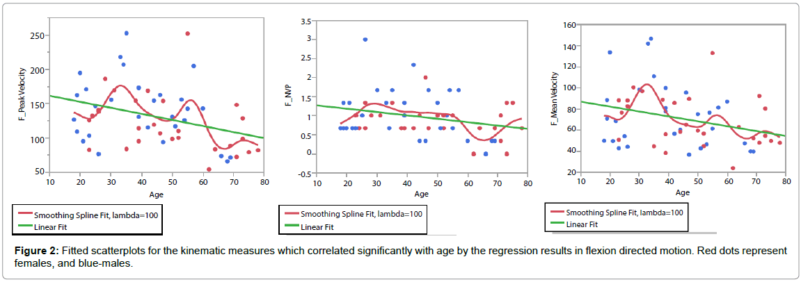
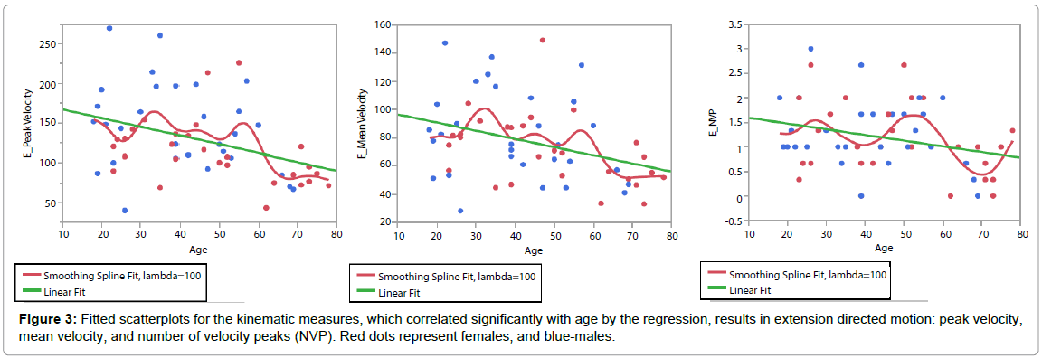
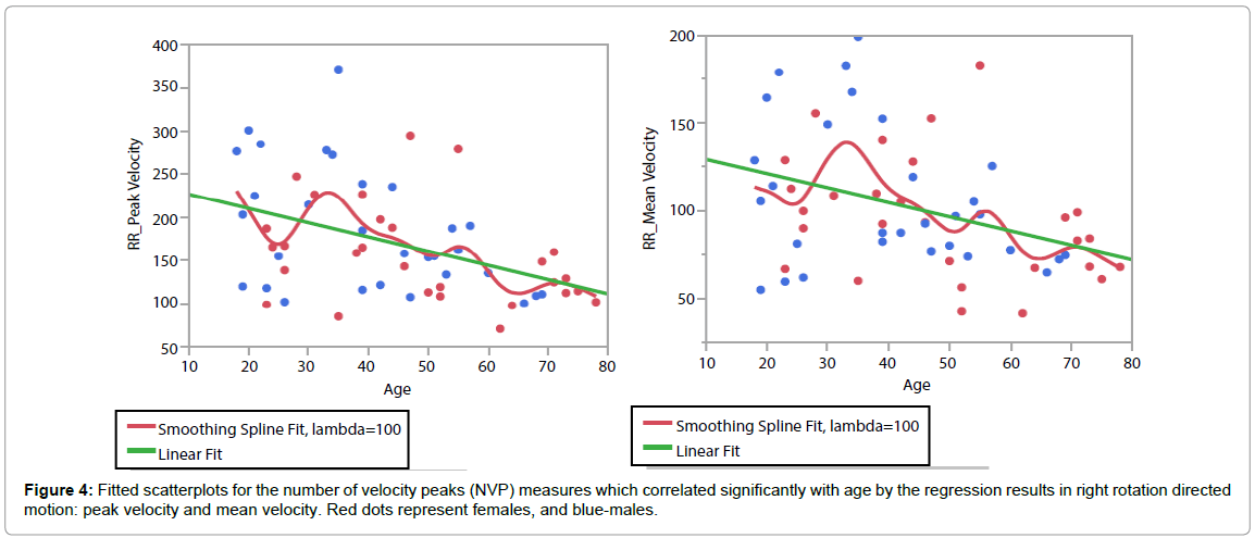
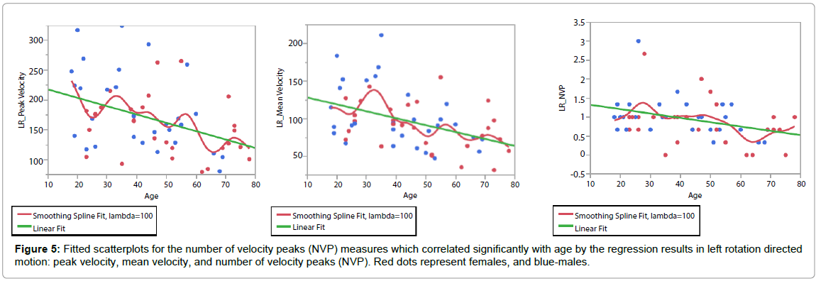
Linear regression analysis of each parameter against age showed significant positive correlations in mean and peak velocity, and in NVP measures with age, excluding NVP in right rotation, as shown in Table 3. No significant correlations were found between TTPP with age in all directions of neck movement (p>0.05). Fitted scatterplots are presented in Figures 2-5 for the variables found significantly related to age.
| Movement direction | Kinematic measure | Regression equation | F df=1,44 | r |
|---|---|---|---|---|
| Flexion | Vpeak (º/sec) | 170.31-0.88*Age | 7.27** | -0.34 |
| Vmean (º/sec) | 91.59-0.46*Age | 5.24* | -0.29 | |
| NVP | 1.36-0.01*Age | 4.64* | -0.28 | |
| TTPP (%) | NS | NS | NS | |
| Extension | Vpeak (º/sec) | 178.01-1.10*Age | 9.08* | -0.37 |
| Vmean (º/sec) | 102.45-0.58*Age | 7.84** | -0.35 | |
| NVP | 1.71-0.012*Age | 4.74* | -0.28 | |
| TTPP (%) | 31.19+0.19*Age | 2.72* | -0.22 | |
| Right rotation | Vpeak (º/sec) | 243.22-1.65*Age | 12.83** | -0.43 |
| Vmean (º/sec) | 137.28-0.81*Age | 8.85** | -0.37 | |
| NVP | NS | NS | NS | |
| TTPP (%) | NS | NS | NS | |
| Left rotation | Vpeak (º/sec) | 231.42-1.40*Age | 11.0** | -0.41 |
| Vmean (º/sec) | 137.38-0.92*Age | 12.64** | -0.43 | |
| NVP | 1.43-0.01*Age | 8.08** | -0.36 | |
| TTPP (%) | NS | NS | NS |
Table 3: Regression analysis results: Listed below are the fast neck motion kinematic measures that were found to be significantly related to age.
Time to peak velocity was the only measure which consistently did not show age group differences nor related to age by both the ANOVA and regression analyses. Similar results were demonstrated by the two methods of analysis, with the regression showing the correlation, while the ANOVA localized where the changes occurred between the age groups.
Discussion
This study showed that age influenced the velocity in which asymptomatic individuals could move their neck, specifically in elders over 60 years of age. A significantly slower motion was demonstrated in all movement directions in the elderly group when compared to the younger asymptomatic individuals. Velocity of motion showed the most powerful age-group differences, followed by smoothness of fast neck motion (represented by NVP), unlike the symmetry of velocity profile (represented by TTP) which did not demonstrate any agegroups differences or association to age.
The strong association with age found for speed of movement (mean and peak velocity), and smoothness (NVP) rather than symmetry of velocity profile (TTP) strengthens the value of these fast neck motion measures. Previous studies supported the reliability and sensitivity of cervical motion velocity and have highlighted velocity as the most valuable diagnostic factor [12,13]. As such, it seems that fast neck motion measures should be applied in clinical assessment and therapeutic methods addressing this factor should be investigated for clinical management. However, the results of the current study suggest that age related decline should be accounted for when assessing this in elders who have neck pain.
These findings seem to be in line with some other age related changes related to the cervical spine as well as various aspects of physiological, biological and anatomical aging processes. Such age related changes may include degenerative changes in the intervertebral discs, the facet joints, and calcifications that may be asymptomatic. Consistent evidence has shown that cervical ROM reduces with age, mostly showing a gradual reduction each decade [16-20]. This study did not evaluate range of motion, which limits the direct comparison to existing ROM studies. However, the current findings demonstrated a decline in fast neck motion specifically in elders and seem to be complimentary to the previous ROM findings. The results are also similar to those investigating age related changes in some but not other impairments associated with neck pain [25-27]. For example, increased joint repositioning error, reflecting reduced cervical proprioceptive ability was found in older subjects (mean age=68 years) as compared to younger ones (mean age=23 years) [28] and also across different ages (20-93 years) [29]. In contrast, one recent study showed no relationship between age and proprioception [25,30] and another found no relationship between age and neuromotor control of the cervical flexor muscles tested using the cranio-cervical flexion test (CCFT) in asymptomatic adults [31].
Nevertheless, increasing age is accompanied with a decline in balance, probably due to normative changes in vestibular, visual and neuromuscular function [32]. Accordingly, greater disturbances in healthy elders when compared to younger individuals have been seen in postural control and gait [33,34]. In addition, aging has been associated with increased muscle fatigue that could potentially alter kinematics of reactive postural control movements [35]. These features of altered fast neck motion, proprioception, muscle function and balance have all been identified as associated impairments in neck pain [6-8,36,37] primarily in younger to middle aged groups with both idiopathic and whiplash associated neck pain [38-41].
A few limitations were identified in this study. Neck movement as assessed using the VR system did not allow measurements of side flexion motion. This compromise appears to be minor as side flexion seems a less functional neck movement, but rather more an associated movement.
This current study was limited to asymptomatic individuals and further research is now needed in symptomatic individuals in matching ages, for comparison.
As neck pain’s prevalence peaks in middle age [1], it would be beneficial to compare mid-age patients with neck pain to both younger and older patients. Interestingly, other studies that have compared cervical impairments in elders with and without neck pain, found that elders with neck pain had greater sensorimotor impairments [42,43] suggesting that the neck pain causes changes above and beyond the normal ageing process. Future research is now required to compare fast cervical motion in elders with and without neck pain.
Until then, the clinical implication of the presented results is that when an individual over 60 years of age is presenting with slow cervical motion, it is probably partly due to aging and possibly presence of neck pain could worsen his performance. This age-related change should be taken into account in the management and expected level of neck motion performance.
Conflict of Interest
None
Funding
No funding was provided for the current research
Acknowledgements
We would like to thank Dr. Elliot Sprecher for his highly professional statistical advice and support.
References
- Hogg-Johnson S, van der Velde G, Carroll LJ, Holm LW, Cassidy JD, et al. (2009) The burden and determinants of neck pain in the general population: results of the Bone and Joint Decade 2000–2010 Task Force on Neck Pain and Its Associated Disorders. J Manipulative PhysiolTher32:S46-S60.
- Bot SD, van der Waal JM, Terwee CB, van der Windt DA, Schellevis FG, et al. (2005) Incidence and prevalence of complaints of the neck and upper extremity in general practice. Ann Rheum Dis 64: 118-123.
- Cassou B, Derriennic F, Monfort C, Norton J, Touranchet A (2002) Chronic neck and shoulder pain, age, and working conditions: longitudinal results from a large random sample in France. Occup Environ Med 59: 537-544.
- Rudolfsson T, Björklund M, Djupsjöbacka M (2012) Range of motion in the upper and lower cervical spine in people with chronic neck pain. Man Ther 17: 53-59.
- SarigBahat H, Takasaki H, Chen X, Bet-Or Y, Treleaven J (2015) Cervical kinematic training with and without interactive VR training for chronic neck pain–a randomized clinical trial. Manual therapy20:68-78.
- Sjölander P, Michaelson P, Jaric S, Djupsjöbacka M (2008) Sensorimotor disturbances in chronic neck pain--range of motion, peak velocity, smoothness of movement, and repositioning acuity. Man Ther 13: 122-131.
- Röijezon U, Djupsjöbacka M, Björklund M, Häger-Ross C, Grip H, et al. (2010) Kinematics of fast cervical rotations in persons with chronic neck pain: a cross-sectional and reliability study. BMC MusculoskeletDisord 11: 222.
- Bahat HS, Weiss PL, Laufer Y (2010)The effect of neck pain on cervical kinematics, as assessed in a virtual environment. Arch Phys Med Rehabil91:1884-1890.
- Woodhouse A, Vasseljen O (2008) Altered motor control patterns in whiplash and chronic neck pain. BMC MusculoskeletDisord 9: 90.
- Röijezon U, Björklund M, Bergenheim M, Djupsjöbacka M (2008) A novel method for neck coordination exercise--a pilot study on persons with chronic non-specific neck pain. J NeuroengRehabil 5: 36.
- Pereira MJ, Jull GA, Treleaven JM (2008)Self-reported driving habits in subjects with persistent whiplash-associated disorder: relationship to sensorimotor and psychologic features. Arch Phys Med Rehabil89:1097-1102.
- Bahat HS, Sprecher E, Sela I, Treleaven J (2016) Neck motion kinematics: an inter-tester reliability study using an interactive neck VR assessment in asymptomatic individuals. European Spine Journal 25: 2139-2148.
- Bahat HS, Chen X, Reznik D, Kodesh E, Treleaven J (2014) Interactive cervical motion kinematics: Sensitivity, specificity and clinically significant values for identifying kinematic impairments in patients with chronic neck pain. Manual therapy 20: 295-302.
- Bahat HS, Weiss PL, Sprecher E, Krasovsky A, Laufer Y (2014) Do neck kinematics correlate with pain intensity, neck disability or with fear of motion? Man Ther 19: 252-258.
- Bahat HS, Weiss PL, Laufer Y (2010) Neck pain assessment in a virtual environment. Spine (Phila Pa 1976) 35: E105-112.
- Dvorak J, Antinnes JA, Panjabi M, Loustalot D, Bonomo M (1992) Age and gender related normal motion of the cervical spine. Spine (Phila Pa 1976) 17: S393-S398.
- Lind B, Sihlbom H, Nordwall A, Malchau H (1989) Normal range of motion of the cervical spine. Arch Phys Med Rehabil 70: 692-695.
- Trott P, Pearcy M, Ruston S, Fulton I, Brien C (1996) Three-dimensional analysis of active cervical motion: the effect of age and gender. Clinical Biomechanics11:201-206.
- Youdas JW, Garrett TR, Suman VJ, Bogard CL, Hallman HO, et al. (1992) Normal range of motion of the cervical spine: an initial goniometric study. Physical Therapy72:770-780.
- Malmström EM, Karlberg M, Fransson PA, Melander A, Magnusson M (2006) Primary and coupled cervical movements: the effect of age, gender, and body mass index. A 3-dimensional movement analysis of a population without symptoms of neck disorders. Spine (Phila Pa 1976)31:E44-E50.
- Salo PK, Häkkinen AH, Kautiainen H, Ylinen JJ (2009) Quantifying the effect of age on passive range of motion of the cervical spine in healthy working-age women. J Orthop Sports PhysTher 39: 478-483.
- Walmsley RP, Kimber P, Culham E (1996) The effect of initial head position on active cervical axial rotation range of motion in two age populations. Spine (Phila Pa 1976) 21: 2435-2442.
- Castro W, Sautmann A, Schilgen M, Sautmann M (2000)Noninvasive three-dimensional analysis of cervical spine motion in normal subjects in relation to age and sex. An experimental examination. Spine 25:443-449.
- Treleaven J, Battershill J, Cole D, Fadelli C, Freestone S, et al. (2015) Virtual reality for neck rehabilitation: Simulator sickness incidence and susceptibility. Virtual Reality 19: 267-275.
- Kristjansson E, Treleaven J (2009) Sensorimotor function and dizziness in neck pain: implications for assessment and management. J Orthop Sports PhysTher 39: 364-377.
- Treleaven J, Jull G, LowChoy N (2006) The relationship of cervical joint position error to balance and eye movement disturbances in persistent whiplash. Man Ther 11: 99-106.
- Jull G, Kristjansson E, Dall'Alba P (2004) Impairment in the cervical flexors: a comparison of whiplash and insidious onset neck pain patients. Man Ther 9: 89-94.
- Vuillerme N, Pinsault N, Bouvier B (2008) Cervical joint position sense is impaired in older adults. Aging ClinExp Res 20: 355-358.
- Lansade C, Laporte S, Thoreux P, Rousseau MA, Skalli W, et al. (2009) Three-dimensional analysis of the cervical spine kinematics: effect of age and gender in healthy subjects. Spine 34:2900-2906.
- Artz NJ, Adams MA, Dolan P (2015) Sensorimotor function of the cervical spine in healthy volunteers. ClinBiomech (Bristol, Avon) 30: 260-268.
- Domenech MA, Sizer PS, Dedrick GS, McGalliard MK, Brismee JM (2011) The deep neck flexor endurance test: normative data scores in healthy adults. PM R 3: 105-110.
- Ahmed AA, Ashton-Miller JA (2005) Effect of age on detecting a loss of balance in a seated whole-body balancing task. ClinBiomech (Bristol, Avon) 20: 767-775.
- Jacobson GP, Shepard NT (2008) Balance function assessment and management. Plural Pub.
- Liaw MY, Chen CL, Pei YC, Leong CP, Lau YC (2009) Comparison of the static and dynamic balance performance in young, middle-aged, and elderly healthy people. Chang Gung Med J 32: 297-304.
- Papa EV, Foreman KB, Dibble LE (2015) Effects of age and acute muscle fatigue on reactive postural control in healthy adults. ClinBiomech (Bristol, Avon) 30: 1108-1113.
- Nordin M, Carragee EJ, Hogg-Johnson S, Schecter Weiner S, Hurwitz EL, et al. (2008) Assessment of Neck Pain and Its Associated Disorders. Results of the Bone and Joint Decade 2000–2010 Task Force on Neck Pain and Its Associated Disorders. Eur Spine J33:S101-S122.
- Woodhouse A, Liljebäck P, Vasseljen O (2010) Reduced head steadiness in whiplash compared with non-traumatic neck pain. J Rehabil Med 42: 35-41.
- Field S, Treleaven J, Jull G (2008) Standing balance: a comparison between idiopathic and whiplash-induced neck pain. Man Ther 13: 183-191.
- Humphreys BK (2008) Cervical outcome measures: testing for postural stability and balance. J Manipulative PhysiolTher 31: 540-546.
- Treleaven J (2008)Sensorimotor disturbances in neck disorders affecting postural stability, head and eye movement control. Manual Therapy13:2-11.
- Treleaven J (2008)Sensorimotor disturbances in neck disorders affecting postural stability, head and eye movement control - Part 2: Case studies. Manual Therapy13:266-275.
- Uthaikhup S, Jull G, Sungkarat S, Treleaven J (2012) The influence of neck pain on sensorimotor function in the elderly. Arch GerontolGeriatr 55: 667-672.
- Poole E, Treleaven J, Jull G (2008) The influence of neck pain on balance and gait parameters in community-dwelling elders. Man Ther 13: 317-324.
Relevant Topics
- Electrical stimulation
- High Intensity Exercise
- Muscle Movements
- Musculoskeletal Physical Therapy
- Musculoskeletal Physiotherapy
- Neurophysiotherapy
- Neuroplasticity
- Neuropsychiatric drugs
- Physical Activity
- Physical Fitness
- Physical Medicine
- Physical Therapy
- Precision Rehabilitation
- Scapular Mobilization
- Sleep Disorders
- Sports and Physical Activity
- Sports Physical Therapy
Recommended Journals
Article Tools
Article Usage
- Total views: 11675
- [From(publication date):
October-2016 - Mar 31, 2025] - Breakdown by view type
- HTML page views : 10766
- PDF downloads : 909


