Mini Review Open Access
Cerebral Hemorrhage in CADASIL: First Report in Entre Ríos
Guarnaschelli Marlen* and Sotelo AndreaNeurology Service, Sanatorio Adventista del Plata, Libertador San Martín, Entre Ríos, Argentina
- *Corresponding Author:
- Guarnaschelli Marlen
Neurology Service, Sanatorio Adventista del Plata
Libertador San Martín, Entre Ríos, Argentina
Tel: 5493434063526
E-mail: marlenguarnaschelli@hotmail.com
Received date: December 06, 2016; Accepted date: December 26, 2016; Published date: December 31, 2016
Citation: Marlen G, Andrea S (2016) Cerebral Hemorrhage in CADASIL: First Report in Entre Ríos. J Alzheimers Dis Parkinsonism 6:296. doi:10.4172/2161-0460.1000292
Copyright: © 2016 Marlen G, et al. This is an open-access article distributed under the terms of the Creative Commons Attribution License, which permits unrestricted use, distribution, and reproduction in any medium, provided the original author and source are credited.
Visit for more related articles at Journal of Alzheimers Disease & Parkinsonism
Abstract
Introduction: CADASIL is a hereditary disease of the cerebral small blood vessels. We describe the case of a patient with diagnosis of CADASIL and intracerebral hemorrhage. Clinical case: A 47-year-old hypertensive male patient treated with antithrombotic agents, who was diagnosed with CADASIL by skin biopsy and admitted at the emergency department for sudden and intense headache. The MRI showed left occipital intracerebral hemorrhage. Conclusion: In patients diagnosed with CADASIL, a positive echo-time for microbleeds could avoid the use of antithrombotic agents given the high risk of ICH. Thus avoid further damage to patients with a disease that currently has no specific treatment.
Keywords
Anatomopathology; CADASIL; Echo-time; Intracerebral hemorrhage; Microbleeds.
Introduction
CADASIL or Cerebral Autosomal Dominant Arteriopathy with Subcortical Infarcts and Leukoencephalopathy is the most frequent hereditary small vessel cerebral arteriopathy. It is caused by mutations in the Notch 3 gene on chromosome 19. It causes an accumulation of the corresponding protein in the smooth muscle cells of the vascular wall, and progressive degeneration of the vessel. This deposit is called GOM in electron microscopy (Globular Osmiophilic Material). The main clinical features of the disease are: recurrent ischemic strokes, migraine and cognitive impairment that evolves into dementia. Other associated symptoms are seizures and psychiatric comorbidities such as: depression, behavioral changes and confusional state episodes. In this work, the case of a CADASIL diagnosed patient with cerebral hemorrhage is described.
Clinical Case
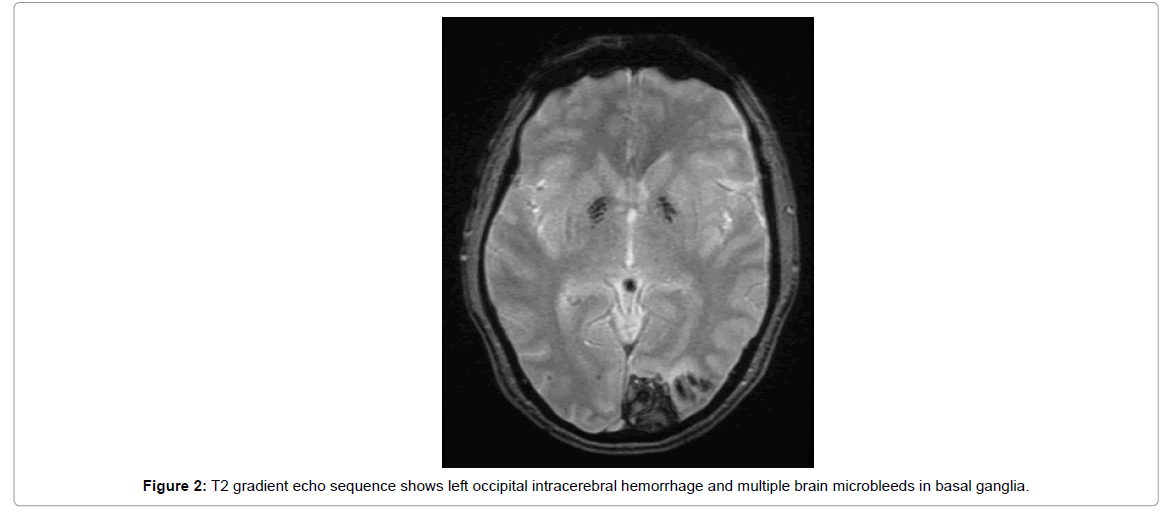
A 47-year-old hypertensive male, medicated with aspirin 100 mg and enalapril 10 mg, having good blood pressure controls since the diagnosis of CADASIL 1 year ago. The diagnosis was performed by skin biopsy showing GOM pathology (Figure 1). The patient was admitted at the emergency department with sudden and intense headache and blurry vision. On the neurological examination, right homonymous hemianopsia was found. Brain MRI in T2 gradient echo sequence shows left occipital intracerebral hemorrhage and multiple microbleeds in basal ganglia (Figure 2).
The patient has as family history associated to CADASIL. His deceased mother, passed away at the age of 65 with a presumptive diagnosis of CADASIL. Six of his eleven maternal uncles had dementia, four of whom died with a diagnosis of Alzheimer's disease. His mother was treated for progressive cognitive impairment, recurrent confusional state episodes and seizures. Prior to her death, a genetic study was performed at The Children's Hospital of Philadelphia, in USA, where exons 2 to 5, 8, 11, 14, 18, 19, 22 and 23 of the Notch 3 gene were amplified by PCR without detecting mutations. The brain MRI of his mother is shown (Figure 3).
The 47-year-old patient evolved favorably with an upper right homonymous quadrantopsia, and migraine is well controlled with Valproate Magnesium at a dose of 800 mg daily.
Comments
In contrast to the vascular disease of small vessels of the hypertensive patient, where the association with ischemic and hemorrhagic strokes is clear, the possibility of intracerebral hemorrhage in CADASIL has not been thoroughly evaluated. However, multiple cerebral microbleeds have been reported in 31-69% of patients with this diagnosis [1]. Microbleeds are small focal cerebral hemorrhage on T2 gradient echo sequence representing old hemosiderin deposits and are associated with an increased risk of intracerebral hemorrhage [1,2].
25% of symptomatic patients with CADASIL have intracerebral hemorrhages as an initial manifestation or during the progression of the disease after its diagnosis. Most of these patients are medicated with antithrombotic agents for secondary prevention of stroke. In a recent work [3], it has been found that patients with CADASIL and hypertension have significantly higher prevalence of cerebral infarctions and hemorrhages, and more microbleeds than patients without hypertension [3]. Lian et al. reported a case with spontaneous intracerebral hemorrhage in CADASIL without hypertension but with microbleeds [4]. Hemorrhages are more frequent in patients using antithrombotic agents and in patients who show a high number of microbleeds in T2 gradient echo sequence. It has also been suggested that the neurological prognosis is worse in patients with hemorrhage than in those without hemorrhage [3-5].
A positive T2 gradient echo sequence for multiple cerebral microbleeds could alert the physician not to prescribe antithrombotic agents in a patient with high risk of cerebral hemorrhage. Thus avoid additional damage to patients with an illness which currently has no specific treatment.
References
- Choi JC, Kang SY, Kang JH, Park JK (2006) Intracerebral hemorrhages in CADASIL. Neurology 67: 2042-2044.
- Fan Y, McGowan S, Rubeiz H, Wollmann R, Javed A, et al. (2012) Acute encephalophaty as initial manifestation of Cadasil. Neurol Clin Pract 2: 359-361.
- Choi JC, Song SK, Lee JS, Kang SY, Kang JH (2013) Diversity of stroke presentation in CADASIL: study from patients harboring the predominant NOTCH3 mutation R544C. J Stroke Cerebrovasc Dis 14: 126-131.
- Lian L, Li D, Xue Z, Liang Q, Xu F, et al. (2013) Spontaneous Intracerebral Hemorrhage in Cadasil. J Headache Pain 14: 98.
- Ramírez-Moreno JM, Gaspar-García E, Gómez-Baquero MJ (2011) Brain microbleeds in a patient with poorly controlled hypertension. A new marker of hypertensive vascular disease. Hipertens riesgo vasc 28: 108-111.
Relevant Topics
- Advanced Parkinson Treatment
- Advances in Alzheimers Therapy
- Alzheimers Medicine
- Alzheimers Products & Market Analysis
- Alzheimers Symptoms
- Degenerative Disorders
- Diagnostic Alzheimer
- Parkinson
- Parkinsonism Diagnosis
- Parkinsonism Gene Therapy
- Parkinsonism Stages and Treatment
- Stem cell Treatment Parkinson
Recommended Journals
Article Tools
Article Usage
- Total views: 4464
- [From(publication date):
December-2016 - Apr 02, 2025] - Breakdown by view type
- HTML page views : 3589
- PDF downloads : 875


