Cell Entry Inhibitor with Sulfonated Colloid Gold as New Potent Broad-Spectrum Virucides
Received: 30-Jun-2021 / Accepted Date: 15-Jul-2021 / Published Date: 22-Jul-2021 DOI: 10.4172/2332-0877.1000469
Abstract
Nontoxic drugs that irreversibly inhibit viruses (virucidal) are postulated to be ideal on this pandemic era against respiratory viral spreading. Unfortunately, all virucidal molecules described to date are cytotoxic. We recently developed nontoxic, broad-spectrum virucidal gold nanoparticles, less than 10 nm sized modified with sulfonic acids (mesilate) to mimic heparan sulfates and to provide the key nontoxic virucidal action. The anti-influenza effects of Camostat, a serine protease inhibitor can introduce the gold nanoparticles to the influenza virus with ionic bonding. In this study, we have examined the ability of a novel sulfonated colloid gold to inhibit the influenza virus A/PR/8/34 (H1N1; PR8) strain of mice infection in vivo. Our data show that intranasal treatment of mice with A/PR/8/34 (H1N1;PR8) fully protected the animals from a lethal infection and significantly decreased the viral titers in the lungs of the infected animals. Thus, camostat-colloid gold is a promising candidate for the development of antiviral drugs for preventing and treating the influenza infection.
Keywords: Camostat-collid gold; Osteltamivir; Influenza; Heparan Sulfate Proteoglycan (HSPG); ACE2 receptor
Introduction
Heparan sulfate proteoglycan (HSPG) are highly sulfated and broadly used by a range of viruses, to attach to the cell surface. Due to the heavily sulfated chains, they present a global negative charge that can interact electrostatically with the basic residues of viral surface glycoproteins. Viruses exploit these weak interactions to increase their concentration at the cell surface and augment their chances of binding a more specific entry receptor such as angiotensin-converting enzyme 2 (ACE2). HSPG-dependent with ACE2 receptor viruses can be grouped as rhino virus, adenovirus, echo virus, Coxsackie virus, corona virus and Respiratory Syncytial Virus (RSV) [1]. Gold nanoparticles coated with sulfonic acid inhibit different strains of influenza virus which do not bind HSPG. The antiviral action is virucidal and irreversible for influenza A (H1N1, H3N2, and H5N1) and B virus strains by the interactions between the influenza virus hemagglutinin protein and sulfated glycan’s [2].
We designed antiviral nanoparticles with flexible linkers mimicking HSPG, allowing for effective viral association with a binding that we simulate to be strong and multivalent units, generating forces that eventually lead to irreversible viral deformation [3].
Materials and Methods
Chemicals
Camostat mesylate powder was purchased from HyperChem (Shanghai, China).Colloid gold (MediGOLD) was purchased from Nutraneering (Irvine, CA, USA). Tamiflu (Oseltamivir Phosphate) was purchased from Hoffmann-La Roche Limited (St. Louis, MO, USA).
Mice and viruses
All research studies involving the use of animals were reviewed and approved by the Institutional Animal Care and Use Committee (IACUC) at the Charles River Laboratory. Six to eight week old female C57BL/6 mice were purchased from Charles River Laboratories (Portage, MO, USA) and housed (n=10). The mouse-adapted influenza strains A/ PR/8/34 (H1N1;PR8) were provided by Charles River Laboratories. The viruses were amplified in 10 day old embryonated hens’ eggs using standard procedures as described previously [4].
Infection and treatment of mice
For primary influenza infection, mice were anesthetized by intraperitoneal injection of Avertin (2,2,2-tribromoethanol;240 mg/kg; Sigma-Aldrich, St. Louis, MO, USA) before intranasal infection with 50 plaque-forming units (PFUs) of PR8 virus in 30 ml PBS (WISENT Incorporated). Mice were weighed daily and assessed for clinical signs of disease. Mice were treated orally 4 h before infection and twice daily (B.I.D.) during 7 day with saline or Tamiflu (Oseltamivir Phosphate; 5 or 50 mg/kg B.I.D.) camostat (30 mg/Kg), colloid gold (3 mL/Kg) to evaluate morbidity and adaptive immune responses or during 3 day for early cytokine, the immune response, and lung viral titers.
Bronchoalveolar lavages
Bronchoalveolar lavages (BALs) were performed to collect airway cells. Lung were recovered as described previously [5]. Briefly, organs were mechanically processed, and cells were isolated through centrifugation and wash steps.
Lung viral titer determination
Lung viral titers were determined from 10 fold dilutions of clarified supernatants of lung homogenates in viral plaque assays in MDCK cells as previously described [6]. The number of viral plaques was counted to determine viral PFUs.
Real-time PCR analyses
RNA isolated from lung homogenates was reverse (rv) transcribed, and real-time PCRs were performed. Primer sets were as follows: polymerase acidic (PA),
forward (fw) 5-CGGTCCAAATTCCTGCTGA-3 and rv 5-CATTGGGTTCCTTCCATCCA-3;
TNF-a, fw 5-CCAAAGGGATGAGAAGTTCC-3 and rv 5-TCCACTTGGTGGTTTGCTA-3;
IFN-g, fw 5-TCAAGTGGCATAGATGTGGAAGAA-3 and rv 5 TGGCTCTGCAGGATTTTCATG-3;
IL6, fw 5-GCTCGCCGGCTTCGA-3 and rv 5-GGTAGGTCTGAAAGGCGAACAG-3
IL10, fw 5-CATGCTGCTGGGCCTGAA-3 and rv 5-CGTCTCCTTGATCTGCTTGATG-3
Fold inductions were calculated using the 2-DDC time method and normalized using ribosomal 18S RNA expression. In all the figures, the group of saline treated and uninfected mice served as the control and was set to 1.
Cell surface staining and flow cytometric analysis
Fc receptors were blocked with anti-CD16/32 antibody (BD Biosciences, Franklin Lakes, NJ, USA) for 20 min at 4°C. Innate immune myeloid cells were stained using the antibodies (BD Biosciences or eBioscience, San Diego, CA, USA). Monocytes/macrophages were identified with surface marker Ly6C and CD11b. Lymphocytes were stained for 30 min at 4°C. After cell staining, acquisitions were performed with a FACSCalibur or a FACS Can to flow cytometer (BD Biosciences). Flow cytometry data were analyzed using Flow Jo 7.6.4 software (Tree Star, Ashland, OR, USA).
Statistical analyses
Results are presented as the mean 6 SEM. Comparison of data from 4 groups of mice was analyzed using the unpaired Student’s t-test with the Welch correction. For comparison of 4 groups, 1-way ANOVA followed by Tukey posttest was performed. Statistical analyses were done using Graph Pad Prism 5 (La Jolla, CA, USA). A P value, 0.05 was considered statistically significant (*P, 0.05, **P, 0.01, and ***P, 0.001).
Results
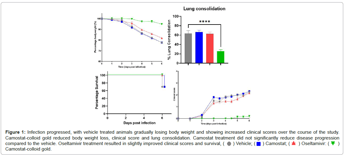
Camostat-colloid gold administration reduces morbidity and decreases influenza replication in mice than ostelmivir. Oseltamivir administration decreases the morbidity and mortality related to influenza and camostat were effective in ameliorating mouse influenza by blocking the hemagglutinin cleavage [7]. In this study, mice were treated with camostat (30 mg/kg b.i.d.), camostat with colloid gold (3 mL/kg b.i.d.), oseltamivir (50 mg/kg b.i.d.) with 50 PFU of PR8 virus. Mice that received camostat-colloid gold suffered no weight loss, whereas ostelmivir-treated mice lost up to 20% of their initial body weight on day 6 postinfection (d6 p.i.) (Figure 1). Lung consolidation was significantly reduced in camostat-colloid gold group compared to ostelmivir-treated mice on d6 d.p.i. There is no difference of the survival rate to d6 p.i. between Ostelmivir-treated and camostat-gold treated group. Showing increased clinical scores over the course of the study in ostelmivir-treated group, but nearly kept on the base line in camostat-colloid gold treated mice (Figure 1). The symptom score is consists of 1) abnormal gesture of the body 2) pilo-erection 3) abnormal respiration 4) eye discharge 5) abnormal activity.
Figure 1: Infection progressed, with vehicle treated animals gradually losing body weight and showing increased clinical scores over the course of the study.
Camostat-colloid gold reduced body weight loss, clinical score and lung consolidation. Camostat treatment did not significantly reduce disease progression
compared to the vehicle. Oseltamivir treatment resulted in slightly improved clinical scores and survival,  Camostat-colloid gold.
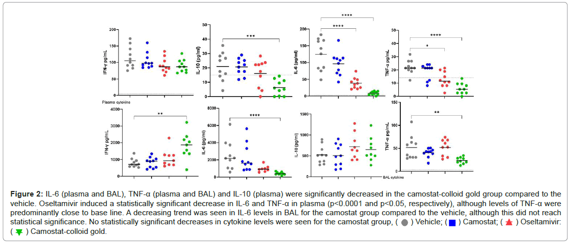
Camostat-colloid gold administration reduces early inflammatory responses in mice during influenza infection than oseltamivir. In general, levels of cytokines were higher in BAL than in plasma. The detection of viral molecules by lung epithelial cells and resident immune cells induces a first wave of cytokine production that generates an antiviral and inflammatory state [8,9] Infection of mice with the PR8 virus induced the expression of several crucial antiviral and inflammatory cytokines in the lungs (Figure 2). Indeed, reductions of 50% for IL6 expression, 45% for TNF-a,the early innate response cytokines, were observed in mice that received camostat-colloid gold treatment compared to infected mice [10,11].
Figure 2: IL-6 (plasma and BAL), TNF-a (plasma and BAL) and IL-10 (plasma) were significantly decreased in the camostat-colloid gold group compared to the
vehicle. Oseltamivir induced a statistically significant decrease in IL-6 and TNF-a in plasma (p<0.0001 and p<0.05, respectively), although levels of TNF-a were
predominantly close to base line. A decreasing trend was seen in IL-6 levels in BAL for the camostat group compared to the vehicle, although this did not reach
statistical significance. No statistically significant decreases in cytokine levels were seen for the camostat group,  Oseltamivir:
 Camostat-colloid gold.
Camostat-colloid gold administration reduces early inflammatory responses in mice during influenza infection than oseltamivir. In general, levels of cytokines were higher in BAL than in plasma. The detection of viral molecules by lung epithelial cells and resident immune cells induces a first wave of cytokine production that generates an antiviral and inflammatory state [8,9] Infection of mice with the PR8 virus induced the expression of several crucial antiviral and inflammatory cytokines in the lungs (Figure 2). Indeed, reductions of 50% for IL6 expression, 45% for TNF-a, the early innate response cytokines, were observed in mice that received camostat-colloid gold treatment compared to infected mice [10,11].
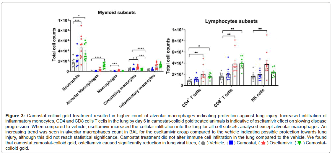
PR8 infection induced a massive recruitment of monocytes/ macrophages, neutrophils in to the lungs and airways of mice. In contrast, in camostat colloid gold and oseltamivir-treated mice, these innate immune cell populations were significantly reduced in the lungs and tended to return to levels observed in uninfected mice (Figure 3).
Figure 3: Camostat-colloid gold treatment resulted in higher count of alveolar macrophages indicating protection against lung injury. Increased infiltration of
inflammatory monocytes, CD4 and CD8 cells T cells in the lung by day 6 in camostat-colloid gold treated animals is indicative of oseltamivir effect on slowing disease
progression. When compared to vehicle, oseltamivir increased the cellular infiltration into the lung for all cell subsets analysed except alveolar macrophages. An
increasing trend was seen in alveolar macrophages count in BAL for the oseltamivir group compared to the vehicle indicating possible protection towards lung
injury, although this did not reach statistical significance. Camostat treatment did not alter immune cell infiltration in the lung compared to the vehicle. We found
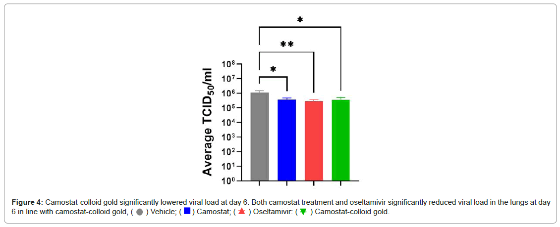
that camostat,camostat-colloid gold, osteltamivir caused significantly reduction in lung viral titres,  Camostat-colloid gold.
The lymphocyte influx into the BALF of influenza-infected mice increased gradually. This was determined by differential cell staining for total lymphocytes. In addition, we observed the CD4+ and CD8+ T lymphocytes of the adaptive immune response (Figure 4) [10].
Discussion
Oseltamivir exerts its antiviral activity by inhibiting the activity of the viral neuraminidase enzyme found on the surface of the virus, which prevents budding from the host cell, viral replication, and infectivity. The clinical benefit of use of oseltamivir is greatest when administered within 48 hours of the onset of influenza symptoms since effectiveness decreases significantly after that point in time; there is generally no benefit in use beyond 48 hours for healthy, low-risk individuals as influenza is a self-limiting illness. Early antiviral treatment can shorten the duration of fever and illness symptoms, and may reduce the risk of some complications (including pneumonia and respiratory failure). Due to the risk of adverse effects such as nausea, vomiting, psychiatric effects and renal adverse events in adults and vomiting in children, the harms are generally considered to outweigh the small clinical benefit of use of oseltamivir.
Camostat mesylate, an orally available well-known serine protease inhibitor, is a potent inhibitor of TMPRSS2. Camostat mesylate inhibits virus-cell membrane fusion and hence viral replication. Camostat mesylate treatment during acute infection with influenza, also dependent on TMPRSS2, leads to a reduced viral load. The decreased viral load may be associated with an improved patient outcome. During infection, the virus may trigger release of pro-inflammatory cytokines including IL-10, IL-6 and TNF-leading to tissue damage with subsequent vascular leakage. If left uncontrolled, this disease process can give rise to a cytokine release storm with lymphocyte infiltration. macrophage activation syndrome (MAS), can thus lead to lung tissue damage and oedema eventually resulting in life-threatening respiratory failure. Camostat mesylate reduced the inflammatory markers IL-6 and TNF- in the cell supernatants compared with non-treated controls. The drug has an excellent safety profile in humans [12].
Nanoparticles (NPs) can closely mimic the virus and interact strongly with its proteins due to their morphological similarities. Hence, NP-based strategies for tackling this virus have immense potential. Gold nanopartilces cause to the blocking by electrostatic interactions between the cationic glycoproteins of the respiratory vituses and the negatively charged colloid [13]. While Gold nanoparticles (AuNPs) themselves have negligible toxicity, Camostat-colloid gold ion complex has a safe theranostics to kill the respiratory viruses strongly with cheap costs.
Conclusion
Camostat as a cell entry inhibitor of TMPRSS on ACE2 receptors of the host cell, displaying a virustatic activity, can help ferry gold nanoparticles from the receptor binding sites to glycoprotein of influenza virus. Gold nanoparticles as the ideal antiviral should target conserved viral domains and be virucidal, i.e., irreversibly inhibit viral infectivity. These properties of gold nanoparticles make them as an effective antiviral inhibitor.
Acknowledgement
The writers would prefer to give their gratitude to the Charles River Laboratory to aid this research.
Disclosure
The authors declared no conflict of interest for this work.
References
- Cagno V, Tseligka ED, Jones ST, Tapparel C (2019) Heparan sulfate proteoglycans and viral attachment: true receptors or adaptation bias? Viruses 11:596.
- Cagno V, Gasbarri M, Medaglia C, Gomes D, Clement S, et al. (2020) Sulfonated nanomaterials with broad-spectrum antiviral activity extending beyond heparan sulfate-dependent viruses. Antimicrob Agents Chemother 64:2001.
- Cagno V, Andreozzi P, D’Alicarnasso M, Silva PJ, Mueller M, et al. (2018) Broad-spectrum non-toxic antiviral nanoparticles with a virucidal inhibition mechanism. Nat Mater 17:195-203.
- Marois I, Cloutier A, Garneau E, Richter MV (2021) Initial infectious dose dictates the innate, adaptive, and memory responses to influenza in the respiratory tract. J Leukoc Biol 92:107-121.
- Cloutier A, Marois I, Cloutier D, Verreault C, Cantin AM, et al. (2012) The prostanoid 15-deoxy-Δ12, 14-prostaglandin-j2 reduces lung inflammation and protects mice against lethal influenza infection. J Infect Dis 205:621-630.
- Lee MG, Kim KH, Park KY, Kim JS (1996) Evaluation of anti-influenza effects of camostat in mice infected with non-adapted human influenza viruses. Arch Virol 141:1979-1989.
- Holt PG, Strickland DH, Wikström ME, Jahnsen FL (2008) Regulation of immunological homeostasis in the respiratory tract. Nat Rev Immunol 8:142-152.
- Sanders CJ, Doherty PC, Thomas PG (2011) Respiratory epithelial cells in innate immunity to influenza virus infection. Cell Tissue Res 343:13-21.
- Marois I, Cloutier A, Garneau E, Lesur O, Richter MV (2015) The administration of oseltamivir results in reduced effector and memory CD8+ T cell responses to influenza and affects protective immunity. FASEB J 29(3):973-87.
- Wong ZX, Jones JE, Anderson GP, Gualano RC (2011) Oseltamivir treatment of mice before or after mild influenza infection reduced cellular and cytokine inflammation in the lung. Influenza other Respir. Viruses 5:343-350.
- Chockalingam AK, Hamed S, Goodwin DG, Rosenzweig BA, Pang E (2016). The effect of oseltamivir on the disease progression of lethal influenza A virus infection: plasma cytokine and miRNA responses in a mouse model. Dis Markers 1-3.
- Breining P, Frolund AL, Hojen JF, Gunst JD, Staerke NB, et al. (2021) Camostat mesylate against SARSâ€CoVâ€2 and COVIDâ€19-Rationale, dosing and safety. Basic Clin Pharmacol Toxicol 128:204-12.
- Medhi R, Srinoi P, Ngo N, Tran HV, Lee TR (2020) Nanoparticle-based strategies to combat COVID-19. ACS Appl. Nano Mater 3:8557-80.
Citation: Chin C (2021) Cell Entry Inhibitor with Sulfonated Colloid Gold as New Potent Broad-Spectrum Virucides. J Infect Dis Ther 4:469. DOI: 10.4172/2332-0877.1000469
Copyright: © 2021 Chin C. This is an open-access article distributed under the terms of the Creative Commons Attribution License, which permits unrestricted use, distribution, and reproduction in any medium, provided the original author and source are credited.
Share This Article
Recommended Journals
Open Access Journals
Article Tools
Article Usage
- Total views: 1743
- [From(publication date): 0-2021 - Nov 21, 2024]
- Breakdown by view type
- HTML page views: 1201
- PDF downloads: 542


