CBCT Evaluation of Inter-Root Distances for Orthodontic Mini-Screws
Received: 17-Jan-2023 / Manuscript No. jdpm-23-88871 / Editor assigned: 19-Jan-2023 / PreQC No. jdpm-23-88871 (PQ) / Reviewed: 02-Feb-2023 / QC No. jdpm-23-88871 / Revised: 06-Feb-2023 / Manuscript No. jdpm-23-88871 (R) / Published Date: 13-Feb-2023
Abstract
Objectives: This study aimed to assess the mandibular and maxillary teeth inter-root distance using a Cone- Beam Computed Tomography (CBCT) image and determine the safe zone to insert mini-screws.
Methods: In this retrospective study, we included 100 subjects that were taken by CBCT in the Department of Orthodontics, School of Dentistry, Mongolian National University Medical Sciences (MNUMS) of Mongolia, from 2014-2021. We used CBCT images in the 100 subjects were obtained with using OnDemand3D software for linear measurements.
Results: The maxillary teeth inter-root distance was analyzed a total 100 (men 30, female 70) CBCT scans. There was no statistically significant difference between the genders. Maximum inter-root distance in maxilla were measured 7 mm above Cementoenamel Junction (CEJ) 1.89 mm between canine and I premolar teeth (p<0.05), 1.73 mm between I and II premolar (p>0.05), 1.79 mm between II premolar and I molar (p>0.05) and 1.59 mm between I and II molar (p<0.001), respectively. Maximum inter-root distance in mandible was measured 7 mm below CEJ, 2.51 mm between I and II premolar (p<0.001), 2.16 mm between II premolar and I molar (p<0.01) and 2.43 mm between I and II molar (p<0.01), respectively.
Conclusion: This suggests that the maxillary, mandibular molar teeth inter-root on the buccal side far from 7mm CEJ is considered to be the safest position to implant mini screws on cortical bone.
Keywords
CBCT; Cortical bone; Bone anchored devices
Introduction
The use of mini screws has increased in recent years because of their role in Temporary Anchorage Devices (TAD`s) for orthodontic treatment. TAD’s allowed orthodontists to perform those difficult movements using its absolute anchorage system without difficulty [1,2].
According to research papers due to incomplete study of dimensions, such a complication can occur including mini screw fracture, nerve damage, maxillary and nasal cavity perforation, to inflammation or mini-screw, loosening and shedding of teeth can occur [2-6]. However, the placement sites may affect the success or failure of the procedure, so it is very important to determine the appropriate and safe location for orthodontic mini implants [7].
Global research has shown that, evaluated and measured anatomical sites for safely and securely place of mini-screws in the inter-root distances of the maxillary and mandibular arches [8- 10]. The inter-root distances have been assessed through the use of panoramic radiography, computed tomography (CT), and Cone-Beam Computed Tomography (CBCT) [11-13]. CBCT is the one of recent technological advantages in clinical dentistry and provides a detailed three-dimensional image of bones as well as accurate measurements of clinical parameters. To date, assessment of tooth inter-root distance has not been investigated in the Mongolian population. The objective of this study was to assess the mandibular and maxillary inter-root distance thickness using a CBCT image and determine the safe zone to insert mini screws.
Materials and Methods
Study design and subjects
In this retrospective study, we included 100 subjects that were taken by CBCT in the Department of Radiology, University Dental Hospital, Mongolian National University Medical Sciences (MNUMS), from 2014-2021. The inclusion criteria were no periodontal disease with no alveolar bone loss, no missing teeth, without root anomalies including severe dilacerations and idiopathic root resorption. Exclusion criteria were fractures and pathological conditions in maxilla and mandible, and root anomalies including severe dilacerations and idiopathic root resorption.
Measurements
We used Free FOV (4cm×5cm) and Full CBCT (16cm×8cm) scans using the target sampling method. All the CBCT images (85kW, 7mA) were obtained with DENTRI (HDX WILL, Seoul, Korea) apparat using on Demand 3D software for linear measurements. All images were observed and evaluated by an expert radiologist. On 10 randomly selected cases, all measurements were made twice to calculate intrarater reliability, 3 weeks apart.
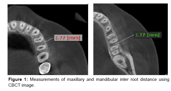
Using the CBCT scan and looking at the axial plane, it was possible to measure the linear measurements in the following maxillary and mandibular teeth: canine, 1st premolar, 2nd premolar, 1st molar, and 2nd molar. Measurements were made between inter-root bi-cortical bones (between the anterior teeth cortical bone and posterior teeth cortical bone) at a distance of 3, 5, and 7mm from the Cementoenamel Junction (CEJ) mesiodistal surface to the root according to the method described by Lee KJ, et al. [12] (Figure 1). Mesiodistal distance was measured parallel to the mean arch forms connecting the mid root portion of each root, at each vertical level on the buccal side. The interroot distance was assessed only on side of the maxilla and mandible.
Statistical analysis
Normal distribution of the measured data was confirmed by using the Kolmogorov-Smirnov method. The mean and standard deviation of inter-root distance in the axial plane were reported based on the patient's gender and age groups. Chi-square (exact test when actual or expected cell filling was low) test was used to analyze differences between inter-root distance, gender and age groups. Statistical significance was set at p≤0.05. Data analyzed using IBM SPSS version 26 software.
Ethical approval
The study was approved by the Research Ethics Committee of Mongolian National University of Medical Sciences on August 03, 2019 (No. 2019/3-08).
Results
Descriptive and reproducibility
In total 100 subjects met the inclusion criteria andinter-root distance were assessed in maxilla and mandible. Mean age 26.7±7.1 years, and 70 were female. There was no statistically significant difference between the gender and age.
Inter root distance in the maxilla
Table 1 the results obtained from the measurements of the maxillary inter-root distance. Maximums maxillaries inter-root distance was measured 7 mm above CEJ between canine and 1st premolar (1.89 mm), followed by between 1st premolar and 2nd premolar (1.73 mm), between 2nd premolar and 1st molar area (1.71 mm), and between 1st molar and 2nd molar (1.34 mm) (Table 1).
| 3 мм | 5 ммb | 7 ммc | Р valueabc | |
|---|---|---|---|---|
| Mean | Mean | Mean | ||
| Canine and I premolar | 1.73±0.42 | 1.83±0.42 | 1.89±0.54 | 0.048 |
| I premolar-II premolar (Pm I-Pm II) |
1.7±0.49 | 1.72±0.49 | 1.73±0.48 | 0.891 |
| II premolar-I molar (Pm II-M I) |
1.79±0.48 | 1.67±0.49 | 1.71±0.6 | 0.284 |
| I molar-II molar (M I-M II) |
1.59±0.54 | 1.31±0.56 | 1.34±0.63 | 0.001* |
Table 1: Maxillaries inter root distance.
Inter root distance in the mandible
Table 2 the results obtained from the measurements of the mandibular inter-root distance. The maximum mandibular inter-root distance was measured 7 mm above CEJ between 1st premolar and 2nd premolar (2.51 mm), between 1st molar and 2nd molar area (2.43 mm) and between 2nd premolar and 1st molar (2.16 mm) (Table 2).
| 3 мм | 5 мм | 7 мм | Р утгаabc | |
|---|---|---|---|---|
| Mean | Mean | Mean | ||
| I premolar-II premolar (Pm I-Pm II) |
1.87±0.39 | 2.29±0.5 | 2.51±0.58 | 0.0001* |
| II premolar-I molar (Pm II-M I) |
1.97±0.48 | 2.02±0.59 | 2.16±0.65 | 0.05 |
| I molar-II molar (M I-M II) |
2.02±0.57 | 2.21±0.74 | 2.43±0.92 | 0.006 |
Table 2: Mandibular inter root distance.
Discussion
In our study, the inter-root distance was observed at the 7 mm from CEJ on the axial plane of CBCT image at maxillary and mandible arch. We are studied morphometric evaluation of maxillary and mandibular teeth inter-root distance using 100 subjects 16-14 aged Mongolian. Also, we compared our results to other researcher’s results.
Lee KJ et al. Computerized tomography of 30 maxillae and mandibles were taken from non-orthodontic treatment adults with normal occlusion [12]. Both mesiodistal inter-root distance and bone thickness over the narrowest inter-root distance (safety depth) were measured at 2, 4, 6, and 8 mm from the CEJ. The widest distance is 3.27 mm on the medial side of the 2nd molar at a distance of 8 mm and 7.26 mm on the distal side of the 2nd molar in the maxillary bone. This is due to the fact that the tooth source was measured at distances 2, 4, 6, and 8 mm from the CEJ to the source.
Chaimanee P, et al. measured the distance between the maxillary and mandibular teeth source at a distance of 3, 5, 7, 9, and 11 mm from CEJ to source of tooth tissue to a total of 60 people with Angle class I, II, and III pendants and determined the optimal distance for implant placement.7 Researchers concluded that maximum inter-root distance were measured between 2nd premolar and 1st molar, 3.9±1.7 mm, II- 4.0±1.8, III-3.8±1.8 mm in person with bite Angle class I minimum inter-root distance were measured between 1st molar and 2nd molar, 5.3±1.8 mm, II 6.0±1.6 mm, III 5.5±1.7 mm in person with bite Angle class I 15 According to Chaimanee P, (2011) measurement of interroot distance 3, 5, and 7 mm distance from CEJ to tooth source has a relative different in the parameters may be due to the fact that the distance between the sources is determined not only by the type of bite , but also by the distance of 11 mm.
Omami G, at al. mentioned that CBCT method for measurement of the inter-root distance is an advanced technology that is very important to select the size of implants [10].
To the best of our knowledge, this study provides the first threedimensional measurement of the inter-root distance of the maxilla and mandible in the Mongolian population. Perhaps, our study provided clinically relevant outcomes to the orthodontists to accurately position the mini screws in their daily practice.
However, the limitations of our study were following. First, our date is based on relatively few samples due to the large number of edentulous and malocclusion patients in our samples. Second, the measurement of maxillary and mandibular inter-root distance is not always done in the same person. Since all our measurements and analysis were achieved separately by maxilla and mandible, this limitation will not affect the quality of the study.
Conclusion
Maximum inter-root distance of Mongolian population was 1.89 mm between maxillary canine and I premolar teeth and 2.51 mm between mandibular I and II premolar, measured 7 mm away from CEJ in maxilla and mandible. For inter-root distance, the most suitable position of orthodontics mini-screws on maxillary and mandibular was 7mm far from CEJ.
Pre-treatment assessment of morphometry of maxillary and mandibular bone in Mongolians using CBCT is important to positively affect the outcome of further treatment. The use of the morphometric dimensions of the study as a reference dimension in the treatment of post orthodontics and orthodontics is important to improve treatment outcomes and to avoid errors during treatment.
References
- Kyung HM, Park HS, Bae SM, Sung JH, Kim IB, et al. (2003) Development of orthodontic micro-implants for intraoral anchorage. J Clin Orthod 37: 321-328.
- Chang HP, Tseng YC (2014) Miniscrew implant applications in contemporary orthodontics. Kaohsiung J Med Sci 30: 111-115.
- Melsen B (2005) Mini-implants: Where are we?. J Clin Orthod 39: 539-547.
- Montes CC, Pereira FA, Thomé G, Alves EDM, Acedo RV, et al. (2007) Failing factors associated with osseointegrated dental implant loss. Implant Dent 16: 404-412.
- Bortoluzzi MC, Cella C, Haus SF (2017) Dentofacial deformity and quality of life: a case control study. RSBO 14: 24-29.
- Papadopoulos MA, Papageorgiou SN, Zogakis IP (2011) Clinical effectiveness of orthodontic miniscrew implants: a meta-analysis. J Dent Res 90: 969-976.
- Chaimanee P, Suzuki B, Suzuki EY (2011) “Safe zones” for miniscrew implant placement in different dentoskeletal patterns. Angle Orthod 81: 397-403.
- Holmes PB, Wolf BJ, Zhou J (2015) A CBCT atlas of buccal cortical bone thickness in interradicular spaces. Angle Orthod 85: 911-919.
- Arai Y, Tammisalo E, Iwai K, Hashimoto K, Shinoda K (1999) Development of a compact computed tomographic apparatus for dental use. Dentomaxillofac Radiol 28: 245-248.
- Omami G, Al Yafi F (2019) Should Cone Beam Computed Tomography Be Routinely Obtained in Implant Planning?. Dent Clin North Am 63: 363-379.
- Ahmad M, Jenny J, Downie M (2012) Application of cone beam computed tomography in oral and maxillofacial surgery. Aust Dent J 57(s1): 82-94.
- Lee KJ, Joo E, Kim KD, Lee JS, Park YC, et al. (2009) Computed tomographic analysis of tooth-bearing alveolar bone for orthodontic miniscrew placement. Am J Orthod Dentofacial Orthop 135: 486-494.
- Baumgaertel S, Hans MG (2009) Buccal cortical bone thickness for mini-implant placement. Am J Orthod Dentofacial Orthop 136: 230-235.
Indexed at, Google Scholar, Crossref
Indexed at, Google Scholar, Crossref
Indexed at, Google Scholar, Crossref
Indexed at, Google Scholar, Crossref
Indexed at, Google Scholar, Crossref
Indexed at, Google Scholar, Crossref
Indexed at, Google Scholar, Crossref
Indexed at, Google Scholar, Crossref
Indexed at, Google Scholar, Crossref
Citation: Rashsuren O, Bold J, Batmunkh E, Sundui E, Zolzaya S, et al. (2023)CBCT Evaluation of Inter-Root Distances for Orthodontic Mini-Screws. J DentPathol Med 7: 140.
Copyright: © 2023 Rashsuren O, et al. This is an open-access article distributedunder the terms of the Creative Commons Attribution License, which permitsunrestricted use, distribution, and reproduction in any medium, provided theoriginal author and source are credited.
Select your language of interest to view the total content in your interested language
Share This Article
Recommended Journals
Open Access Journals
Article Usage
- Total views: 2736
- [From(publication date): 0-2023 - Nov 21, 2025]
- Breakdown by view type
- HTML page views: 2366
- PDF downloads: 370

