Case Report Open Access
Case Report of Pelvic Actinomycosis Presenting as a Complex Pelvic Mass
| Pandian Radha*, Amy Forsberg Condon and Timothy Lim Yong Kuei | |
| K K Women’s & Children’s Hospital, 100 Bukit Timah Road, Singapore | |
| Corresponding Author : | Pandian Radha K K Women’s & Children’s Hospital 100 Bukit Timah Road, Singapore Tel: 65 6394-1067 Fax: 65 6298- 6343 E-mail: radhahk@gmail.com |
| Received September 06, 2014; Accepted December 12, 2014; Published December 17, 2014 | |
| Citation: Radha P, Condon AF, Yong Kuei TL (2015) Case Report of Pelvic Actinomycosis Presenting as a Complex Pelvic Mass. J Preg Child Health 2:125. doi: 10.4172/2376-127X.1000125 | |
| Copyright: © 2015 Radha P, et al. This is an open-access article distributed under the terms of the Creative Commons Attribution License, which permits unrestricted use, distribution, and reproduction in any medium, provided the original author and source are credited. | |
Visit for more related articles at Journal of Pregnancy and Child Health
Abstract
Pelvic actinomycosis is a chronic granulomatous infection caused by Gram- positive anaerobic bacteria, Actinomycosis israeli, which constitutes 3% of actinomycosis infections. Pelvic actinomycosis is insidious in onset, often presenting as a firm mass that appears fixed to the surrounding tissues, mimicking malignancy. It is also mistaken for other conditions such as diverticulosis and inflammatory bowel disease, which presents a diagnostic challenge pre-operatively. It has been recognized that pelvic actinomycosis is associated with long term use of an intrauterine contraceptive device (IUCD). Here we present a case which initially presented as a complex pelvic mass concerning for malignancy and was diagnosed as pelvic actinomycosis post-operatively on histopathology.
| Keywords |
| Pelvis; Actinomycosis; Infection; Tumour |
| Introduction |
| Pelvic actinomycosis is a chronic granulomatous infection caused by Gram- positive anaerobic bacteria, Actinomycosis israeli, which constitutes 3% of actinomycosis infections. Pelvic actinomycosis is insidious in onset, often presenting as a firm mass that appears fixed to the surrounding tissues, mimicking malignancy. It is also mistaken for other conditions such as diverticulosis and inflammatory bowel disease, which presents a diagnostic challenge pre-operatively. It has been recognized that pelvic actinomycosis is associated with long term use of an intrauterine contraceptive device (IUCD). Here we present a case which initially presented as a complex pelvic mass concerning for malignancy and was diagnosed as pelvic actinomycosis postoperatively on histopathology. |
| Case presentation |
| A 45 year old Vietnamese woman was referred to our hospital with left illac fossa pain of two months duration, which had worsened over the past four days. She also complained of fever, dysuria, vomiting, and weight loss. Her gynaecological history was unremarkable except for IUCD use for the past six years. She had no menstrual complaints. On physical examination she was clinically stable without any sign of febrile toxicity. The abdomen was soft and revealed a tender, 10 cm mass filling the left lower quadrant. Left costovertebral angle tenderness was present. There were no signs of an acute abdomen. On pelvic examination the cervix appeared normal with the IUCD string visualized. No vaginal discharge or cervical motion tenderness. On digital rectal examination a large, firm mass was palpable. Biochemical and haematological investigations demonstrated an elevated C-reactive protein of 78.8 [0.0- 9.0mg/L] and leukocytosis of 20.72 [4.50-11 10[9]/L] with a left shift, normochromic normocytic anaemia [7.9 g/dl], and thrombocytosis [platelets 607 10[9]/L]. The renal panel and liver function tests were normal. Leukocytes were present in the urine. Her ovarian tumour markers [CA-125, B-HCG, AFP, and CEA] were normal. |
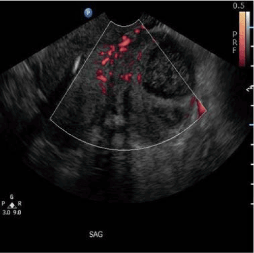
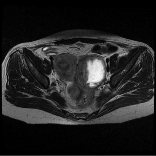
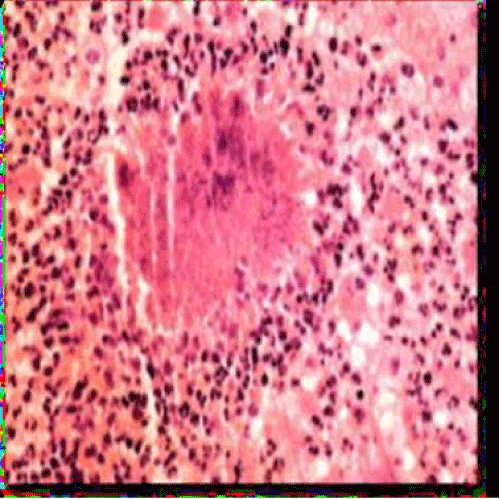
| On endovaginal sonogram, the IUCD was seen in satisfactory position. The endometrial thickness was 6mm. A 7.0 cm X 5.7 cm complex mass containing a cystic avascular area with internal echoes was present in the left adnexa (Figure 1). A large bridging vessel was visualized between uterus and the mass. The left ovary was not seen. MRI showed a left adnexal mass extending into the left perirectal space without any plane of separation seen from the rectum (Figure 2). Hydroureter and a prominent left pelvicalyceal system were noted. Despite multiple imaging studies, the origin of the mass remained uncertain. The differential diagnosis included an exophytic uterine fibroid vs. an ovarian tumour, vs. a colon tumour. Broad spectrum IV antibiotics were started for suspected pyelonephritis and possible pelvic inflammatory disease. The IUCD was removed. The leukocytosis and C-reactive protein normalized after 4 days of antibiotic therapy. The urine culture revealed no growth. Exploratory laparotomy was performed on hospital day number four when the patient had improved clinically and was afebrile. Pre-operative colonoscopy was done by colorectal team and reported normal. Gynaecological oncology and colorectal surgeons were available for back up. The intra-operative findings were as follows: An inflamed 8 cm left tubo-ovarian complex was adherent to the pelvic side wall and rectum. The left ureter was noted to be adhered to the mass. The uterus, cervix, right ovary, right fallopian tube, and omentum appeared normal. As the mass was densely adherent to rectum and ureter, it required sharp dissection to start, but then peeled off once the proper surgical plane was identified. Lysis of adhesions, total abdominal hysterectomy with bilateral salpingoohporectomy and left ureteral lysis were performed. In view of the persistent pain and discomfort the patient was keen for permanent surgical option of total abdominal hysterectomy with bilateral salpingo-oophorectomy. We proceeded for surgery which was feasible with proper plane of cleavage without any visceral damage. Moreover, histological confirmation was possible to rule out malignancy. Frozen section was obtained, which confirmed the diagnosis of tubo-ovarian abscess with actinomycosis organisms. No malignant cells were present. IV ceftriaxzone, metronidazole, and gentamycin were continued for 7 days post-operatively. The final histology of the left ovary and tube showed a tuboovarian abscess with an actinomycosis like organism (Figure 3). The pathology of the uterus was remarkable for a fibroid uterus with an actinomycosis like organism with suppurative myometritis. Gram stain showed radiating filament of gram positive organism rimmed by acute inflammatory cells. Her Pap smear result, which returned post-operatively, was remarkable for an actinomycosis like organism without evidence of malignancy. Her postoperative recovery was uneventful. She received one unit of packed red blood cells. She remained afebrile and her pain resolved. She was started on oral Penicillin VK 500 milligrams four times daily for six months and discharged on post-operative day number ten. |
| Discussion |
| Actinomycosis is a chronic infection characterized by the presence of dense fibrous tissue and purulent fluid. Approximately 20% of the actinomycosis infections occur in the abdomen and pelvis. The infection does not invade intact mucosa and it can be also be introduced by oralgenital contact. Most of the cases are diagnosed only post-operatively or intra-operatively. Pelvic actinomycosis is difficult to differentiate from pelvic malignancy. It presents as a firm mass that appears to be fixed to the surrounding structures and mistaken for a pelvic tumour. Abdominal surgery, ruptured viscus, tubo ovarian abscess and presence of an IUCD are recognized risk factors. Actinomycosis israeli infects 1.65% to 11.6% of IUCD users and it is common in women who have an IUCD in place for more than four years [1]. Pelvic actinomycosis causes endometritis, salpingo-ooporitis, tubo-ovarian abscess, and pelvic masses suspicious for pelvic malignancy. It extends to the abdominal wall and deep pelvic structures. Although primary bowel involvement is rare, the most commonly affected sites are the transverse colon, cecum, and appendix. |
| Diagnosis of pelvic actinomycosis can be difficult pre operatively due to the insidious onset and lack of signs of obvious pelvic inflammation. Pelvic exam is often concerning for malignancy. Actinomycosis should be included in the differential diagnosis when the CT scan shows bowel wall thickening and a regional pelvic or peritoneal mass with extensive infiltration, especially in a patient with abdominal pain, fever, leucocytosis with the long term use of an IUCD [2]. Diagnostic imaging may be non-specific and misleading unless the possibility of actinomycois is considered. The CT scan appearance of abdominopelvic actinomycosis possibly includes the presence of a solid mass with a focal area of reduced attenuation or thick- walled cystic mass [3]. Dense inhomogeneous contrast enhancement in the solid component is noted in two-thirds of the patients [3]. Lymph node enlargement is not a prominent feature. Infiltration of the soft tissue and loss of the normal tissue planes is frequently noted with Actinomycosis infection [4,5]. |
| MRI appearance of pelvic actinomycosis possibly includes the presence of a tubo-ovarian mass of relatively low signal intensity on T2 weighted sequences with extensive pelvic infiltration. Any pelvic mass with a high signal cystic area but lacks high signal intensity in the solid tumour on T2 weighted sequences is atypical for malignancy and co-existence of infiltrative changes in parametrium, bladder and bowel should raise the suspicion of pelvic actinomycosis [6]. An infiltrative mass with unusual aggressiveness is an important radiological finding, but still can be confused with inflammatory disease and malignancy. If a patient with an IUCD presents with symptoms of pelvic pain and a pelvic mass not resolving with antibiotics, pelvic actinomycosis should be considered. An ultrasound guided biopsy or CT guided drainage with cytology may be helpful in confirmation of diagnosis. Although ultrasound guided or CT guided drainage and cytology help to confirm diagnosis, recurrence after antibiotic treatment is possible, surgery may be ultimately necessary [7]. After definitive surgery, complete resolution is expected. |
| In general, primary antimicrobial therapy should be considered as first choice in patient who present with pelvic pain, pelvic mass with long term IUCD use, considering pelvic actinomysocis as a possible diagnosis. Surgery is required for draining the abscesses, but because actinomycosis infection does not follow tissue planes, surgery may be complicated and if possible, should delayed until completion of a long term course of Penicillin. Surgeon should be aware of this infection in order to avoid excessive surgical procedure. |
| Diagnosis pre-operatively can be difficult using standard imaging techniques. Computed tomography or ultrasound guided biopsy can be used to obtain a specimen for diagnosis. Although in our case the actinomycosis presented with fever and pelvic pain which raised the suspicion of pelvic inflammatory disease, the appearance of the pelvic mass increased the suspicion of malignancy. Despite widespread availability of diagnostic imaging, the true diagnosis often remains obscured until exploratory laparotomy is performed and histological diagnosis is confirmed. Penicillin is the drug of choice and it should be given for a three to six month course. |
| Beta lactum antibiotic combined with a beta lactamase inhibitor should be the first choice of antibiotics [8]. In patient with showed penicillin allergy, clindamycin or tetracycline can be used with good results [9]. Parenteral therapy is required only for severe infection. IM Ceftriaxone therapy can facilitate outpatient management instead of IV Penicillin which often requires long term hospitalization [10]. Generally the treatment is continued until evidence of complete resolution. |
| Conclusion |
| Pelvic actinomycosis is often mistaken for pelvic malignancy. Pelvic actinomycosis should be always considered in the differential diagnosis of a patient with long term use of IUCD and a pelvic mass. Post-operative histology and cytology confirms the diagnosis in most of the cases. Primary antimicrobial therapy should be attempted. If the condition is unresponsive to antibiotic, surgery should be performed for complete cure. Moreover, exploratory laparotomy helps in definitive diagnosis, as pre-operative diagnosis remain obscured in most of cases, as is consistent with our case report .Removal of the infected pelvic mass with an appropriate six month course of Penicillin renders cure in most cases. |
| Conflict of Interest Statement |
|
The authors declare that there are no conflicts of interest. |
References
- McCormick JF, Scorgie RD (1977) Unilateral tubo-ovarian actinomycosis in the presence of an intrauterine device. American Journal of Clinical Pathology 68 (5): 622-626.
- Pusiol T, Morichetti D, Pedrazzani C, Ricci F (2011) Abdominal-Pelvic Actinomycosis Mimicking Malignant Neoplasm. Infectious Diseases in Obstetrics and Gynecology.
- Ha HK, Lee H J ,Kim H, Ro HJ, Park YH, et al. (1993) Abdominal actinomycosis: CT findings in 10 patients. American Journal of Roentgenology 161: 791-794.
- Oslon MC, Demos TC, Tamayo JP (1993) Actinomycosis of the retroperitoneum and an extremity: CT features. Abdominal Imaging 18: 295-297.
- Shah HR, Williamson MR, Boyd CM, Balachandran S, AnqtuacoTL, et al. CT findings in abdominal actinomycosis. Journal of Computer Assisted Tomography 11: 466-469.
- Hawnaur JM, Reynolds K, McGettigan C (1999) Magnetic resonance imaging of actinomycosis presenting as pelvic malignancy. The British Journal of Radiology 72:1006-1011.
- Marella VK, Hakimian O, Wise GJ, Silver DA (2004) Pelvic actinomycosis: Urological Perspective. International Braz J Urol 30:367-376.
- Smith AJ,Hall V,Thakker B, Gemmell CG (2005) Antimicrobial susceptibility testing of Actinomyces species with 12 antimicrobial agents. Journal of Antimicrobial Chemotherapy 56: 407-409.
- Wagenlerhner FME, Mohren B, Naber KG, Mannl HFK (2003) Abdominalactinomycosis.Clinical Microbiology and Infection 9:881-885.
- Onal ED, Altinbas A, Onal IK, Ascioglu S, Akpinar MG, et al. (2009) Successful Outpatient management of Pelvic actinomycosis by ceftriaxone: A report of 3 cases. The Brazilian Journal of Infectious Diseases, 13: 391-393.
Figures at a glance
 |
 |
 |
| Figure 1 | Figure 2 | Figure 3 |
Relevant Topics
Recommended Journals
Article Tools
Article Usage
- Total views: 14489
- [From(publication date):
February-2015 - Dec 18, 2024] - Breakdown by view type
- HTML page views : 9760
- PDF downloads : 4729
