Case Report Open Access
Case Report: Closed Posteromedial Dislocation of the Ankle without Medial Malleolar Fracture
Jakrapong Orapin1, Paphon Sa-ngasoongsong1, Sorawut Thamyongkit1,2, Noratep Kulachote1, Sukij Laohajaroensombat1*, Chanyut Suphachatwong1 and Pornchai Mulpruek1
1Department of Orthopaedic Surgery, Ramathibodi Hospital, Faculty of Medicine, Mahidol University, Bangkok, Thailand
2Chakri Naruebodindra Medical Institute, Ramathibodi Hospital, Faculty of Medicine, Mahidol University, Bangkok, Thailand
- *Corresponding Author:
- Laohajaroensombat S
Department of Orthopaedic Surgery, Ramathibodi Hospital
Faculty of Medicine, Mahidol University, Bangkok, Thailand
Tel: 02-201-1589
Fax: 02-201-1599
E-mail: lasukij@hotmail.com
Received date: Jul 07, 2016; Accepted date: Aug 05, 2016; Published date: Aug 16, 2016
Citation: Orapin J, Sa-ngasoongsong P, Thamyongkit S, Kulachote N, Laohajaroensombat S, et al. (2016) Case Report: Closed Posteromedial Dislocation of the Ankle without Medial Malleolar Fracture. Clin Res Foot Ankle 4:197. doi:10.4172/2329-910X.1000197
Copyright: © 2016 Orapin J, et al. This is an open-access article distributed under the terms of the Creative Commons Attribution License, which permits unrestricted use, distribution, and reproduction in any medium, provided the original author and source are credited.
Visit for more related articles at Clinical Research on Foot & Ankle
Abstract
Closed ankle dislocation without associated fractures of malleoli is a very rare condition and has been reported sparsely in literatures. We present two cases of this type of injury. All patients were treated conservatively with immediate closed reduction and immobilization in a short leg slab. Mechanism of injury, MRI findings, management, functional outcome and possible complications were discussed. Sufficient immobilization period with gradual ankle joint motion exercise with brace support resulted in good to excellent clinical outcomes. A review of the literature is also presented in this paper.
Keywords
Ankle; Dislocation; Closed dislocation; Tibiotalar dislocation; Posteromedial dislocation
Introduction
Closed ankle dislocation without associated fracture of malleoli is a very rare condition and only a few literatures have been reported. The most common type of the closed ankle dislocation was posteromedial direction [1-3].
In 1939, Wilson and colleagues reported a case series of this type of injury with good clinical outcome after conservative treatments [4].
Closed reduction and immobilization in this type of injury result in good clinical outcome [5].
In contrast, operative method is a preferred treatment in open injury which can perform both joint debridement and repair of the disrupted ligaments [6,7].
We have presented here 2 cases of closed posteromedial ankle dislocation while playing basketball.
Mechanism of injury, MRI findings, management, functional outcome and possible complications have been discussed.
Case Reports
Case 1
A 29-year–old male accidentally injured his left ankle while playing basketball. He landed on his left ankle.
The ankle was swollen and deformed with tenderness and ecchymosis around the joint line. There was no open wound nor signs of neurovascular injury.
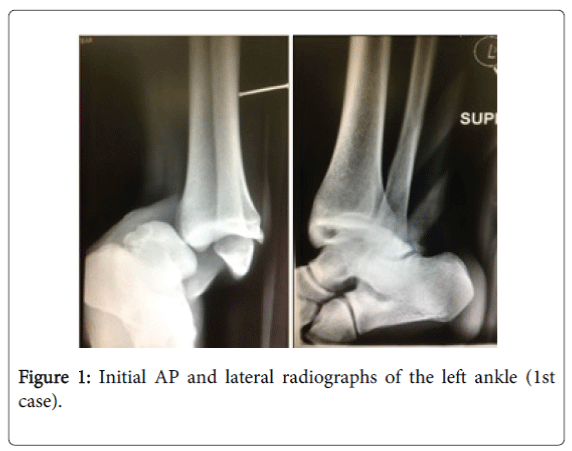
Initial radiographs showed posteromedial dislocation of the tibiotalar joint without any fracture (Figure 1).
Closed reduction was performed under intravenous analgesia and sedation for adequate relaxation.
Patient was positioned in knee flexion and ankle in plantar flexion. Longitudinal manual traction was done gently and carefully.
After reduction, his left ankle was then immobilized in a short leg slab.
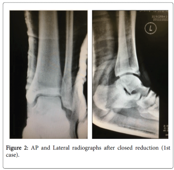
Post-reduction radiographs were obtained and no fracture fragment or residual subluxation were found (Figure 2).
The short leg slab was retained for 3 weeks. Then, the patient was allowed to gradually increase his range of motion and weight-bearing with pneumatic walking boots.
After 12 weeks, patient was allowed full ROM and weight-bearing.
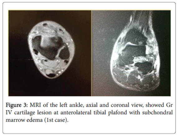
MRI images of this patient showed Gr IV cartilage lesion with subchondral marrow edema at anterolateral of tibial plafond (Figure 3), complete tear of ATFL, PTFL, CFL with intact AITFL.
Complete tear of tibiotalar part and partial tear of tibiocalcaneal part of deltoid ligament were also noted.
Case 2
An 18-year-old man sustained a left ankle injury while playing basketball.
His ankle was injured while the left ankle was in plantar flexion, inversion.
Deformity of the left ankle was found with no loss of foot sensation or distal pulse.
His skin was tent on anterior side due to anterior edge of tibial plafond. He had just 1-cm abrasion at ankle without any open wound.
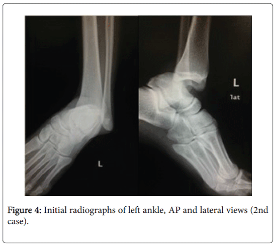
Initial X-ray images showed posteromedial dislocation of left ankle with no malleolar fracture (Figure 4).
Reduction was achieved under intravenous sedation and analgesia. The patient was positioned in knee flexion and ankle in plantar flexion.
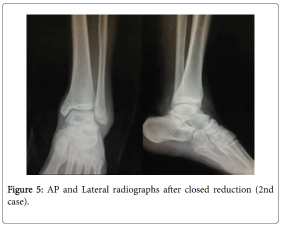
Post-reduction radiographs showed talus in normal mortise without any bony fragment (Figure 5).
After reduction his ankle was immobilized for 3 weeks in a short leg slab followed by wearing walking boots with progressive weight bearing until full weight bearing was reached.
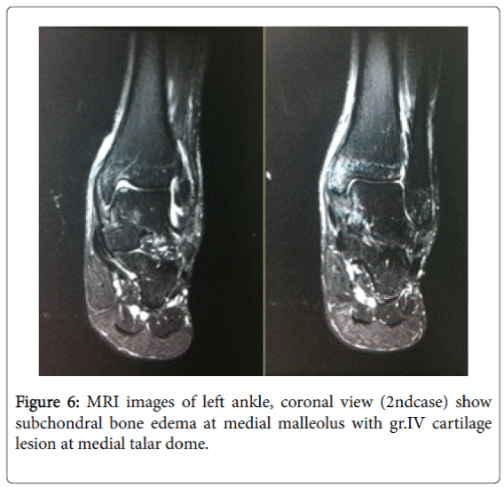
MRI images after injury showed Gr IV cartilage lesion at medial talar dome with subchondral marrow edema (Figure 6).
Intact CFL, partial tear of ATFL, partial tear of deep deltoid, Gr I-II sprain PTFL and bimalleolar marrow edema.
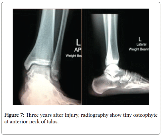
Three-year follow up images showed a tiny spur at talar neck with no ankle arthritic change (Figure 7).
Discussion
Closed ankle dislocation without fracture is a rare injury with a number of case reports in literature. This rarity could be explained by the stronger ligaments around the ankle mortise and the weaker malleoli. Therefore, there was a chance of malleoli to be broken before ligament tearing. According to a study of Rasmussen et al. ankle stability is created by congruency of talus in ankle mortise which is decreased when foot is in plantar flexed position. More stress occurred to anterior joint capsule and anterior talofibular ligament [8]. Moreover, the injury to the structures also depends on the direction of talar displacement and severity of injury [9-14]. Predisposing factors of ankle dislocation without malleolar fracture were generalized ligamentous laxity, weakness of the peroneal muscles, hypoplasia of the medial malleolus and repeated ankle sprain [15]. There are some open posteromedial dislocations in higher energy injury because anterior soft tissue is stretched when ankle is in plantar flexion position.
Although motor vehicle accident is the most common cause of ankle dislocation, the second is sports trauma. It occurs especially when jumping which is a fundamental component of sports such as volleyball and basketball [16]. Forty-five percent of basketball injuries, 25% of volleyball injuries, and 31% of soccer injuries are to the ankle [17].
Complications are rarely seen in this type of injury. However, vascular injuries and neurological injuries are occasionally seen. Due to high energy mechanism and many important structures are close to the ankle, careful physical examination should be done especially in open dislocation [18,19]. A slight loss of dorsiflexion, joint stiffness, joint instability, degenerative arthritis, and capsular calcification can be seen as late complications.
To our knowledge, closed reduction under adequate analgesia and sedation followed by immobilization for 4-6 weeks are still the treatment of choice for closed dislocation as mentioned in many literatures [2,3,20]. After full weight bearing of the injured ankle is allowed, residual pain, discomfort, and instability have to be reevaluated. Further investigation and treatment might be required [21,22] in case of established residual ankle instability (occurs in approximately 20% of all acute ankle injuries) that may lead to posttraumatic osteoarthritis. Lateral ligament repair by widely used modified Brostrom technique can be performed with either open or arthroscopic technique if clinically indicated [23,24,25].
The authors have not found predisposing factors for ankle dislocation in these patients. Mechanism of injury was axial loading on foot in plantar flexion and inversion position. This force put the narrowest part of talus into mortise and stretching of capsule and collateral ligaments occurred, which resulted in dislocation of the ankle [19]. From MRI images, these 2 patients similarly had chondral lesion, ligament injuries and subchondral marrow edema. However, the location of chondral lesion and severity of the injured ligaments were different. The first case had chondral lesion at anterolateral plafond and complete tear of deltoid, CFL and ATFL ligament. Whereas the second case had subchondral lesion on medial talar dome and partial tear of deltoid. This might be due to the difference of position of the foot combined with higher degree of rotational force and tilting of the ankle joint at the time of injury.
After 2-year period of follow up, the first patient was able to have pain-free daily activities, walked without difficulty on any surfaces, and even ran for 30 minutes with minimal or no pain. The second patient was training in Royal Military Academy and he could perform longdistance running without ankle pain. However, his last radiographic image showed tiny osteophyte at talar neck (Figure 7). Functional outcome assessment was performed by using AOFAS Ankle-Hindfoot score and foot and ankle VAS questionnaire. Their final radiographic and functional outcomes were good to excellent.
Conclusion
Sport related ankle injury can result in various degrees of injuries to bones, cartilage, and ligaments. If this injury is suspected, bone and ligaments should be carefully examined. Even though MRI images showed no obvious fracture of malleoli, exhibited marrow edema was evidence of some variable degree of ligament and bone injuries. Closed reduction under an adequate intravenous sedation and analgesia is still the treatment of choice for this injury. Sufficient immobilization period with gradual ankle joint motion exercise with brace support results in good to excellent clinical outcomes.
References
- Wang YT, Wu XT, Chen H (2013) Pure closed posteromedial dislocation of the tibiotalar joint without fracture. Orthopsurg 5:214-218.
- Qayyum F, Qayyum AA, Sahlstrum SA (2014) Dislocation of the ankle without simoustaneously fracture of the bones. UgeskrLaeger 176:1628-1683.
- Karabila MA, Kharmaz M (2016) Dislocation tibio-talar pure in a young athlete. Pan Afrmed J 23:17.
- Wroble RR, Nepola JV, Malvitz TA (1988) Ankle Dislocation without Fracture. Foot Ankle Int 9:64-74.
- Wight L, Owen D, James D (2015) Pure ankle dislocation: management with early weight bearing and mobilization. ANZ JSurg.
- Krishnamurthy S, Schultz RJ (1985) Pure posteromedial dislocation of the ankle joint. A case report. ClinOrthopRelate Res201:68-70.
- Daruwalla JS(1974) Medial dislocation of the ankle without fracture: a case report. Injury 5:215-216.
- Rasmussen O (1985) Stability of the ankle joint. Analysis of the function and traumatology of the ankle ligaments. ActaOrthopScandSuppl 211:1-75.
- D'Anca AF (1970) Lateral rotatory dislocation of the ankle without fracture: A case report. J Bone J Surg Am52:1643-1646.
- Agrawal AC, Raza HKT, Haq RU (2008) Closed posterior dislocation of the ankle without fracture. Indian J Orthop 42:360-362.
- Lertwanich P, Santanapipatkul P, Harnroonroj T (2008) Closed posteromedial dislocation of the ankle without fracture: a case report. J Med Assoc of Thai91:1137-1140.
- Lui TH, Chan KB (2012) Posteromedial ankle dislocation without malleolar fracture: a report of six cases. Injury 43:1953-1977.
- Jerbi M, Grissa Y, Weslati A, Naouer N, Ben Maitigue M, et al.(2015) Anterior pure tibiotalar dislocation: A rare lesion. Pressemedicale 44:544-546.
- Wilson AB, Toriello EA (1991) Lateral rotatory dislocation of the ankle without fracture. JOrthopTrauma 5:93-95.
- Rivera F, Bertone C, De Martino M, Pietrobono D, Ghisellini F (2001) Pure dislocation of the ankle: three case reports and literature review. ClinOrthopRelat Res382:179-184.
- Uyar M, Tan A, IÅÂ?ler M, Cetinus E (2004) Closed posteromedial dislocation of the tibiotalar joint without fracture in a basketball player. British Journal of Sports Medicine 38:342-343.
- Garrick JG (1977) The frequency of injury, mechanism of injury, and epidemiology of ankle sprains. Am JSports Med 5:241-242.
- Kelly PJ, Peterson LF (1962) Compound dislocation of the ankle without fracture. Am JSurg103:170-172.
- Colville MR, Colville JM, Manoli A (1987) Posteromedial dislocation of the ankle without fracture. J Bone Joint Surg Am69:706-711.
- Toohey JS, Worsing RA (1989) A long-term follow-up study of tibiotalar dislocations without associated fractures. ClinOrthopRelat Res239:207-210.
- Carbone A, Rodeo S (2016)Review of current understanding of post-traumatic osteoarthritis resulting from sports injuries. JOrthop Res.
- Lofvenberg R, Karrholm J, Lund B (1994) The outcome of nonoperated patients with chronic lateral instability of the ankle: a 20-year follow-up study. FootAnkle Int15:165-169.
- Hamilton WG, Thompson FM, Snow SW (1993) The modified brostrom procedure for lateral ankle instability. Foot Ankle Int 14:1-7.
- Maffulli N, Ferran NA (2008) Management of acute and chronic ankle instability. J Am AcadOrthopSurg 16:608-615.
- Acevedo JI, Mangone P (2015) Ankle instability and arthroscopic lateral ligament repair. FootAnkle Clinics North America20:59-69.
Relevant Topics
Recommended Journals
Article Tools
Article Usage
- Total views: 14206
- [From(publication date):
September-2016 - Apr 07, 2025] - Breakdown by view type
- HTML page views : 13305
- PDF downloads : 901

