Case Report: A Rare Case of Appendicitis Incarcerated in an Inguinal Hernia in an 8 Year Old Male Child
Received: 15-Jan-2018 / Accepted Date: 26-Jan-2018 / Published Date: 02-Feb-2018 DOI: 10.4172/2376-127X.1000365
Abstract
Introduction: Amyand’s hernia may present with the inflammation of the appendix vermiform is enclosed in its sac. Its diagnosis is very difficult in the pre-operative period, it is usually an incidental finding.
Case presentation: We present a case of Amyand’s hernia in an 8 year-old male who presented with an obstructed hernia having acute appendicitis within the hernia sac.
Conclusion: Amyand’s hernia is a rare condition yet represents two of the most common diseases a general surgeon has to face. It is difficult to diagnose preoperatively via clinical examination. Diagnosis can be aided by a high index of suspicion, and accurate interpretation of a Computed Tomography.
Keywords: Appendix; Inguinal hernia; Amyand; Acute abdomen
Introduction
An Inguinal hernia containing appendix is termed an Amyand’s hernia. The presence of the vermiform appendix within an inguinal hernia was first described by Amyand in 1736 [1]. Case reports in the literature indicate that about 1% of the inguinal hernias contain a portion of the vermiform appendix and its complicated by acute appendicitis in 0.08% of cases [2,3]. Furthermore, it is three times more likely in children than in adults [4,5]. We report a rare case of Amyand’s henia occurring in a male who presented with an acute irreducible erythematous mass located in the right inguinal area.
Case Presentation
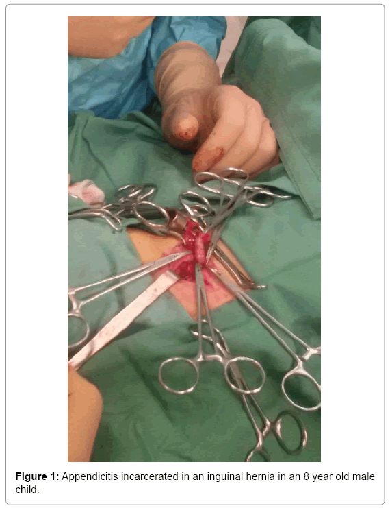
An 8 year old Caucasian male was admitted to our Emergency department suffering from pain in the right inguinal area. Physical examination revealed an acute irreducible erythematous mass located in the right inguinal region. Laboratory analysis showed leukocytosis. All the other routine laboratory investigation included urinalysis and abdominal radiograph were normal. Presumptive diagnosis was of an incarcerated indirect congenital inguinal hernia and the patient was scheduled for surgery. Under general anesthesia, an incision was made in the skin crease of the right inguinal region and inguinal canal was opened. Hernia sac was identified and opened. A small amount of clear fluid was noted in the peritoneum. The inflamed appendix was found in the hernia sac with some adhesions. The cecum was freed from the flimsy adhesions to the sac. The base of the appendix was free of inflammation, so a classic appendicectomy was performed and the cecum was reduced in the abdominal cavity. Then we performed an immediate herniorrhaphy. Histopathology revealed an inflamed appendix with dilated serosal capillaries. The patient was given intravenously cefotaxime and metronidazole, the postoperative course was uneventful and the patient was discharged from the clinic, on the 6th postoperative day, in a good general condition [6,7].
Discussion
Amyand Hernia is a rare condition and represents two of the most common diseases a general surgeon has to face. It was first described by Amyand in 1736, which performed his first appendectomy during an inguinal repair in an 11 year old male [1]. Amyand Hernia is most frequently reported in men and almost exclusively on the right side, probably due to the common anatomical position of the appendix [8]. Although it has been reported the presence of Amyand’s hernia on the left side in cases of situs inversus, mobile cecum or intestinal malrotation [9]. There have been fewer than 200 cases of Amyand’s hernia in the literature, two of which presenting as scrotal swelling [7].
The disorder comprises less that 1% of all inguinal hernia-cases and 0.2% of appendicitis cases. The incidence of the presence of a uninflammed appendix vermiform is in the sac of an inguinal hernia is estimated to be 0.13%, but it is even rarer to find an inflamed appendix in an inguinal hernia sac. It is most common within the sac, omentum, small intestine or urinary bladder to be found. Aside from these conditions, Meckel’s diverticulum (Littre hernia), part of the intestinal wall (Richter’s hernia) or inflamed or uninflamed appendix vermiform (Amyand’s hernia) [6,7].
Amyand’s hernia is difficult to diagnose clinically and is rarely diagnosed preoperatively. A preoperative ultrasonography and computed tomography scanning of the abdomen could be helpful for diagnosis, but this is not a routine practice after the clinical suspicion of a complicated hernia. Still one case of a 3 month old male has been reported in which a right-sided sliding appendiceal inguinal hernia was diagnosed preoperatively with sonography [2,10] (Figure 1).
The difficulty in diagnosis appears due to considerable variations of symptoms that patients present with, depending on whether there is inflammation of the appendix, perforation or even normal texture. The symptoms vary from minor discomfort in the inguinal area, to painful inguinal or inguinoscrotal welling. Fever and leukocytosis are incontinent findings. Medical history and clinical examination usually point to an incarcerated hernia [6,11]. Preoperative Computed Tomography is useful in establishing the diagnosis early but is not routinely used in clinical situations where complicated hernia is suspected [6]. Computed Tomography combined with multi-planar reconstruction is the most useful technique as described by Laermants et al. [12] as it visualizes better the appendix and its relationship with the surrounding structures, thus the correct diagnosis of Amyand’s hernia might be found preoperatively.
The treatment of the Amyand’s hernia depends on the inflammatory state of the appendix. In cases of non-inflammation of the appendix, simple hernia repair is being performed either using a mesh or not. In the presence of inflammation and contamination of the local area, it is absolutely contraindicated the use of synthetic mesh [13]. In details, in the cases where an inflamed, suppurative or perforated appendicitis is encountered, no prosthetic mesh should be used because of the increased risk of surgical site infection, as well as fistula formation from the appendicular stump. In such cases, Should ice technique should be considered due to its lower recurrence rate. In general each surgeon must perform the technique mostly known to them due to experience [8]. Vermillion et al. described, also, the laparoscopic reduction of Amyand’s hernia [14].
Conclusion
Amyand’s hernia is a rare condition yet represents two of the most common diseases a general surgeon has to face. It is difficult to diagnose preoperatively via clinical examination. Diagnosis can be aided by a high index of suspicion and accurate interpretation of a Computed Tomography.
Ethics Approval
All procedures performed in studies involving human participants were in accordance with ethical standards of the institutional and/or national research committee and with the 1964 Helsinki declaration and its later amendments or comparable ethical standards.
References
- Amyand C (1735) Of an inguinal rupture, with a pin in the appendix coeci, incrusted with stone and some observations on wounds in the guts. Phil Trans Royal Soc 39: 329-342.
- Tsang WK, Lee KL, Tam KF, Lee SF (2014) Acute appendicitis complicating amyand’s hernia: Imaging features and literature review. Hong Kong Med J 20: 255-257.
- Ivashchuk G, Cesmebasi A, Sorenson EP, Blaak C, Tubbs SR, et al. (2014) Amyand’s hernia: A review. Med Sci Monit 20: 140-146.
- Gurer A, Ozdogan M, Ozlem N, Yildirim A, Kulacoglu H, et al. (2006) Uncommon content in groin hernia sac. Hernia 10: 152-155.
- Yazici M, Etensel B, Gursoy H, Ozkisacik S, Erkus M, et al. (2003) Infantile amyand’s hernia. Pediatr Int 45: 595-596.
- Dange A, Gireboinwad S (2013) Case report: A rare case of Amyand’s hernia presenting in a 3 year old male child. Indian J Surg 75: 332-333.
- Sun XF, Cao DB, Zhang T, Zhu YQ (2013) Amyand’s hernia in a neonate: A case report. J Res Med Sci 19: 193-195.
- Morales-Cardenas A, Ploneda-Valencia CF, Sainz-Escárrega VH, Hernández-Campos AC, Navarro-Muñiz E, et al. (2015) Amyand hernia: Case report and review of the literature. Ann Med Surg 4: 113-115.
- Gupta S, Sharma R, Kaushik R (2005) Left-sided amyand hernia. Singapore Med J 46:424-425.
- Celik A, Ergun O, Ozbek SS, Dokumcu Z, Balik E (2003) Sliding appendiceal inguinal hernia: Preoperative sonography diagnosis. J Clin Ultrasound 31: 156-158.
- Sharma H, Gupta A, Shekhawat NS, Memon B, Memon MA (2007) Amyand’s hernia: A report of 18 consecutive patients over a 15 year period. Hernia 11: 31-35.
- Laermants S, Aerts R, De Man R (2007) Amyands hernia: Inguinal hernia with acute appendicitis. JBR-BTR 90: 524-525.
- Solecki R, Matyja A, Milanowski W (2002) Amyand’s hernia: A report of two cases. Hernia 7: 50-51.
- Vermillion JM, Abernathy SW. Snyder SK (1999) Laparoscopic reduction of Amyand’s hernia. Hernia 3: 159-160.
Citation: Blevrakis E, Xenaki S, Chrysos E (2018) Case Report: A Rare Case of Appendicitis Incarcerated in an Inguinal Hernia in an 8 Year Old Male Child. J Preg Child Health 5: 365. DOI: 10.4172/2376-127X.1000365
Copyright: © 2018 Blevrakis E, et al. This is an open-access article distributed under the terms of the Creative Commons Attribution License, which permits unrestricted use, distribution, and reproduction in any medium, provided the original author and source are credited.
Share This Article
Recommended Journals
Open Access Journals
Article Tools
Article Usage
- Total views: 4777
- [From(publication date): 0-2018 - Feb 22, 2025]
- Breakdown by view type
- HTML page views: 4069
- PDF downloads: 708

