Calcific Tendinopathy of the Rotator Cuff (CTRC): The Combined Effects of Extracorporeal Shock Wave Therapy (ESWT) and the Frequency Rhythmic Electrical Modulation System (FREMS) - A Case Report
Received: 10-Nov-2022 / Manuscript No. jnp-22-79458 / Editor assigned: 14-Nov-2022 / PreQC No. jnp-22-79458 (PQ) / Reviewed: 28-Nov-2022 / QC No. jnp-22-79458 / Revised: 05-Dec-2022 / Manuscript No. jnp-22-79458 (R) / Published Date: 12-Dec-2022
Abstract
Purpose of the article: To demonstrate the application of synergistic treatments by means of extracor-poreal shockwave therapy (ESWT) and the Frequency Rhythmic Electrical Modulation System (FREMS) in the treatment of a calcific tendinopathy of the rotator cuff monitored through ultrasound imaging.
Materials and Methods: The patient was a 52-year-old female PC operator with a sedentary lifestyle. She complained of intermittent left shoulder pain which had begun two years previously, with episodes of pain becoming more frequent in recent months. Pain was present in abduction and elevation of the left arm, and ultrasound imaging supported a diagnosis of a calcific tendinopathy of the supraspinatus. The combined application of extracorporeal shockwave therapy and Frequency Rhythmic Electrical Modulation System was monitored from diagnosis to resolution by functional evidence and ultrasound imaging.
Results and conclusions: Recovery was completed in 4 weeks shortening the originally planned program by at least one week. Based on our professional experience the application of a combined treatment under clinical and instrumental evaluation could justifiably be considered a “fast track rehabilitation” of this clinical condition.
Keywords
Calcific tendinopathy; Extracorporeal shock wave; FREMS; Ultrasound imaging
Introduction
Calcific tendinopathy of the rotator cuff (CTRC) is a frequent condition caused by calcium deposits over the fibrocartilaginous metaplasia of the tenocytes, often affecting the supraspinatus tendon. Its prevalence has been reported in the range of 2.7-10.3% of the healthy population , 50% of which will go on to develop chronic symptoms [1,2]. While the causes of this condition are still unclear , a recent investigation supported by artificial intelligence has found that female sex, hyperlipidemia, diabetes mellitus, and hypothyroidism are independent risk factors for this condition [3,4].
CTRC is a cell-mediated process with three well-defined stages [5]:
• The pre-calcific phase with fibrocartilaginous transformation of the tendon tissue.
• The calcific phase with the deposit of calcium crystals, subsequently reabsorbed by macrophagic activity. Transportation of the crystals into the sub-acromial bursa is accompanied by edema, with a consequent increase in intrabursal pressure and pain.
• The post-calcific phase with a re-modeling of the extracellular matrix of the tendon by fibroblasts, and substitution of the crystals with granulation tissue until tendon recovery is complete.
Several treatments are available to treat this condition, ranging from surgery, iontophoresis with acetic acid extracorporeal shockwave therapy (ESWT) ultrasound-guided corticosteroid injections ultrasound guided percutaneous lavage ESWT plus kinesio taping and the Frequency Rhythmic Electrical Modulation System (FREMS) [6- 12].
The use of ultrasound imaging as a tool to monitor the rehabilitation process, and its recent incorporation into the practice of physiotherapy, has led to tremendous advances in the understanding of CTRC and the monitoring of its development [13,14].
Materials and Methods
Ultrasound imaging
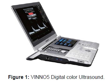
VINNO5 is a professional digital color ultrasonic apparatus (VINNO Technology, Jiangsu, China). It transmits ultrasound waves into the body tissues and displays the echo images of the tissues and blood flow on the screen (Figure 1). A linear probe (frequency 10Mhz) in B-Mode was used for the ultrasound study.
In physiotherapy, ultrasound is used as a support device for specific treatments (ESWT, lasertherapy, FREMS) and to optimize the rehabilitation program. The physiotherapist does not use ultrasound imaging for diagnostic purposes, which is a specific medical competence [15].
ESWT
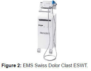
A radial shockwave device (EMS Swiss Dolor Clast, EMS Electro Medical Systems, Nyon, Switzerland) was used (Figure 2). A projectile inside a handpiece is accelerated by a pressurized air source, striking the 15mm-diameter metal applicator, and the generated energy transmitted to the patient’s skin as a shock wave through a standard ultrasound gel. The wave then disperses radially from the application site into the targeted tissue. The energy generated depends considerably on the working pressure set by operator.
At each session, 2500 pulses were applied at a pressure of 2.4 bars (approximate energy flux density of 0.08 mJ/mm²) and a frequency of 12Hz, and the area was treated under ultrasound guidance to ensure all stimulation was directed to the injured tissue.
ESWT has been shown to be a safe and effective noninvasive treatment for patients with calcific tendinopathy of the shoulder, increasing VEGF, PCNA and eNOS expression and modulating substance P in the targeted area [16-20].
FREMS
FREMS (Fremslife Srl Genoa – Italy) is an internationally patented electrostimulation device, classified as an “electroceutical” device as its stimuli is not limited to nerve stimulation but have a specific therapeutic effect: vasomotor activity H-reflex modulation (anticontraction) and vascular endothelial growth factor (VEGF) synthesis have all been demonstrated [21-23].
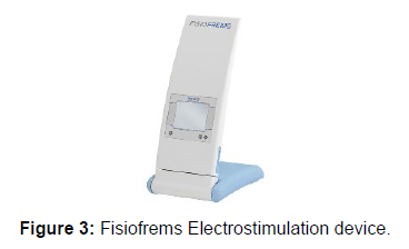
A biphasic balanced current stimulus is modulated in frequency, pulse width and intensity in a pseudorandom way, exerting a physiological stimulus on tissue metabolic activity. In addition to involving selectively different frequency-specific activities, this variability also prevents neural response habituation. FREMS devices are available in two versions which differ only in the number of output dipoles (16 or 32) and in the number and type of treatment protocols, selectable via a user-friendly GUI (Graphic User Interface). (Figure 3).
The Patient
The patient was a 52-year-old female PC operator with a sedentary lifestyle. She was a non-smoker, had a regular menstrual cycle, and followed a varied diet. She complained of intermittent left shoulder pain which had begun two years previously, with episodes of pain becoming more frequent in recent months. No nocturnal pain. Pain was present in abduction and elevation of the left arm, and ultrasound imaging [24] (Figure 4) supported the diagnosis of a calcific tendinopathy of the supraspinatus (0.98 cm in resting phase) performed by an Orthopedist MD.
Clinical assessment
Lax subject with generalized joint hypermobility. Anteposed left shoulder with hypotonotrophy of the periscapular muscles, rotator cuff and deltoid from underuse. Range of movement (ROM) was limited and painful at maximal elevation and abduction. Painful arc test [25] Jobe test [26] and Yocum test [27] were positive.
Rehabilitation program
Three once-weekly ultrasound-guided rESWT sessions. Tissues treated with rESWTs undergo cavitation “build-up”, Mechan transduction and a biological alteration which “kick-starts” the healing response [28]. The treatments should be followed by a controlled program of therapeutic exercises.
Rehabilitation Progress
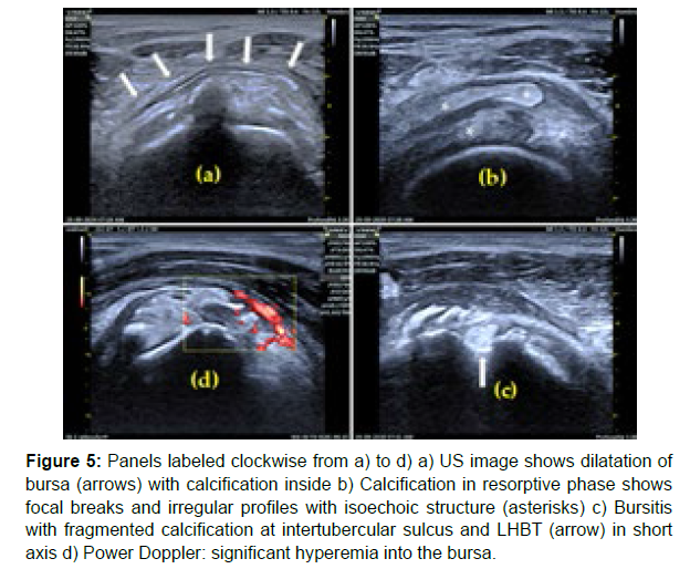
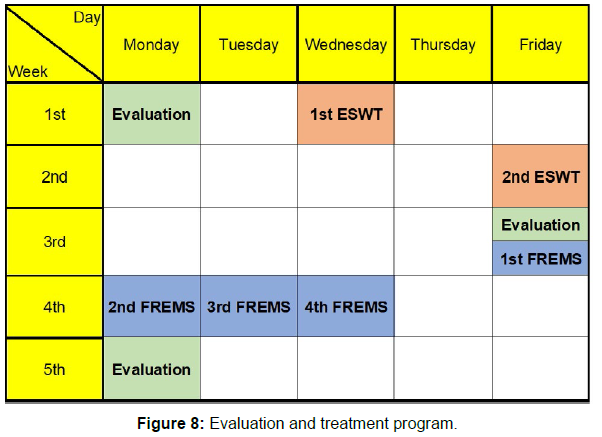
Seven days after the second ESWT treatment, the patient reported a worsening of pain and a reduction in joint mobility of the left shoulder. Pain was also present at rest. Ultrasound imaging showed the beginning of the resorptive phase and migration of the calcification into the subacromial bursa. The bursa appeared thickened and filled with nonhomogeneous hyperechoic fluid containing calcium and debris, in association with edema in the surrounding space [29,30] (Figure 5).
Figure 5: Panels labeled clockwise from a) to d) a) US image shows dilatation of bursa (arrows) with calcification inside b) Calcification in resorptive phase shows focal breaks and irregular profiles with isoechoic structure (asterisks) c) Bursitis with fragmented calcification at intertubercular sulcus and LHBT (arrow) in short axis d) Power Doppler: significant hyperemia into the bursa.
Based on the patient’s clinical condition, we decided to forgo the third ESWT application, since the desired outcome (the start of the resorptive phase) had already been achieved, and four sessions of treatment using the Frequency Rhythmical Electrical Modulation System (FREMS) were planned.
FREMS is known for its effectiveness at modulating the Hoffmann reflex [31] promoting vasomotor activity [32] and treating pain [33] .The selected program (Shoulder – Flogistic lesions) was applied daily for four days (three consecutive) with seven pairs of electrodes applied as shown in Figure 6.
Results
At the follow-up appointment one week after completion of the FREMS treatment cycle, the patient re-ported a significant reduction in pain symptoms and a remarkable improvement in clinical signs (painful arc, Jobe and Yocum tests), with a significant increase in shoulder ROM for both elevation and abduction, and no signs of subacromial impingement.
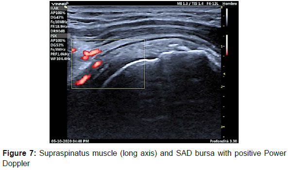
Ultrasound imaging showed an almost complete reabsorption of the calcification, almost complete re-gression of the SAD calcific bursitis, and slight distension of the SAD bursa, while the power Doppler image was still positive (Figure 7), with a high probability of structural remodeling processes in the post calcific phase.
Post treatment activities
The patient was taught to perform a series of exercises at home using an elastic resistance band to pro-mote trophism and support the recovery of muscular proprioception for the muscles involved in the stabilization of the shoulder (scapula and rotator cuff).
General comments
The synergy between ESWT and FREMS with ultrasound monitoring has proven to be successful at providing a rapid solution to a painful condition as part of a four-week treatment program (Figure 8). ESWT stimulates the metabolism of the tendon, initiating the healing process, while FREMS promotes the correct progression of flogosis in the resorptive phase and supports microcirculation in the area.
Discussion
Three important aspects emerge from this case study:
• The combination of different physical agents in this case ESWT and FREMS offer a synergistic approach for a “fast track rehabilitation”, with better and faster results. Time is a crucial factor in reducing needless patient discomfort and the risk of comorbidities, as well as overall healthcare costs.
• The combination of conventional functional evaluation tests and ultrasound imaging to obtain evidence-based information is of immense value for guiding and tailoring the rehabilitation program. In this case, ultrasound imaging permitted us to exclude a session of ESWT which would have been pointless (and contraindicated, increasing pain and inflammation, while reducing the total treatment time by at least one week (or 25%). Our experience suggests that the use of ultrasound imaging by the physiotherapist as a monitoring instrument would improve the overall results of rehabilitation programs in most musculoskeletal conditions normally treated by physiotherapists.
• Although this is just a case report, it reflects the physiotherapy experience gained at the clinical re-habilitation centers where we operate. In our shared vision and experience, patient-centered rehabilitation begins with the evaluation process, essential for achieving the best possible objectivity and reliability. The process then continues in the form of a combination of rehabilitation techniques based on EBR (evidence-based rehabilitation), or KBR (knowledge-based rehabilitation) to be more accurate; a body of knowledge which grows from our training, experiences and a critical appraisal of the results we observe day by day. We believe that it is very difficult sometimes we are tempted to say “almost impossible” – to design and perform a statistically sound trial due to the wide variabilities inherent in the practice of physiotherapy.
Funding: This research received no external funding
Ethics: review and approval were waived for this study that was performed during routine rehabilitation activities in the NF Rehab clinic
Patient Consent: The patient gave his informed written consent to the treatment and to the anonymous publication his treatment data
Conflict of Interest: MG is a minority shareholder of Fremslife, that manufactures the FREMS device. NF and GB declare the absence of any conflict of interests.
CARE Check List has been performed and is attached
References
- De Carli A, Pulcinelli F, Rose GD, Pitino D, Ferretti A (2014) Calcific tendinitis of the shoulder. Joints 2: 130-136.
- Oliva F, Via AG, Maffulli N (2011) Calcific tendinopathy of the rotator cuff tendons. Sports Med Arthrosc Rev 19: 237-243.
- Darrieutort-Laffite C, Blanchard F, Le Goff B (2018) Calcific tendonitis of the rotator cuff: From formation to resorption. Jt Bone Spine 85: 687-692.
- Dong S, Li J, Zhao H (2022) Risk Factor Analysis for Predicting the Onset of Rotator Cuff Calcific Tendinitis Based on Artificial Intelligence. Comput Intell Neurosci 8978878.
- Uhthoff HK, Loehr JW (1997) Calcific Tendinopathy of the Rotator Cuff: Pathogenesis, Diagnosis, and Management. J Am Acad Orthop Surg 5: 183-191.
- Medina-Gandionco M, Briggs RA (2020) Calcific Tendinopathy of the Rotator Cuff Treated With Acetic Acid Iontophoresis. J Orthop Sports Phys Ther 50: 650.
- Magosch P, Lichtenberg S, Habermeyer P (2003) Radiale Stosswellentherapie der Tendinosis calcarea der Rotatoren-manschette--Eine prospektive Studie [Radial shock wave therapy in calcifying tendinitis of the rotator cuff--a prospective study]. Z Orthop Ihre Grenzgeb 141: 629-636.
- Farr S, Sevelda F, Mader P, Graf A, Petje G, et al. (2011) Extracorporeal shockwave therapy in calcifying tendinitis of the shoulder. Knee Surg Sports Traumatol Arthrosc 19: 2085-2089.
- Louwerens JKG, Sierevelt IN, Kramer ET (2020) Comparing Ultrasound-Guided Needling Combined with a Sub-acromial Corticosteroid Injection Versus High-Energy Extracorporeal Shockwave Therapy for Calcific Tendinitis of the Rotator Cuff: A Randomized Controlled Trial. Arthroscopy 36: 1823-1833.e1.
- Zhang T, Duan Y, Chen J, Chen X (2019) Efficacy of ultrasound-guided percutaneous lavage for rotator cuff calcific tendinopathy: A systematic review and meta-analysis. Medicine (Baltimore) 98: e15552.
- Frassanito P, Cavalieri C, Maestri R, Felicetti G (2018) Effectiveness of Extracorporeal Shock Wave Therapy and kinesio taping in calcific tendinopathy of the shoulder: a randomized controlled trial. Eur J Phys Rehabil Med 54: 333-340.
- Masini A, Momoli F Novelli M, Salvi V (2004) Nuovi orizzonti nel trattamento conservativo della spalla dolorosa: studio multicentrico sull’utilizzo della FRE.M.S. (Frequency Modulated Neural Stimulation). Lorenz Therapy. Eura Medicophys 40: 433-5.
- Ponti F, Parmeggiani A, Martella C, Facchini G, Spinnato P (2022) Imaging of calcific tendinopathy in atypical sitesy ultrasound and conventional radiography: A pictorial essay. Med Ultrason 24: 235-241.
- Papalexis N, Ponti F, Rinaldi R (2022) Ultrasound-guided Treatments for the Painful Shoulder. Curr Med Imaging 18: 693-700.
- Wang CJ, Yang KD, Wang FS, Chen HH, Wang JW (2003) Shock wave therapy for calcific tendinitis of the shoulder: a prospective clinical study with two-year follow-up. Am J Sports Med 31: 425-430.
- Kuo YR, Wang CT, Wang FS, Chiang YC, Wang CJ (2009) Extracorporeal shock-wave therapy enhanced wound healing via increasing topical blood perfusion and tissue regeneration in a rat model of STZ-induced diabetes. Wound Re-pair Regen17: 522-530.
- Meirer R, Brunner A, Deibl M, Oehlbauer M, Piza-Katzer H (2007) Shock wave therapy reduces necrotic flap zones and induces VEGF expression in animal epigastric skin flap model. J Reconstr Microsurg 23:231-236.
- Calcagni M, Chen F, Högger DC (2011) Microvascular response to shock wave application in striated skin muscle. J Surg Res 171:347-354.
- Maier M, Averbeck B, Milz S, Refior HJ, Schmitz C 2003) Substance P and prostaglandin E2 release after shock wave application to the rabbit femur. Clin Orthop Relat Res (406): 237-245.
- Famm K, Litt B, Tracey KJ, Boyden ES, Slaoui M (2013) Drug discovery: a jump-start for electroceuticals. Nature 496: 159-161.
- Popa SO, Ferrari M, Andreozzi GM, Martini R, Bagno A (2015) Wavelet analysis of skin perfusion to assess the effects of FREMS therapy before and after occlusive reactive hyperemia. Med Eng Phys 37: 1111-1115.
- Bevilacqua M, Dominguez LJ, Barrella M, Barbagallo M (2007) Induction of vascular endothelial growth factor release by transcutaneous frequency modulated neural stimulation in diabetic polyneuropathy. J Endocrinol Invest 30: 944-947.
- Albano D, Coppola A, Gitto S, Rapisarda S, Messina C (2021) Imaging of calcific tendinopathy around the shoulder: usual and unusual presentations and common pitfalls. Radiol Med 126: 608-619.
- Flynn TW, Cleland J, Julie Whitman (2008) Users' Guide to the Musculoskeletal Examination: Funda-mentals for the Evidence-Based Clinician. Buckner, KY: Evidence in Motion, Print
- Jain NB, Fan R, Higgins LD, Kuhn JE, Ayers GD (2018) Does My Patient With Shoulder Pain Have a Rotator Cuff Tear ? A Predictive Model From the ROW Cohort. Orthop J Sports Med 6: 2325967118784897.
- Ferenczi A, Ostertag A, Lasbleiz S (2018) Reproducibility of sub-acromial impingement tests, including a new clinical manoeuver. Ann Phys Rehabil Med 61:151-155.
- Császár NB, Angstman NB, Milz S (2015) Radial Shock Wave Devices Generate Cavitation. PLoS One 10:e0140541.
- Della Valle V, Bassi EM, Calliada F (2015) Migration of calcium deposits into subacromial-subdeltoid bursa and into humeral head as a rare complication of calcifying tendinitis: sonography and imaging. J Ultrasound 18: 259-263.
- Barrella M, Toscano R, Goldoni M, Bevilacqua M (2007) Frequency rhythmic electrical modulation system (FREMS) on H-reflex amplitudes in healthy subjects. Eura Medicophys 43: 37-47.
- Conti M, Peretti E, Cazzetta G (2009) Frequency-modulated electromagnetic neural stimulation enhances cutaneous microvascular flow in patients with diabetic neuropathy. J Diabetes Complicat 23(1): 46-48.
- Bosi E, Bax G, Scionti L (2013) Frequency-modulated electromagnetic neural stimulation (FREMS) as a treatment for symptomatic diabetic neuropathy: results from a double-blind, randomised, multicentre, long-term, placebo controlled clinical trial. Diabetologia 56: 467-475.
- Crasto W, Altaf QA, Selvaraj DR(2022) Frequency Rhythmic Electrical Modulation System (FREMS) to alleviate painful diabetic peripheral neuropathy: A pilot, randomised controlled trial (The FREMSTOP study). Diabet Med 39: e14710.
- Cook DJ, Griffith LE, Sackett DL (1995) Importance of and satisfaction with work and professional interpersonal issues: a survey of physicians practicing general internal medicine in Ontario. CMAJ 153: 755-764.
Indexed at, Google Scholar, Crossref
Indexed at, Google Scholar, Crossref
Indexed at, Google Scholar, Crossref
Indexed at, Google Scholar, Crossref
Indexed at, Google Scholar, Crossref
Indexed at, Google Scholar, Crossref
Indexed at, Google Scholar, Crossref
Indexed at, Google Scholar, Crossref
Indexed at, Google Scholar, Crossref
Indexed at, Google Scholar, Crossref
Indexed at, Google Scholar, Crossref
Indexed at, Google Scholar, Crossref
Indexed at, Google Scholar, Crossref
Indexed at, Google Scholar, Crossref
Indexed at, Google Scholar, Crossref
Indexed at, Google Scholar, Crossref
Indexed at, Google Scholar, Crossref
Indexed at, Google Scholar, Crossref
Indexed at, Google Scholar, Crossref
Indexed at, Google Scholar, Crossref
Indexed at, Google Scholar, Crossref
Indexed at, Google Scholar, Crossref
Indexed at, Google Scholar, Crossref
Indexed at, Google Scholar, Crossref
Indexed at, Google Scholar, Crossref
Indexed at, Google Scholar, Crossref
Indexed at, Google Scholar, Crossref
Indexed at, Google Scholar, Crossref
Indexed at, Google Scholar, Crossref
Indexed at, Google Scholar, Crossref
Citation: Fortis N, Bernabei G, Gallamini M (2022) Calcific Tendinopathy of the Rotator Cuff (CTRC): The Combined Effects of Extracorporeal Shock Wave Therapy (ESWT) and the Frequency Rhythmic Electrical Modulation System (FREMS) – A Case Report. J Nov Physiother 12: 551.
Copyright: © 2022 Fortis N, et al. This is an open-access article distributed under the terms of the Creative Commons Attribution License, which permits unrestricted use, distribution, and reproduction in any medium, provided the original author and source are credited.
Share This Article
Recommended Journals
Open Access Journals
Article Usage
- Total views: 3701
- [From(publication date): 0-2022 - Mar 29, 2025]
- Breakdown by view type
- HTML page views: 3258
- PDF downloads: 443

