Breast Tomosynthesis Equipment: Revolutionizing Breast Cancer Detection
Received: 01-Jun-2023 / Manuscript No. roa-23-103646 / Editor assigned: 03-Jun-2023 / PreQC No. roa-23-103646 (PQ) / Reviewed: 17-Jun-2023 / QC No. roa-23-103646 / Revised: 22-Jun-2023 / Manuscript No. roa-23-103646 (R) / Published Date: 29-Jun-2023 DOI: 10.4172/2167-7964.1000460
Image Article
Millions of women worldwide are affected by breast cancer, a serious health issue. Early detection is crucial for improving survival rates, and advancements in medical technology have played a pivotal role in enhancing breast cancer screening methods. One such groundbreaking innovation is breast tomosynthesis equipment, also known as 3D mammography. In this article, we will explore the benefits, working principles, and potential impact of breast tomosynthesis equipment in the field of breast cancer diagnosis.
Understanding breast tomosynthesis
Breast tomosynthesis is an advanced imaging technique that combines traditional digital mammography with 3D imaging technology. Unlike conventional mammography, which captures twodimensional images of the breast, breast tomosynthesis produces a series of high-resolution images, known as slices or layers. These images are obtained by taking multiple low-dose X-ray projections from different angles around the compressed breast.
Working principles
The process begins with the patient positioned similarly to a traditional mammogram, where the breast is compressed between two plates. The X-ray tube moves in an arc over the breast, capturing a series of images from different angles. These images are then reconstructed by a sophisticated computer algorithm, creating a three-dimensional representation of the breast tissue.
Benefits of breast tomosynthesis
Improved detection: Breast tomosynthesis equipment offers enhanced visualization of breast tissue by reducing the overlapping of structures. This enables radiologists to detect smaller lesions and distinguish between benign and malignant abnormalities more accurately.
Increased sensitivity: Studies have demonstrated that breast tomosynthesis has a higher sensitivity for detecting breast cancers compared to traditional mammography. It is particularly effective for women with dense breast tissue, where the risk of missing abnormalities is higher.
Lower recall rates: One of the primary challenges in breast cancer screening is the occurrence of false positives, which can lead to unnecessary anxiety and additional testing. Breast tomosynthesis has shown to significantly reduce recall rates by providing more precise information and reducing false alarms.
Enhanced diagnostic accuracy: The 3D imaging capability of breast tomosynthesis allows radiologists to view the breast tissue layer by layer, improving their ability to evaluate the extent and characteristics of abnormalities. This leads to more accurate diagnoses and personalized treatment plans.
Patient comfort: Despite the need for breast compression during the imaging process, breast tomosynthesis equipment offers a more comfortable experience for patients. The procedure is quick, typically taking only a few seconds per image, resulting in reduced discomfort and anxiety.
The future of breast cancer screening
Breast tomosynthesis equipment represents a significant leap forward in breast cancer screening technology. Its superior imaging capabilities and increased diagnostic accuracy have the potential to revolutionize breast cancer detection. As the technology continues to evolve, further enhancements and refinements are expected, including reduced radiation doses, faster imaging times, and improved image quality [1,2].
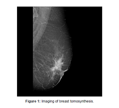
Breast tomosynthesis equipment has emerged as a game-changer in the field of breast cancer screening. By providing three-dimensional imaging and reducing the limitations of traditional mammography, this innovative technology offers improved detection, increased sensitivity, and enhanced diagnostic accuracy. As breast tomosynthesis becomes more widely available, it holds the promise of saving more lives through early detection and targeted treatment (Figure 1).
Acknowledgement
None
Conflict of Interest
None
References
- Thomassin-Naggara I, Cornelis F, Kermarrec E (2019) Breast interventional imaging. Presse Med 48: 1169-1174.
- Whitman GJ (2021) Breast Ultrasound: An Essential Tool. Ultrasound Q 37: 1-2.
Indexed at, Google Scholar, Crossref
Citation: Patel P (2023) Breast Tomosynthesis Equipment: Revolutionizing Breast Cancer Detection. OMICS J Radiol 12: 460. DOI: 10.4172/2167-7964.1000460
Copyright: © 2023 Patel P. This is an open-access article distributed under the terms of the Creative Commons Attribution License, which permits unrestricted use, distribution, and reproduction in any medium, provided the original author and source are credited.
Select your language of interest to view the total content in your interested language
Share This Article
Open Access Journals
Article Tools
Article Usage
- Total views: 1511
- [From(publication date): 0-2023 - Oct 26, 2025]
- Breakdown by view type
- HTML page views: 1213
- PDF downloads: 298

