Research Article Open Access
Brain Gliomas CT-MRI Image Fusion for Accurate Delineation of Gross Tumor Volume in Three Dimensional Conformal Radiation Therapy
| Khalid Iqbal1,2*, Saima Altaf1,2, Muhammad Aqeel3, Muhammad Akram1, Kent A Gifford4 and Saeed Ahmad Buzdar1 | |
| 1Department of physics, The Islamia University of Bahawalpur, Pakistan | |
| 2Department of Radiation Oncology, Shaukat Khanum Cancer Hospital & Research Center, Lahore, Pakistan | |
| 3North West General Hospital and Research Centre, Peshawar, Pakistan | |
| 4Department of Radiation Physics, The University of Texas M D Anderson Cancer Center, Houston, TX, USA | |
| Corresponding Author : | author: Khalid Iqbal Department of Radiation Oncology Shaukat Khanum Cancer Hospital &Research Center Lahore, Pakistan Tel:0092-42-35945100 Fax: 0092-42-35945206 E-mail: Khalid_phy@yahoo.com |
| Received date: December 03, 2014; Accepted date: April 24, 2015; Published date: April 27, 2015 | |
| Citation: Iqbal K, Altaf S, Aqeel M, Akram M, Gifford KA, et al. (2015) Brain Gliomas CT-MRI Image Fusion for Accurate Delineation of Gross Tumor Volume in Three Dimensional Conformal Radiation Therapy. OMICS J Radiol 4:184. doi: 10.4172/2167-7964.1000184 | |
| Copyright: © 2015 Iqbal K, et al. This is an open-access article distributed under the terms of the Creative Commons Attribution License, which permits unrestricted use, distribution, and reproduction in any medium, provided the original author and source are credited.. | |
Visit for more related articles at Journal of Radiology
Abstract
Image fusion has become a widely used tool for enhancing the quality of images in medical applications. This article examines the utility of integrating images from Computed Tomography (CT) and Magnetic Resonance Imaging (MRI) and compare Gross tumor volume (GTV) delineated in CT and CT-MRI fused image data sets separately. Delineation of Gross Tumor volume defined by fusing the two imaging modalities is an important step and could provide significant difference using CT and CT-MRI fused image data sets. In this study twenty two patients suffering from Brain Gliomas were evaluated using CT and CT/MRI images. Match points Registration method is used for the fusion of CT and MRI image data sets. The GTV of CT-MRI fused images was large as compared to CT images with mean volume of (Mean ± SD: 112.67 ± 74.73cc) and 102.15 ± 64.88cc respectively. Difference observed for GTV is 10.52 ± 10.05cc (p=0.004). After taking CT-MRI fused image as reference, calculated mean percentage difference in volumes was about 12%. It was found that GTV was larger on CT-MRI image for brain Gliomas than CT image. Therefore, the fusion of two imaging modalities is recommended for accurate delineation of GTV in radiation therapy treatment planning of brain tumor.
| Keywords |
| CT-MRI fusion; Computed tomography; Multimodality image fusion; Brain gliomas |
| Introduction |
| Quality of image is one of the major component in Three Dimensional Radiotherapy (3DCRT) as well as in Intensity Modulated Radiotherapy (IMRT).Target volumes including gross tumor volume (GTV) and clinical tumor volume (CTV) can be delineated according to the International Commission On Radiation Units (ICRU)-report 50 [1,2]. Marking of the target volume can be done on images from computed tomography (CT) and Magnetic Resonance Imaging (MRI) which really helps to investigate the anatomy of the brain with high precision [3,4]. CT images can easily recognize anatomy of the brain because it provides spatial accuracy and electron density information for heterogeneity correction in Treatment Planning Systems (TPS) for dose calculation [5-9,10]. At the same time poor soft tissue contrast is one of the major disadvantages to outline the normal organs in CT images. |
| Since MRI provides better contrast than CT while differentiating soft tissues delineation of target volumes in MRI images is more accurate and precise but at the same time it has some disadvantages [6]. An MRI image does not provide the exact physical information than the CT. This physical information given in MRI data set can be used into CT data sets by fusing both CT and MRI images [4]. Registration of CT images with MRI images is more effective method in delineation of target volume and organ at risk [5,6,9,11-16]. This integration of combined information of images from two different modalities into a single consistent image is known as fusion of images. CT and MRI registration can be done with different software. A CTMRI fused image really helps the radiation oncologists as well as physicists to outline the tumor and OARs for the better treatment in radiotherapy. Moreover CT-MRI fusion results in improving the delineation of target volumes in brain Gliomas [3,4,10,13]. The aim of this study is to compare the GTV and to find the percentage volume differences when target is separately marked on CT image and CTMRI fused image. Also the importance of the better quality of the image and new modalities used in the Medical imaging which will really helpful in radiotherapy planning is investigated. |
| Materials and Methods |
| Total 22 patients with confirmed stage III/IV Gliomas were selected in this study for examination. All the patients were immobilized with thermoplastic face mask and the whole job was performed for contrast CT and MRI at department of radiation oncology, Shaukat Khanum Memorial Cancer Hospital & Research Center (SKMCH & RC) Lahore Pakistan. CT scans of the patients were obtained on the flat couch of CT scanner (Toshiba, Tsx-021B, JAPAN) with a field of view (FOV) 720 mm and matrix size of 512 × 512. All images were taken from vertex to the 2 cm below the cervical spine. Axial images were reconstructed to 5 mm and transferred to Eclipse treatment planning system (TPS), version 6.5 (Varian Medical System). |
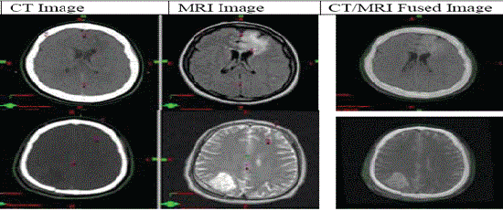
| MRI scans were taken on the 1.5 Tesla (GE medical system, USA) with standard head coil. Axial images were obtained with field of view 480 mm and matrix size 512 × 512; Axial MRI images were reconstructed to 5 mm and transferred to the Eclipse Treatment Planning System (TPS). For the registration of CT and MRI images both T1/T2 weighted images were imported to treatment planning system through DICOM. There are four methods for registration of images in Eclipse treatment planning system named as point matching, DICOM coordinates, image pixel data (automatic registration), and manual registration. We used the method of match points for registration of CT/MRI images. Four match points are required to register two image sets as shown in Figure 1. System requires a reasonable distance between the planes of the match points and position of the match points and it should be same for both image sets. Mean registration error while matching the points was 0.04mm. Registration was rechecked by seeing the anatomical matched markers on fused image by using the option of view blended image. |
| Target volumes and organ at risk were outlined by the radiation oncologists as guided by ICRU report 50. Postoperative planning CT images and pre MRI images were used for target volume delineation. During the marking of gross tumor volume (GTV) the images were seen three dimensionally such as by viewing sagittal, coronal and axial cuts of the image to achieve the high precision. Marked contours of GTV and OAR’s can be edit either on the CT or MRI images by using the editing tools provided in the TPS contouring section. For each patient gross tumor volume was marked on CT images as well as on CT-MRI images (fused image) separately on the treatment planning system. Since all the selected patients had passed through the process of surgery which includes biopsy and resection of residual mass, GTV and pritumoral edema were estimated adequately on each image modality. |
| Results and Discussion |
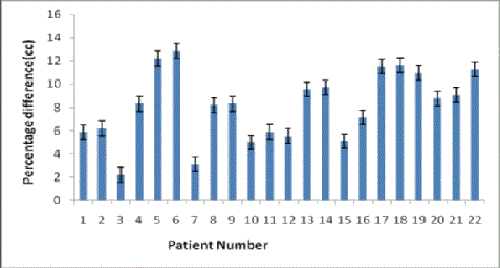
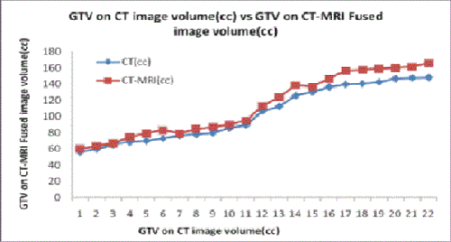
| The GTV was drawn (outlined) on both enhanced contrast CT images and CT-MRI fused images by the experienced radiation oncologist. In all the patients GTV was comparatively large on CTMRI fused images as compared to CT images and this difference was a little large for high grade Gliomas. Mean volume delineated on CTMRI fused image is Mean ± SD: 112.67 ± 74.73 cc and on CT images it is 102.15 ± 64.68cc. Mean Difference observed for GTV is 9.87 ± 10.09cc as shown in Figure 2. |
| Percentage difference calculated taking the CT-MRI fused images volume as reference [CT-MRI fused volume – CT volume/CT-MRI fused volume × 100] and mean percentage volume difference is 8.56%, (SD:3.80)as shown in Table 1. Observed difference in volumes really play a key role to control the under dosage of target volume as well as to control the high dose of critical organs in treatment planning for radiotherapy. |
| Gliomas which include astrocytoma and oligodendroglioma are the most occurring brain tumors which are mostly seen in adults. Gliomas are slowly growing in low grades and rapidly growing in high grades. Tumor marking using only CT images leads to whole brain radiation. Many clinical trials refers the postoperative radiotherapy in such patients, Instead CT/MRI provides helpful information for diagnosis at earlier stage because of good image quality. Especially contrast enhancement in imaging is useful in identifying the isodense lesion from the surrounding normal parenchyma with an area of edema [3]. Malignant Gliomas show less intensity on T1 weighted images and have high intensity on T2 weighted images. Delineation of target volumes is discussed in number of studies by inter-observer and intraobserver variability [3,17,18]. So this study did not emphasize on the observer variability but rather on the importance of target volume difference when two different image modalities was used. |
| CT and CT-MRI images illustrate the clear difference in GTV as each one was obtained from different image modality as shown in Figure 3. This difference is because of difference in mechanism used in both modalities. If we talk about the treatment planning, it can be divided into two phases. For phase-I T2-weighted MRI images can be used, as we can see the edema clearly to mark clinical tumor volume CTV and for boost or phase-II T1-weighted MRI images are used as we concern about the GTV delineation. |
| Conclusion |
| This study highlights the importance of integration of multimodality imaging fusion in conformal radiotherapy for treatment planning of brain Gliomas. This GTV localization and delineation could be expected to facilitate precise and concise radiation therapy treatment planning for brain tumors. This so called Image fusion technique in turn leads to minimum dose to normal tissues and maximum dose to the target tissues. Therefore, ultimately, the fusion of two imaging modalities is recommended for accurate delineation of GTV in conformal radiation therapy treatment planning of brain tumors. |
| Acknowledgements |
| We are thankful to Department of Radiation Oncology, Shaukat Khanum Cancer Hospital and Research Center Lahore, Pakistan for providing facilities to conduct this research work. |
References |
|
Tables and Figures at a glance
| Table 1 |
Figures at a glance
 |
 |
 |
| Figure 1 | Figure 2 | Figure 3 |
Relevant Topics
- Abdominal Radiology
- AI in Radiology
- Breast Imaging
- Cardiovascular Radiology
- Chest Radiology
- Clinical Radiology
- CT Imaging
- Diagnostic Radiology
- Emergency Radiology
- Fluoroscopy Radiology
- General Radiology
- Genitourinary Radiology
- Interventional Radiology Techniques
- Mammography
- Minimal Invasive surgery
- Musculoskeletal Radiology
- Neuroradiology
- Neuroradiology Advances
- Oral and Maxillofacial Radiology
- Radiography
- Radiology Imaging
- Surgical Radiology
- Tele Radiology
- Therapeutic Radiology
Recommended Journals
Article Tools
Article Usage
- Total views: 16999
- [From(publication date):
April-2015 - Jul 06, 2025] - Breakdown by view type
- HTML page views : 12252
- PDF downloads : 4747
