Make the best use of Scientific Research and information from our 700+ peer reviewed, Open Access Journals that operates with the help of 50,000+ Editorial Board Members and esteemed reviewers and 1000+ Scientific associations in Medical, Clinical, Pharmaceutical, Engineering, Technology and Management Fields.
Meet Inspiring Speakers and Experts at our 3000+ Global Conferenceseries Events with over 600+ Conferences, 1200+ Symposiums and 1200+ Workshops on Medical, Pharma, Engineering, Science, Technology and Business
Short Communication Open Access
Bone Infection: Therapeutical Pearls Based on Evidence of 458 Cases on Infected Pseudoarthrosis and Other Cases of Bone Infection in the Department of Orthopedics of Lecco from 1981 to 2011
| Maurizio A Catagni* | ||
| Milano University, Medical School, Italy | ||
| Corresponding Author : | Maurizio A Catagni Professor at the Milano University Medical School, Lecco, Italy Tel: +39-0341-364662 Fax: +39-0341-630630 E-mail: maurizio@catagni.it |
|
| Received August 28, 2014; Accepted October 30, 2014; Published November 06 2014 | ||
| Citation: Catagni MA (2014) Bone Infection: Therapeutical Pearls Based on Evidence of 458 Cases on Infected Pseudoarthrosis and Other Cases of Bone Infection in the Department of Orthopedics of Lecco from 1981 to 2011. J Infect Dis Ther 2:183. doi:10.4172/2332-0877.1000183 | ||
| Copyright: © 2014 Catagni MA. This is an open-access article distributed under the terms of the Creative Commons Attribution License, which permits unrestricted use, distribution, and reproduction in any medium, provided the original author and source are credited. | ||
Related article at Pubmed Pubmed  Scholar Google Scholar Google |
||
Visit for more related articles at Journal of Infectious Diseases & Therapy
| Short Communication |
| Substantially what is bone infection? It’s a bacterial colonization of a tissue which presents scarce or no humoral immunity as it has lost vascularity and has become necrotic. |
| If vascularity is lost, the tissue becomes like a foreign body which is conducive to a surrounding inflammatory granuloma which aims at isolating the necrotic area. However, the microbes remain in situ and proliferate, being essentially isolated from the natural defense system. Any specific antibiotic, which destroys the microbes in vitro where there is ample contact between pathogen and drug, cannot reach the pathogen in a way to be effective in vivo, as the vascularity which is the delivery vehicle does not extend into the necrotic bone and therefore stops in the inflammatory area without reaching the necrotic bone. |
| In the years past, and unfortunately, to this date, there was, and still remains, a school of thought (purely philosophical in my opinion), which considers bone infection in the same line of any other localized infection where vascularity is maintained and the possibility that it can be reached by drugs and humoral defensive is real. |
| Even from a diagnostic point of view, it is currently observed (in portion due to defensive medicine) that there is a waste of tests which are not at all useful from a diagnostic point of view and even less from a therapeutic standpoint. They are also highly invasive (CT and Scintiography). A diagnosis of bone infection is relatively straightforward if the clinician has the patience to examine the patient and order a simple plain film radiograph; inflammatory blood markers are useful to better assess although not indispensable. When a patient presents in tumefaction, pain and fever make the diagnosis highly probable. Certainly, the differential diagnosis should include other pathologies, perhaps, metastatic, but patient history is highly indicative. If, for example, a leg fracture is treated with a internal plate and presents with an inflammatory reaction, with fever, tumefaction and perhaps a fistula, a tumor should be ruled out easily, but an infection is obviously likely possible and it will not be scintiography to enlighten the physician to reach the diagnosis! it would be as if on a rainy day, one would call a chemist to analyze a strange substance falling from the sky!!! |
| Accepting that bone infection is made in a context of necrotic tissue , where bone is necrotic and does not resorb like any necrotic soft tissue but becomes sclerotic and can remain in situ for decades like a foreign body, maintaining an ideal environment for proliferation, it becomes obvious that medical treatment cannot be effective. |
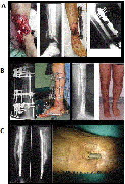
| A bone infection is a complication, in the majority of cases, of high or low energy trauma which results in a fracture with periosteal stripping of the bone, comminution with devitalized fragments and perhaps contaminated from exposure to the environment. It can also occur from a closed, simple fracture turned open for surgical fixation (Figure 1). A closed, low energy fracture will rarely if ever become infected due to preexisting comorbidities (diabetes etc.). |
| Evaluating a closed fracture to be surgically fixated, risk factors for surgical fixation related to internal fixation are poor stability of the fixation construct, excessive periosteal stripping of devitalized fragments which can become completely necrotic and therefore easily succumb to infection. Also, it should not be forgotten that it is the soft tissue that nourish the periosteum and careless dissection, perhaps to apply 2-3 plates, inevitably leads to necrosis of the overlying soft tissue with interruption of the periosteal nourishment, bone exposure and fixation, and therefore necrosis and infection (Figure 2). |
| Even in the case of less aggressive surgical exposure, as in intramedullary nailing, stability and anatomy of fracture segments should be respected, without forgetting that reaming with instruments that are not sharp, lead to accelerated burn necrosis of the medullary canal and of the overlyingsoft tissue, which can lead to bone exposure. The fact that this leads to exposure of bone and necrosis in only a few hours is universally accepted. Therefore, to avoid infection and bone necrosis, it is necessary to follow a few rules so surgery can be performed in a logic manner and by expert hands. If one has never treated bone infection they will never identify it, treat it effectively and will ultimately lose the patient. It should not be forgotten that bone infection carries an extremely high public health cost not only for direct treatment related costs, but more importantly for the quality of life of the patient which drastically deteriorates. |
| However, when an bone infection presents itself, treatment choice becomes for some really difficult and complicated, especially if one does not apply a simple and logical approach: determine why the infection developed to begin with. |
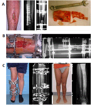
| If the patient was treated for a high energy fracture with external fixator and the bone fragments are devitalized and more importantly exposed, a reconstructive effort is useless and perhaps dangerous, as if burying a cadaver under a thin layer of dirt, worms would come out immediately! The solution in these cases should be to remove all devitalized fragments and devitalized soft tissue and the process with soft tissue coverage procedures (Figure 3). |
| Such step has always been successfully one the bone fragments have been stabilized and all necrotic tissue debrided. Once the patient has been stabilized, skeletal reconstruction can be carried out with whatever procedure the surgeon is comfortable with. If familiar with bone transport with external fixation, the surgeon can apply it for large bone defects, whereas for small defects, other methods are available (i.e. Masquele’, vascularized fibular transplant). |
| In the case of infection over hardware, it is absolutely paramount and sine qua non to success to remove all indwelling hardware which represents a nidus for infection and at the same time a careful examination of the bone. If soft tissue coverage is adequate and the bone doesn’t present any necrotic foci, the simple removal of hardware and application of antibiotic beads can cure the infection. However, it is still necessary to maintain skeletal stability, obviously external, to encourage healing of the fracture. In cases of necrotic bone more or less associated with soft tissue loss, bone debridement should be performed without hesitation, visually inspecting the bone. At the same time, reconstructive planning should be carried out. |
| The surgeons who use bone transport has a lesser necessity of involving the plastic surgeon, as soft tissue loss can be corrected during bone transport, thus re establishing the bone and soft tissue continuity. |
| In any case, clinical examination has to be the pinnacle for diagnosis. CT is not better than plain film radiographs to evaluate bone loss, fixation position and stability; for an evaluation of the extent of bone to be debrided, only direct visual inspection can reveal the level of vascularized bone. A bone scan, however invasive, is easily positive even in the presence of a hypertrophic pseudoarthrosis where there is active repair process, even if it does not lead to bone consolidation, it is still a continued inflammatory process and the radioactive material will tend to deposit with the tagged white blood cells. |
| RMN never gives, in our extensive experience, any indication of either extent of infection or extent of bone to be debrided. |
| Now, in the case of bone loss after resection and temporary fixation, the question remains, what is the percentage and quality of healing that can be expected. Ideally, beyond eliminating the infection, and obtain bony consolidation, it is absolutely important to obtain axial alignment of the bone segments. This is necessary to avoid loss of symmetry and to restore function of adjacent joints. |
| In our experience of treatment with circular fixators and bone transport, the achievement of the goals set close to 99% with treatment times vary based on the amount of the osteo-cutaneous loss, between 7 and 18 months with circular external fixator (average 11 months). |
| Probably in future the reconstruction with bone grafts, autologous mesenchymal cells, BMP osteoinductive and drugs, maybe will be able to shorten the time of treatment then the result is always a bone morphologically and mechanically valid with function as normal as possible and hoping that the health system can withstand the extremely high costs currently of these new procedures. |
Figures at a glance
 |
 |
 |
||
| Figure 1 | Figure 2 | Figure 3 |
Post your comment
Relevant Topics
- Advanced Therapies
- Chicken Pox
- Ciprofloxacin
- Colon Infection
- Conjunctivitis
- Herpes Virus
- HIV and AIDS Research
- Human Papilloma Virus
- Infection
- Infection in Blood
- Infections Prevention
- Infectious Diseases in Children
- Influenza
- Liver Diseases
- Respiratory Tract Infections
- T Cell Lymphomatic Virus
- Treatment for Infectious Diseases
- Viral Encephalitis
- Yeast Infection
Recommended Journals
Article Tools
Article Usage
- Total views: 14259
- [From(publication date):
December-2014 - Apr 09, 2025] - Breakdown by view type
- HTML page views : 9674
- PDF downloads : 4585
