Research Article Open Access
Bond Strength Comparison of One-Step/Two-Step Self-Etch Adhesives to Cavity Floor Dentin after 2.5 Years Storage in Water
Satoki Kawashima, Koichi Shinkai*, Masaya Suzuki and Shiro SuzukiDepartment of Operative Dentistry, The Nippon Dental University School of Life Dentistry at Niigata, Chuo-ku, Niigata, Japan
- *Corresponding Author:
- Koichi Shinkai
Department of Operative Dentistry
The Nippon Dental University School of Life Dentistry at Niigata
Chuo-ku, Niigata, Japan
Tel: +81-25-267-1500
Fax: +81-25-265-7259
E-mail: shinkaik@ngt.ndu.ac.jp
Received date: May 16, 2017; Accepted date: June 12, 2017; Published date: June 16, 2017
Citation: Kawashima S, Shinkai K, Suzuki M, Suzuki S (2017) Bond Strength Comparison of One-Step/Two-Step Self-Etch Adhesives to Cavity Floor Dentin after 2.5 Years Storage in Water. J Interdiscipl Med Dent Sci 5: 213. doi:10.4172/2376-032X.1000213
Copyright: © 2017 Kawashima S, et al. This is an open-access article distributed under the terms of the Creative Commons Attribution License, which permits unrestricted use, distribution, and reproduction in any medium, provided the original author and source are credited.
Visit for more related articles at JBR Journal of Interdisciplinary Medicine and Dental Science
Abstract
In recent years, decrease in dentin bond strength of one-step self-etch adhesives after long-term water storage has been reported; however, the flat dentin surface of the extracted tooth has been used to carry out the bond strength tests in the majority of the previous studies. Objectives: The purpose of this study was to compare the micro-tensile-bond-strength (μTBS) of three one-step self-etch adhesives and a two-step self-etch adhesive on the cavity floor dentin after 2.5 years storage in distilled water. Materials and Methods: Adhesives were used in this study: three one-step self-etch adhesives (Primefil, GBOND PLUS and Beauti Bond), and one two-step self-etch adhesive (Clearfil SE Bond), which was used as the control. Bowl-shaped cavities of extracted human incisors were treated with each adhesive and filled with each flowable resin composite from the same manufacturer. The specimens were stored in distilled water at room temperature for two and a half years. The beam samples were made and subjected to μTBS test using the tabletopmaterial- tester. The data were statistically analyzed using one-way ANOVA. Results: The μTBS value of G-BOND PLUS was higher than that of other adhesives; however, the results of ANOVA showed no significant differences in μTBS values among all adhesives. Conclusion: There was no significant difference between one-step and two-step on the bond strength of selfetch adhesives to the cavity dentin floor after a storage period of 2.5 year.
Keywords
Micro tensile bond strength; One-step self-etch adhesive; Long-term water storage; Cavity dentin floor
Introduction
Flowable resin composites have been used frequently in clinics due to improvements in bond strength, physical properties, esthetics, and ease of handling. Recently, the successful use of flowable resin composites on the occlusal surface has been reported owing to their superior mechanical properties and ease of handling [1,2]. In addition, the strong bonding between flowable resin composites and the tooth surface is also a key-factor for the success of the restoration. Various adhesive systems have been developed to improve the bonding between the tooth and the resin composite [3-7]. A basic dentin surface treatment consists of three steps including, etching, priming and bonding [3,5]. The introduction of the priming procedure led to significant improvements in dentin bond strength [3,5]. Subsequently, to simplify the bonding procedure, a two-step self-etch or etch-andrinse adhesive system was developed [4,6,7]. This was then replaced by a one-step treatment method [6]. However, several studies have reported the demerits of the one-step adhesive system [8,9]. Unfavourable factors including phase separation of the liquid, transpiration of the solvent, low efficacy of tooth decalcification, and low content of adhesive monomer were recognized to affect dentin bonding in the one-step adhesive system [10-12]. The acidity of the liquid appeared to lower the efficacy of decalcification in the one-step adhesive system when compared with the two-step adhesive system [4]. The presence of a smear layer on prepared dentin treated using the one-step adhesive system has been reported in several studies [11,12].
In recent years, the deterioration of bonding interfaces and the decrease in bond strength after long-term storage have been discussed [7,11,13-17]. Loguercio AD et al. reported that bond strength decrease was observed in one-step self-etch systems after three years; however, degradation rates after three years were reduced when the adhesives were appliedactivelytodentin [15]. Itoh S et al. examined the microtensile bond strength (μTBS) of three HEMA-containing one-step selfetch adhesives after water storage for 24 h, 6 months, and 1 year, and the amounts of initial water sorption and solubility were measured [14]. Their results showed that the μTBS of all adhesives decreased over time; however, water sorption and solubility were not related to the dentin bonding durability of the one-step self-etch adhesives tested [14]. Osorio R et al. evaluated the effects of different application parameters on μTBS of a one-step self-etch adhesive to dentin after storage for 24 h, 6 months, and 1 year in water at 37°C; they concluded that all groups showed a decrease in μTBS after water storage for 6 months and 1 year, (but not 24 h) [16].
Several studies have reported that deterioration of adhesive-dentin interface occurred due to hydrolysis of the dentin collagen [9,10,13]. Fukuoka A et al. evaluated the hydrophilic stability of three one-step self-etch adhesives bonded to dentin through interfacial morphological analysis before and after long-term thermo-cycling by TEM observation, and found many voids as well as degradation of collagen fibrils at the interface after 100,000 thermo-cycles [13]. Hashimoto M et al. examined the morphological aspects of adhesive interface using four one-step self-etch adhesives after water storage for 24 h, and 100, 200, and 300 days by SEM observation [8]. The observation revealed an oxygen-inhibition zone at the adhesive-composite border after 24 hours, which resulted in a decrease in the dentin bond strength of the adhesives after water storage for 100 days [8]. Thus, degradation of the adhesive-dentin interface due to denaturation of collagen fibrils may be associated with the decrease in the dentin bond strength of the onestep self-etch adhesives.
Although many studies have described the dentin bond strengths of one-step self-etch adhesives after short-term and long-term storage [7,9-11,13-17], most of them used specimens in which the resin composite was bonded to the prepared flat dentin surface. Very few studies have evaluated the dentin bond strength of one-step self-etch adhesives on the cavity floor [18-20].
The purpose of this study was to compare the micro-tensile bond strength (μTBS) of three one-step self-etch adhesives and a two-step self-etch adhesive on the cavity floor dentin after 2.5 years storage in distilled water. The null hypothesis was that the dentin bond strengths of one-step self-etch adhesives would not be significantly different from that of a two-step self-etch adhesive on the cavity floor after longterm water storage.
Materials and Methods
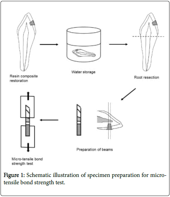
This study was approved by the Ethics Committee of The Nippon Dental University School of Life Dentistry at Niigata, Japan (approval No.131). Figure 1 presents a schematic illustration of the specimen preparation and μTBS test used in this study.
Materials
As shown in Table 1, three one-step self-etch adhesives including Primefil (PF), Beauti Bond (BB), and G-BOND PLUS (GB) were used in this study. A two-step self-etch adhesive (Clearfil SE Bond, CS) was used as a control. The flowable resin composite from the same manufacturer was used for each adhesive.
| Materials | Abb. | Lot no. | Composition | Manufacturer |
|---|---|---|---|---|
| Primefil (Primer) | PF | 002B | Acetone, HEMA, Bis-GMA, Peroxide, Phosphoric acid monomer, Purified water, CQ | Tokuyama Dental |
| Primefil (Paste) | J204 | Silica zirconia filler, Purified water, Bis-GMA, TEGDMA, Bis-MPEP | ||
| BeautiBond | BB | 41173 | Acetone, Purified water, Bis-GMA, Carboxylic acid monomer, Phosphoric acid monomer, TEGDMA | |
| Beautifil Flow Plus (F00) | 11116 | S-PRG filler, MF glass filler, Pigment, Ultra fine filler, Bis-GMA, TEGDMA, Long-chain crosslinking monomer, Photo-initiator | Shofu | |
| G-BOND PLUS | GB | 1009101 | Methacrylate, Phosphoric acid esters, 4-MET, UDMA, Acetone, water, Silica, Zirconia, UDMA, Fluoroaluminatesilicate | GC |
| Unifil Lo Flo Plus | CS | 1005191 | Primer:HEMAJO-MDP, Hydrophilic aliphatic dimethacrylate, CQ, Water, Accelerators, Dyes | |
| Clearfil SE Bond | 01373A | Bond:Bis-GMA, HEMA, 10-MDP, Hydrophobic aliphatic dimethacrylate,Colloidal silica, CQ, Initiators, Accelerators | ||
| Cleafil Majesty LV | 00303B | TEGDMA, CQ, Hydrophobic aromatic dimethacrylate, Silanated barium glass filler, Silanated colloidal silica | Kuraray Noritake Dental |
HEMA, 2- hydroxyethyl methacrylate; Bis-GMA, 2,2-Bis[4-(2-hydroxy-3-methacryloxypropoxy) phenyl]propane,
Bis-MPEP, Bisphenol A polyethoxymethacryhte; TEGDMA, Triethylene glycol dimethacrylate S-PRG, surface pre-reacted glass; 4-MET, 4-mathacryloxyetyl trimellitic acid.
UDMA, Urethane dimethacrylate; 10-MDP, 10-methacryloxydecyl dihydrogen phosphate.
Table 1: Materials used in the present study.
Specimen preparation
Forty extracted human incisors were cleaned and stored in 0.01% thymol solution at 4°C until further use. The labial surfaces of the extracted teeth were ground with 120-grit silicon carbide paper (Carbimet, Buehler Ltd., Lake Bluff, IL, USA) in order to obtain a flat enamel surface, and finished with 600-grit paper using a polishing machine (Lewel specimen polisher, Kasai Co. Ltd., Yokohama, Japan) under water irrigation. Bowl-shaped cavities, 4 mm in diameter and 2 mm in depth, were prepared on the cervical area of the flat labial surfaces using a barrel-shaped diamond point (Bur No.149, Shofu, Kyoto, Japan). Each cavity was randomly assigned to one of four adhesives (n=10).
The adhesives were separately applied to each cavity according to the manufacturers’ instructions. After the application of the all-in-one adhesive the cavity was gently air-blown for 3s, and intensively for another 3s. The bonding agents of GB, BB and CS were photopolymerized for 10s using a light-curing unit (Candelux, Morita Corp., Tokyo, Japan). The bonding agents in PF were not irradiated with light and left for 20s. Each flowable resin composite was placed into the cavity and photopolymerized for 30s through a 0.04 mm thick transparent plastic matrix (matrix tape, 3 M ESPE, St Paul, MN). Thereafter, the specimens were stored in distilled water at room temperature for two and a half years.
Microtensile bond strength test
After storage, the roots were removed using a diamond point (Bur No.105R, ISO size 22; Shofu Inc., Kyoto, Japan). The exposed pulpchamber wall was filled with flowable resin composite (Cleafil Majesty LV) after the application of an accompanied adhesive system (Clearfil SE Bond). The specimens were perpendicularly sectioned into 1 mm thick slabs along the tooth axis through the center of the resin composite restorations using a low-speed diamond saw (Isomet, Buehler Inc., Lake Bluff, IL, USA) under water cooling. The slab obtained from each specimen was then sectioned into a beam (crosssectional area, approximately 1 mm2) using a low-speed diamond saw. The beam samples were attached to the testing device (Bencor-multi-T, Danville Engineering Inc., San Ramon, CA, USA) with cyanoacrylate (Model Repair Pink, Dentsply-Sankin Inc., Tochigi, Japan), which was placed onto the Tabletop material tester (EZ test, Shimadzu Corp., Kyoto, Japan), and subjected to the μTBS test at 0.5 mm/min crosshead speed (Figure 1).
Failure mode analysis
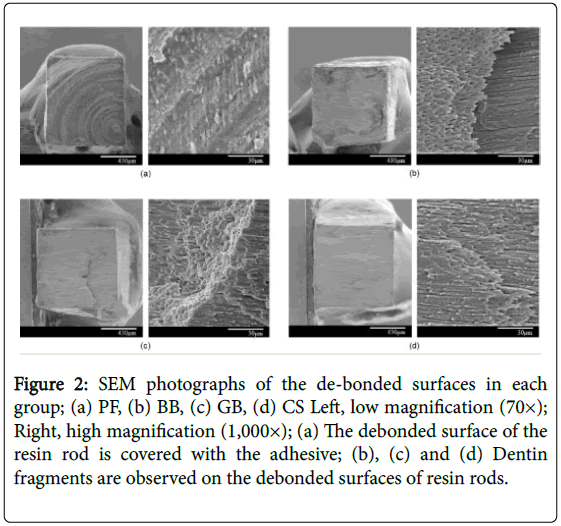
Fractured surfaces of the specimens were examined with a stereomicroscope (Leica EZ4D, Leica Microsystems, Heerbrugg, Switzerland) at 35x magnification, and the fracture modes were determined. Modes of failure were classified as follows: adhesive, when the failure occurred entirely within the adhesive area; cohesive in resin composites, when the failure occurred exclusively within the resin composite area; cohesive in dentin, when the failure occurred exclusively within the dentin area; and mixed, when the failure extended from the adhesive into either the resin composite or the dentin area. Several representative samples were selected from each group for precise analysis of the fractured surfaces using a scanning electron microscope (SEM, S-800, Hitachi Ltd., Tokyo, Japan) at an accelerating voltage of 15 kV after being sputter-coated with palladium and platinum.
Statistical Analysis
The data of the μTBS test were statistically analyzed using one-way ANOVA, followed by Tukey Kramer’s post-hoc test to compare the μTBS values among the four groups (PF, BB, GB and CS) at a 95% confidence level. Statistical analysis was carried out using a statistical analysis add-in software package for Microsoft Excel (BellCurve for Excel, Social Survey Research Information Co., Ltd., Tokyo, Japan). We calculated the power of the one-way ANOVA at an effect size of 0.45 (Cohen’s large effect size), an alpha error probability of 0.05, a total sample size of 40, and four groups using Power analysis software (G Power software version 3.1.9.2). The power of the one-way ANOVA performed in this study was 0.61.
Results
Table 2 presents the results of the μTBS test. The number of detachment between the adhesive and the cavity floor dentin in specimens with PF, BB, GB and CS during beam preparation (pre-test failure) was 4, 4, 2 and 3, respectively. The bond strength of these specimens was considered 0 MPa. The number of detachment at adhesive/pulp chamber dentin interface with PF, BB, GB and CS was 0, 2, 2 and 2, respectively. The bond strengths of these specimens were excluded from the data. The minimum mean value of μTBS was 4.6 MPa in CS, and the maximum mean value was 12.4 MPa in GB. Oneway ANOVA revealed no significant differences in μTBS values among all adhesives (p>0.05) (Figure 1).
| Group | ilTBS* (MPa) | Failure mode** (number of specimens) | Number of detached | |||
|---|---|---|---|---|---|---|
| Mean (SD) | A | Cd | Cr | M | specimens*** | |
| PF | 5.7 (5.2) a | 7 | 0 | 0 | 3 | 4 |
| BB | 5.6 (6.9) ab | 6 | 0 | 0 | 2 | 4 |
| GB | 12.4 (8.3) b | 5 | 1 | 0 | 2 | 2 |
| CS | 4.6 (4.2) a | 6 | 0 | 0 | 2 | 3 |
Table 2: Results of ilTBS test and failure mode analysis (n=10), *Value with the same superscript letters indicates no significant difference (p>0.01);**A: Adhesive failure, Cd: Cohesive failure in dentin, Cr: Cohesive failure in resin, M: Mixed failure; ***Detachment occurred during preparation of specimens for ilTBS test.
All pre-test failures demonstrated adhesive failures. The results of the failure mode analysis revealed adhesive failure (70.6%) as the predominant failure mode, which was followed by mixed failure (26.5%). Figure 2 shows representative SEM photographs of the debonded surfaces of the resin rods after μTBS test in each group. In the PF specimen, the de-bonded surface of the resin rod was covered with the adhesive, thus, it was considered as an adhesive failure (Figure 2a). The SEM photographs of BB, CS, and GB specimens partially demonstrated flat and fractured dentin surfaces on the resin rods, and were therefore, labelled as mixed failures (Figures 2b-2d).
Figure 2: SEM photographs of the de-bonded surfaces in each group; (a) PF, (b) BB, (c) GB, (d) CS Left, low magnification (70×); Right, high magnification (1,000×); (a) The debonded surface of the resin rod is covered with the adhesive; (b), (c) and (d) Dentin fragments are observed on the debonded surfaces of resin rods.
Discussion
The flat dentin surface of the extracted tooth has been used to carry out bond strength tests in the majority of the previous studies. Hence, the findings of those studies may not simulate phenomena occurring in clinical situations. It is well known that the adhesive interface between the resin composite restoration and the dentin is stressed due to contraction during the polymerization. Makishi P et al. evaluated the sealing ability of resin composite restorations using optical coherence tomography (OCT) in order to assess the micro-tensile bond strength of different adhesive systems on the dentin in Class I cavities [21]. They concluded that there was a significant correlation between sealing performance and the bond strength of the adhesives in the whole cavity [21]. In another study, Yoshikawa et al. reported that the bond strength of resin composites to dentin decreased with the increase in polymerization shrinkage [22]. In the present study, Class V resin composite restorations were used to evaluate the micro-tensile bond strength between the one-step self-etch adhesive and the cavity floor dentin. The results of the present study showed high pre-test failure rates (25% to 50%) and lower bond strength values in all groups when compared with the values reported in previous studies after short-term storage. This may be attributed to stress caused during polymerization shrinkage of flowable resin composites. Bakhsh TA et al. investigated class I cavity floor adaptation by OCT in combination with μTBS using different filling methods, and reported that a value of zero MPa was recorded in case a beam failed prior to μTBS testing [19]. Moreover, they mentioned that specimens prepared from areas containing interfacial gaps predominantly failed prior to the test, and were recorded as null bond strength [19]. In the present study, the pretest failure beams were excluded and zero MPa values were not recorded because of the possibility of beam detachment owing to external stress during preparation, thereby compromising the bond strength of the beam.
An adhesive system aids in removal of the smear layer and penetration of the adhesive resin monomer into the dentin surface. The simultaneous progression of the three steps (etching, priming, and bonding) in the one-step self-etch adhesive system is thought to weaken their effects. The two-step self-etch adhesives have shown significantly higher bond strengths when compared with the one-step self-etch adhesives following short-term storage [6,7]. In the present study, the dentin bond strengths of GB were higher than that of CS, whereas, no significant difference in all adhesives after long-term water storage. Therefore, the null hypothesis, which the dentin bond strengths of one-step self-etch adhesives would not be significantly different from that of a two-step self-etch adhesive on the cavity floor after long-term water storage, was accepted.
Previous studies have reported several disadvantages of the onebottle adhesive system, which include the transpiration of the solvent and phase separation within the bottle [9,23]. These shortcomings were found to occur because of an increase in the quantity of water and solvents such as acetone or ethanol [23,24]. The one-step self-etch adhesive contains HEMA and acetone or alcohol to dissolve TEGDMA and Bis-GMA [17]. HEMA is a hydrophilic monomer, which promotes the diffusion of the resin monomer into the demineralization layer of the dentin surface [5,11,25]. The dentin bond strength of the one-step self-etch adhesive containing 10% HEMA is reported to be higher than that of the non-HEMA one-step self-etch adhesive [25]. On the other hand, HEMA has been shown to decrease the physical properties of the hybrid layer by increasing the water absorption due to its hydrophilic nature [25,26]. Therefore, GB, a non-HEMA self-etching adhesive used in the present study exhibited the highest dentin bond strength among the four adhesives tested. It is speculated that the bonding interface of the GB specimens might have retained the physical properties of the hybrid layer when compared with the other adhesives, because of the absence of poly-HEMA.
PF is a new adhesive system that is capable of adhering to the tooth substance via a dedicated bonding agent and the resin composite. This adhesive system does not require the application of the light-curing process because chemical polymerization begins at the bonding interface when the catalyst in the resin composite paste comes in contact with the adhesive [12]. The results of the present study showed that there were no significant differences in dentin bond strengths among PF, BB, and CS, implying that this unique adhesive system, which does not require light-curing for polymerization, may be as effective as other adhesive systems which require light-curing after the application of adhesives.
Many studies have reported significant improvements in the dentin bond strength of resin composites owing to recent developments in adhesive systems [3-7]. Recently, many studies have demonstrated the durability of resin composite bonding to the dentin after long-term storage [4,6-10,15-17,27]; however, the majority of these studies used the self-etch adhesive system with a storage period of less than 2 years. The study by Hashimoto et al. reported disorganization of dentin collagen, a nanoleakage in the hybrid layer, and water tree and spot mode formation in the bonding layer following the use of the onebottle self-etch adhesive [8]. In another study, increased nanoleakage in the hybrid layer was reported after one year of storage with an etchand- rinse adhesive that contained a hydrophilic resin monomer [28]. These results indicate that the dentin bond strength of the self-etch adhesive system decreases in a sequential manner. Although physical factors including occlusal stresses are thought to deteriorate dentin bond strength, recent studies have shown that water sorption by the resin composite and disorganization of the collagen are also associated with the decrease in dentin bond strength [8-10]. Moreover, matrix metalloproteinases (MMPs) released from the dentin matrix contributes to the degradation of the adhesive interface. In this study, the dentin bond strength of three one-step self-etch adhesives were compared with that of a two-step self-etch adhesive after 2.5 years of storage in distilled water. Specimen detachment during beam preparation occurred in almost half of the specimens from each adhesive. The mean values of all adhesives tested in the present study were similar to or lower than those reported in other studies [15-17], which were conducted using specimens with 1-3 year storage periods. Therefore, it was speculated that long-term storage in water may have resulted in the disorganization of collagen fibrils in the hybrid layer for both one-step and two-step self-etch adhesives.
Conclusion
There was no significant difference between one-step and two-step on the bond strength of self-etch adhesives to the cavity dentin floor after a storage period of 2.5 year.
References
- Clelland NL, Pagnotto MP, Kerby RE, Seghi RR (2005) Relative wear of flowable and highly filled composite. J Prosthet Dent 93: 153-157.
- Gallo JR, Burgess JO, Ripps AH, Walker RS, Bell MJ, et al. (2006) Clinical evaluation of 2 flowable composites. Quintessence Int 37: 225-231.
- Ferrari M, Yamamoto K, Vichi A, Finger WJ (1994) Clinical and laboratory evaluation of adhesive restorative systems. Am J Dent 7: 217-219.
- Marchesi G, Frassetto A, Visintini E, Diolosa M, Turco G, et al. (2013) Influence of ageing on self-etch adhesives: one-step vs. two-step systems. Eur J Oral Sci 121: 43-49.
- Munksgaard EC, Asmussen E (1984) Bond strength between dentin and restorative resins mediated by mixtures of HEMA and glutaraldehyde. J Dent Res 63: 1087-1089.
- Sezinando A, Perdigao J, Regalheiro R (2012) Dentin bond strengths of four adhesion strategies after thermal fatigue and 6-month water storage. J Esthet Restor Dent 24: 345-355.
- Takamizawa T, Barkmeier WW, Tsujimoto A, Scheidel DD, Watanabe H, et al. (2015) Influence of water storage on fatigue strength of self-etch adhesives. J Dent 43: 1416-1427.
- Hashimoto M, Fujita S, Endo K, Ohno H (2009) In vitro degradation of resin-dentin bonds with one-bottle self-etching adhesives. Eur J Oral Sci 117: 611-617.
- Hashimoto M, Ohno H, Sano H, Kaga M, Oguchi H (2003) In vitro degradation of resin-dentin bonds analyzed by microtensile bond test, scanning and transmission electron microscopy. Biomaterials 24: 3795-3803.
- Hashimoto M, Fujita S, Kaga M, Yawaka Y (2007) In vitro durability of one-bottle resin adhesives bonded to dentin. Dent Mater J 26: 677-686.
- Shinoda Y, Nakajima M, Hosaka K, Otsuki M, Foxton RM, et al. (2011) Effect of smear layer characteristics on dentin bonding durability of HEMA-free and HEMA-containing one-step self-etch adhesives. Dent Mater J 30: 501-510.
- Suyama Y, Luhrs AK, De Munck J, Mine A, Poitevin A, et al. (2013) Potential smear layer interference with bonding of self-etching adhesives to dentin. J Adhes Dent 15: 317-324.
- Fukuoka A, Koshiro K, Inoue S, Yoshida Y, Tanaka T, et al. (2011) Hydrolytic stability of one-step self-etching adhesives bonded to dentin. J Adhes Dent 13: 243-248.
- Itoh S, Nakajima M, Hosaka K, Okuma M, Takahashi M, et al. (2010) Dentin bond durability and water sorption/solubility of one-step self-etch adhesives. Dental Materials Journal 29: 623-630.
- Loguercio AD, Stanislawczuk R, Mena-Serrano A, Reis A (2011) Effect of 3-year water storage on the performance of one step self-etch adhesives applied actively on dentine. J Dent 39: 578-587.
- Osorio R, Osorio E, Aguilera FS, Tay FR, Pinto A, et al. (2010)Toledano M. Influence of application parameters on bond strength of an ‘‘all in one’’ water-based self-etching primer/adhesive after 6 and 12 months of water aging. Odontology 98: 117-125.
- Walter R, Swift EJ Jr., Nagaoka H, Chung Y, Bartholomew W, et al. (2012) Two-year bond strengths of "all-in-one" adhesives to dentine. J Dent 40: 549-555.
- Almeida e Silva JS, Rolla JN, Baratieri LN, Monteiro S Jr (2011) The influence of different placement techniques on the microtensile bond strengthof low-shrink silorane composite bonded to Class I cavities. Gen Dent 59: 233-237.
- Bakhsh TA, Sadr A, Shimada Y, Mandurah MM, Hariri I, et al. (2013) Concurrent evaluation of composite internal adaptation and bond strength in aclass-I cavity. J Dent 41: 60-70.
- Han B, Dong Y, Gao X, Wang X, Tian F (2012) Effect of filler content on the microtensile bond strength of composite resin anddentin in Class I cavities.Quintessence Int 43: 16-22.
- Makishi P, Thitthaweeratb S, Sadr A, Shimada Y, Martinse AL, et al. (2015) Assessment of current adhesives in class I cavity: Nondestructive imaging using optical coherencetomography and microtensile bond strength. Dent Mater 31: e190-200.
- Yoshikawa T, Sano H, Burrow MF, Tagami J, Pashley DH (1999) Effects of dentin depth and cavity configuration on bond strength. J Dent Res 78: 898-905.
- Tay FR, King NM, Chan KM, Pashley DH (2002) How can nanoleakage occur in self-etching adhesive systems that demineralize and infiltrate simultaneously? J Adhes Dent 4: 255-269.
- Lai SC, Tay FR, Cheung GS, Mak YF, Carvalho RM, et al. (2002) Reversal of compromised bonding in bleached enamel. J Dent Res 81: 477-481.
- Van Landuyt KL, Snauwaert J, Peumans M, De Munck J, Lambrechts P, et al. (2008) The role of HEMA in one-step self-etch adhesives. Dent Mater 24: 1412-1419.
- Tay FR, Pashley DH (2003) Water treeing-a potential mechanism for degradation of dentin adhesives. Am J Dent 16: 6-12.
- Blunck U, Zaslansky P (2007) Effectiveness of all-in-one adhesive systems tested by thermocycling following short and long-term water storage. J Adhes Dent 9: 231-240.
- Tay FR, Hashimoto M, Pashley DH, Peters MC, Lai SC, et al. (2003) Aging affects two modes of nanoleakage expression in bonded dentin. J Dent Res 82: 537-541.
Relevant Topics
- Cementogenesis
- Coronal Fractures
- Dental Debonding
- Dental Fear
- Dental Implant
- Dental Malocclusion
- Dental Pulp Capping
- Dental Radiography
- Dental Science
- Dental Surgery
- Dental Trauma
- Dentistry
- Emergency Dental Care
- Forensic Dentistry
- Laser Dentistry
- Leukoplakia
- Occlusion
- Oral Cancer
- Oral Precancer
- Osseointegration
- Pulpotomy
- Tooth Replantation
Recommended Journals
Article Tools
Article Usage
- Total views: 3935
- [From(publication date):
August-2017 - Apr 03, 2025] - Breakdown by view type
- HTML page views : 3113
- PDF downloads : 822


