| Karski Tomasz* | ||
| Department of the Medical University in Lublin, Vincent Pol University, Lublin, Poland | ||
| Corresponding Author : | Karski Tomasz Department of the Medical University in Lublin Vincent Pol University, Lublin, Poland E-mail: tmkarski@gmail.com |
|
| Received February 05, 2013; Accepted March 25, 2013; Published March 27, 2013 | ||
| Citation: Tomasz K (2013) Biomechanical Etiology of the So-called Idiopathic Scoliosis (1995–2007). Three Groups and Four Types in the New Classification. J Nov Physiother S2:006. doi:2165-7025.S2-006 | ||
| Copyright: © 2013 Tomasz K. This is an open-access article distributed under the terms of the Creative Commons Attribution License, which permits unrestricted use, distribution, and reproduction in any medium, provided the original author and source are credited. | ||
Related article at Pubmed Pubmed  Scholar Google Scholar Google |
||
Visit for more related articles at Journal of Novel Physiotherapies
The so-called idiopathic scoliosis [adolescent idiopathic scoliosis-AIS] is very common in Poland (till 8%) and in every county in Europe and in America. Many children are operated, mostly because of cosmetic reasons and curve more that 40 degree. Adults suffer because of spinal pain. All previous hypothetic causes about etiology were never confirmed, so causal prophylactics were never introduced. In Lublin, during the years 1995–2007 “the biomechanical causes of the development of scoliosis” was described. We introduced new exercises suitable for every type of scoliosis.
| Keywords |
| Etiology of idiopathic scoliosis; Syndrome of contractures; Gait and standing at ease; New classification |
| Introduction |
| Throughout many centuries the etiology of the so-called idiopathic scoliosis (adolescent idiopathic scoliosis-AIS) was unknown [1]. During the examination of children in the years 1984–1995 (during and after my scholarship at the Invalid Foundation in Helsinki, Finland 1984) and next in years (1984–1995) it was found that scoliosis develops due to the “biomechanical causes”. The development is connected with the asymmetry of movements of the left and the right hip. The limited movement of the right hip (restricted adduction, internal rotation and extension) changes the gait and changes the time of standing ‘at ease’ on the left versus the right leg [2]. Due to the limited adduction of the right hip, standing ‘at ease’ on the right leg is more stable and more comfortable. Initially, it deviated a movement of spine to one side. Next after 8-10 years of such standing this position is fixed in form of scoliosis. |
| The asymmetries of movement and asymmetry function are connected to the “syndrome of seven contractures” (Originally in German-“Siebenersyndrom”-Mau). In 2006 we add to this syndrome the “excessive varus shank deformity”–leading in some children to Blount diseases. |
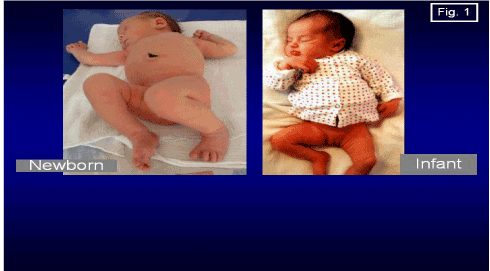
| ��?Syndrome of Contractures�. Our Observations 1974��? 2012 (Figure 1) |
| The “syndrome of contractures” has been described by many authors in Europe and in the United States [3]. Prof. Hans Mau from Tübingen, Germany, has described it in further detail, as “Siebener [Kontrakturen] Syndrom” (syndrome of seven contractures). |
| It is mostly observed on the left sided syndrome as a result of the left positioning of the fetus in the mother’s uterus in 80%-90% of cases. The list of deformities according to Mau, are the following: |
| 1. Skull deformity (plagiocephaly), the flattening mostly of the left side of the forehead and os temporalis. |
| 2. Torticollis muscularis (wry neck), shortening of sternocleidomastoid muscle. |
| 3. Scoliosis infantilis (infantile scoliosis), other than idiopathic scoliosis. |
| 4. Contracture (shortening) of the adductor muscles of the left hip. |
| 5. Haltungsschwäche (weak posture) described by Mau, informs about the ability for deformation but does not give the precise description of “deformity”. In 1995, we established the restricted adduction of the right hip and the even abduction contracture (shortening) of the right hip as an equal for Haltungsschwache. The examination of the adduction movement of the hips should be in the extension position of the joint and it was the secret to examinations for many years, whatever is similar to “Ober test”. The asymmetry of movement of the hip with time makes the asymmetry during gait for loading and for growing, is also the reason of permanent “standing ‘at ease’ on the right leg” [4]. |
| 6. Pelvic bone asymmetry-the oblique positioning of the pelvis, visible during an X-ray examination for hip joint screening due to contracture of adductors of the left hip and contracture of abductors of right hip. |
| 7. Foot deformities such as, pes equino-varus, pes equino-valgus, pes calcaneo-valgus. |
| 8. As mentioned above in 2006 I added the excessive varus deformity (crura vara) of both shanks to the “syndrome of contractures”. This excessive deformity, under special pathological conditions can lead to Blount disease. The development of this varus deformity was described in “Orthopädische Praxis” [5]. |
| Clinical Signs of the ��?Syndrome of Contractures� in Children with the So-called Idiopathic Scoliosis in Literature |
| In the past twentieth century, some authors have observed the symptoms of asymmetry in different parts of the body in patients with “idiopathic scoliosis”. Following is the list of the authors and their observations: |
| a) “In general the left leg tends to be shorter than the right in childhood and this leads to development of the left convex lumbar curve. Pelvic obliquity has been observed in structural scoliosis” [6]. |
| b) “Asymmetry of temporal bones has also been associated with scoliosis” [7]. |
| c) “Plagiocephaly has been considered to be closely related to infantile idiopathic scoliosis” [8]. |
| d) “The difference in the length of extremities, pelvic tilt–secondary scoliosis” [9]. |
| e) “Tilt of pelvis is important sign of development of scoliosis” [10]. |
| f) “So-called idiopathic scoliosis commonly occurs in combination with a characteristic pattern of soft tissue asymmetries in the hip and pelvis region” [11]. |
| Other Influences in the Development of the So-called Idiopathic Scoliosis |
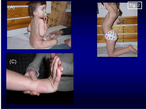
| Independent of causes coming from the asymmetry of function (gait, standing) following illnesses and deformations are the additional causes for development of so-called idiopathic scoliosis. These are: rickets, pelvis and lumbar spine anatomy anomalies (spina bifida occulta), chest and ribs deformities (pectus infundibiliforme) [12]. There is also a great indirect influence from central nervous system (Figure 2). These are: a) straight position of the spine in babies and small children because of spastic or sub spastic contracture of the extensors of the trunk, b) anterior tilt of the pelvis due to spastic or sub spastic contracture of m. rectus, and c) joint laxity, described by Prof. Harald Thom (1972–1973). These three factors exist in children with minimal brain dysfunction MBD or by ADHD (attention deficit and hyperactivity disorder) children. In Poland such children are in 5% to 8% of the whole population [13]. |
| Material |
| Materials in this study consist on the group of children and adults with scoliosis (N=1950 from 1985-2012). The age of patients ranged from 2 to 70. Children and youth presented all four types of scoliosis. Adult patients presented back pain, because of “degenerative scoliosis” as a result of idiopathic scoliosis. The control group, ca. 150 children from the period of 27 years included children examined because of spinal problems in the opinion of their parents, but without scoliosis or without danger of oncoming scoliosis. |
| Short History of Discoveries about Scoliosis (1995��? 2007) |
| In 1995, first presentation of biomechanical etiology of so-called idiopathic scoliosis during the orthopedic congress in Szeged/ Hungary. |
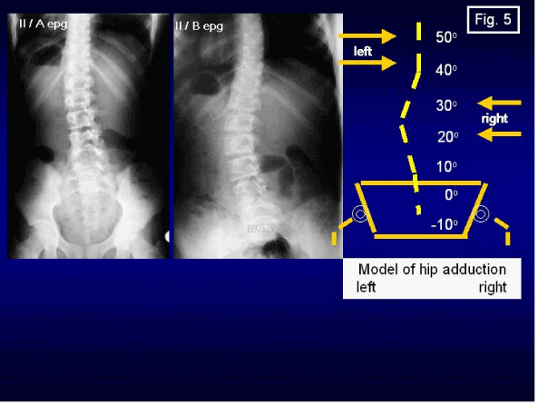
| In 2001, was presented, new classification of scoliosis: “S” scoliosis in I epg, “C” scoliosis in II/A and “S” scoliosis in II/B epg (explanation: epg=etio-pathological group) |
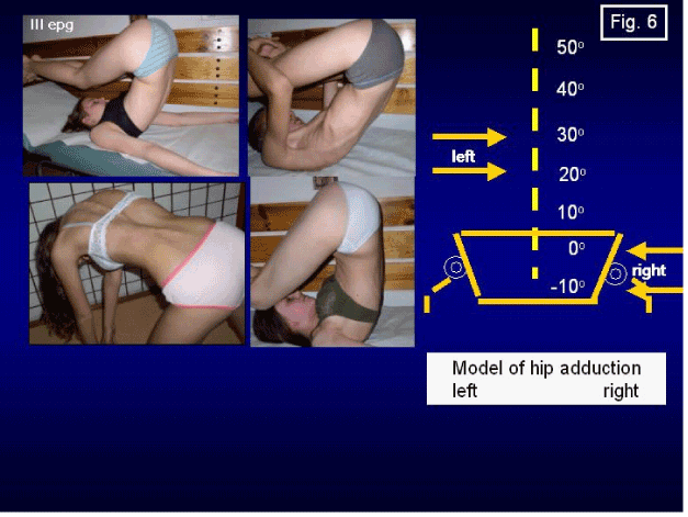
| In 2004, was described, “I” scoliosis in III epg. In these type of scoliosis is only stiffness of spine, but without curves and gibbous or with very small one. |
| In 2006, it was given the description of model of hip movements and the adequate type of scoliosis. |
| In 2007, was presented, the description of indirect influences of the central nervous system to scoliosis (explanation in next chapter) and the answer to why the blind children do not have scoliosis. Explanation: the walking without lifting of legs protect before spine deformity. |
| New Classification-Three Etiopathological (Epg) Groups, Four Types of Scoliosis. Connection with Adequate ��?Model of Hips Movements� |
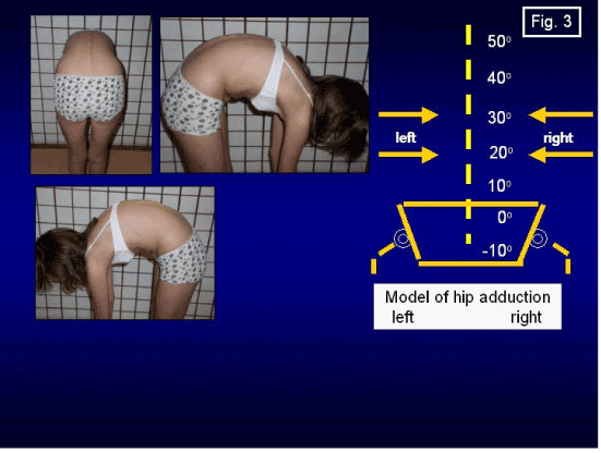
| A) The normal spine (Figure 3). Before presenting the new classification of the so-called idiopathic scoliosis, it is better to remind that the scoliosis depends on “biomechanical influence” and in healthy children this asymmetrical function does not exist. The movement of left and right hip is the same or very similar and standing (important cumulative time) left/right leg is the same and also there are not influences (asymmetries left/right side) coming from “gait”. |
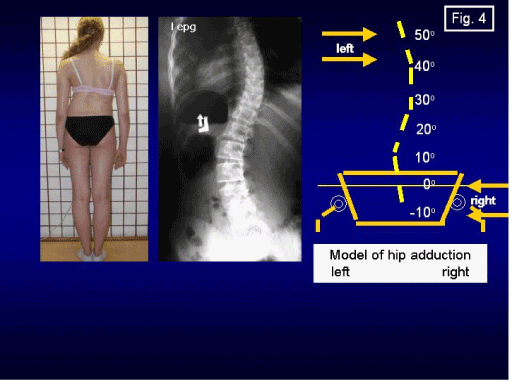
| B) Scoliosis in the form of “S” in I-st etiopathological group (I epg) [14]. [“S” deformity=double curve scoliosis] (Figure 4). |
| The children in this group have the specific “model of hip movements” (Table 1). There is a big difference in movement of both hips. To check this difference of movements the examination should be performed in the extension position of the hip joint. The development of scoliosis is connected with functions: “gait” and “the permanent standing ‘at ease’ on the right leg”. The initial sign of scoliosis is the rotation deformity which is confirmed by computer gait analysis (previous publications). Hyper-rotation movement in the inter-vertebral joints results in permanent distortion and later stiffness of the spine with a flat back. The scoliosis in this group is progressive (Table 1). |
| C) Scoliosis in the form of “C” and “S” in II-nd etiopathological group (“C” II/A epg and “S” II/B epg) (Figure 5). The children in this group have the specific “model of hip movements” (Table 2). There is a small deference in movement of both hips. The examination should be performed in the extension position of the hip joints. Due to permanent standing on the right leg after 8 to 10 years, gradual fixation of the axis of the spine in the shape of “C” occurs. Some cases from “S” II/B epg are kyphoscoliosis. The spine in this group is flexible. The scoliosis II/A epg is not progressive, II/B epg can be progressive (Table 2). |
| D) Scoliosis in the form of “I” III-rd etiopathological group of scoliosis (Figure 6) [15]–it is spine deformity in form of stiffness with little or no curvature. The children in this group have the specific “model of hip movements” (Table 3). It is a real abduction contracture of the right hip from 5 to 10 degrees or adduction 0 degree and the adduction of the left hip is also small from 20 to 30 degrees. The examination should be performed with the joint in a straightened position. Spinal deformity as mentioned above is in “character of stiffness” and connected only with “walking function”. These patients were not treated previously and for many years did not know about their spinal problem. Young patients present problems during sport activities, adult patients present with a range of back pain? (Table 3). |
| Discussion about the ��?Syndrome of Contractures� to Clarify the Character of Scoliosis and to Inform about the Necessity of Prophylactics |
| To understand the etiology and necessity of new therapy of the so-called idiopathic scoliosis we would like to recall that the aspect of scoliosis and its magnitude is connected not only with the “biomechanical causes/etiology” but also with the incorrect and harmful exercises of the therapy. Unfortunately, incorrect therapy was preformed over many years and in many countries all over on the world. The Lublin material during demonstrates exactly that past strengthening exercises in the treatment of scoliosis outside of our Department were incorrect, leading to severe deformity of the spine. After 18 years of teaching (lectures, articles, TV, radio) about the improved point of view on scoliosis, in Poland, many departments and rehabilitation centers rejected the incorrect exercises. Few rehabilitation centers (mostly private) unfortunately, continued with the past method of the therapy. |
| “The syndrome of contractures” alone provides an explanation to some previously unanswered questions about the etiology of the idiopathic scoliosis and provides information about the treatment. |
| • The development of scoliosis is connected with the growth period, gait, and standing at ease on the right leg” [4]. |
| • Scoliosis is a product of the asymmetrical movement of the hips, asymmetry of loading of the right and left leg (pelvis and spine) during walking and asymmetrical time of standing on the left leg versus the right leg-standing on the right leg more. The cumulative time of standing ‘at ease’ on the right leg plays an influential role. These asymmetries are connected with “the syndrome of contractures” as mentioned above. |
| • Scoliosis occurs mostly in girls because “syndrome of contractures” occurs mostly in girls (ratio between boys and girls is 1: 5). |
| • Lumbar left convex, thoracic right convex scoliosis, and rib hump on the right side are connected with the left-sided “syndrome of contractures” which occurs in 85%-90% of pregnancies [16]. |
| • The new classification of the so-called idiopathic scoliosis–“S” I epg, “C” II/A epg, “S” II/B epg and “I III epg is connected with the adequate “model of movement of hips” and other causes described in the article. |
| • The progression of scoliosis during the accelerated growth period in children is related to the asymmetry of bigger bone growth and smaller soft tissues growth. The abduction contracture of right hip is very often connected with flexion and external rotation contracture. Faster growth of bones is the cause of progression especially in I epg. In children with the greatest progression of curves, we observe faster growth of the legs than the trunk, which was also observed by other researchers [10]. |
| • Blind children do not develop scoliosis, which confirms the biomechanical influences (gait) in the development of scoliosis. The different mannerism of gait without lifting the legs protects against scoliosis. It has been observed that blind children use shuffling gait [17]. |
| • In some countries such as Mongolia, there are no cases of scoliosis which confirms the biomechanical influences of gait in the development of scoliosis. Explanation: many Mongolian children ride horses, which greatly protects them against scoliosis [18-24]. |
| • In Okinawa, Japan, there are also no cases of scoliosis (information gathered from Prof. A. Staniszew, president of Karate Okinawa Association in Poland). In this particular district of Japan, 50%-70% of primary schools perform karate stretching exercises. These stretching exercises lead to the symmetries of movement, function, development of the growth and body of the spine, and the entire trunk [24-30]. |
| • In orthopedic literature, it is written that scoliosis develops from the apex of the curve. It is now clear that the deformity of scoliosis begins and progresses from the base of the spine, that is, the pelvis and sacro-lumbar region towards the upper portion of the spine [31,32]. |
| • The new rehabilitation and stretching exercises which remove contractures (contracture referring to the asymmetrical shortening of soft tissues) confirms the biomechanical origin of the etiology [33-38]. |
| • The biomechanical etiology of the so-called idiopathic scoliosis answers all questions given over the years, regarding problems of idiopathic scoliosis. Also the term: “the natural history of scoliosis” (bigger curves, bigger rip hump) will have only historical value [39]. |
| Conclusions |
| 1. The etiology of the so-called idiopathic scoliosis (AIS) is strictly biomechanical and primarily based on the asymmetrical movement of the hip joints. |
| 2. The development of scoliosis is dependent on consequences connected with function, such as “gait” and “the standing position ‘at ease’ mostly on the right leg”. In the absence of these factors, the AIS can not come. |
| 3. There are three groups and four types of scoliosis in this new classification [40-52] and they are determined by an adequate model of hip movements. |
| 4. Children with spinal problems between the ages of 3-5 should be examined with a variety of test such as, the Adams test–“bending test for scoliosis”, the Lublin test–“side bending test for scoliosis”, the Elly Duncan test, and finally the “standing test of the right leg versus on the left leg” |
| 5. The asymmetry of adduction of the hips and continual habit of standing in the ‘at ease’ position on the right leg should be an alarming sign to doctors of oncoming scoliosis. These groups of children should undergo periodical spinal examinations by fully trained specialist [53]. |
| 6. Children who are at risk should be included in an early prophylactic program. The recommended prophylactic exercises are as follows: |
| a) Sit in a physiologically relaxed position, never sitting up straight, |
| b) Sleep in fetal position, |
| c) Stand ‘at ease’ on the left leg or on both legs, |
| d) Practice sport or a physical activity on a daily basis. Above all, stretching exercises at school or at home and taking practice in activities like karate, kung fu, taekwondo, tai chi, aikido, and yoga on a regular basis play an essential role in prophylactics. |
| Acknowledgements |
| I would like to thank MA Katarzyna Karska (Poland) and Claudia Ramirez (USA) for their help in proofreading of the article. |
References |
|
| Table 1 | Table 2 | Table 3 |
 |
 |
 |
| Figure 1 | Figure 2 | Figure 3 |
 |
 |
 |
| Figure 4 | Figure 5 | Figure 6 |
Make the best use of Scientific Research and information from our 700 + peer reviewed, Open Access Journals