Research Article Open Access
Beneficial Effect of a Multifunctional Polyphytocompound in Experimental Prostatic Hyperplasia in Rats
Makoto Kantah1, Birbal Singh2, Hala Sweed3, Geraldo Balieiro Neto4,5, Nikhil Kumar6, Fernando Bahdur Chueire4, Francesco Marotta1*, Aldo Lorenzetti1, Richard Bellow1 and Umberto Solimene71ReGenera Research Group for Aging Intervention and San Babila Clinic, Milano, Italy
2ICAR-Indian Veterinary Research Institute, Regional Station, Palampur, Himachal Pradesh, India
3Department of Geriatrics and Gerontology, Ain Shams University, Cairo, Egypt
4Department of Agriculture and Food Supply, São Paulo Agency for Agribusiness Technology, University of São Paulo, São Paulo, Brazil
5Department of Nutrition, Ribeirão Preto Medical School, University of São Paulo, São Paulo, Brazil
6Department of Life Sciences, Shri Venkateshwara University, JP Nagar, Uttar Pardesh, India
7WHO-Centre for Traditional Medicine and Biotechnology, University of Milano, Italy
- *Corresponding Author:
- Francesco Marotta
ReGenera Research Group for Aging Intervention and San Babila Clinic
Corso Matteotti, 1/A 20121 Milano, Italy
Tel: +39-024077243
E-mail: fmarchimede@libero.it
Received Date: February 27, 2017; Accepted Date: March 24, 2017; Published Date: March 27, 2017
Citation: Kantah M, Singh B, Sweed H, Neto GB, Kumar N, et al. (2017) Beneficial Effect of a Multifunctional Polyphytocompound in Experimental Prostatic Hyperplasia in Rats. Clin Pharmacol Biopharm 6:169. doi: 10.4172/2167-065X.1000169
Copyright: © 2017 Kantah M, et al. This is an open-access article distributed under the terms of the Creative Commons Attribution License, which permits unrestricted use, distribution, and reproduction in any medium, provided the original author and source are credited.
Visit for more related articles at Clinical Pharmacology & Biopharmaceutics
Abstract
The aim of the present study was to assess the efficacy of a poly-phytocompound in a model of experimental BPH. Adult 8 weeks male Wistar rats were subjected to complete orchiectomy under anesthesia (i.p. injection of 100 mg/kg body weight of sodium pentobarbital). After castration, experimental BPH was reproduced by subcutaneous injection of testosterone (20 mg/kg) for 4 weeks and, at the same time, rats randomly divided in 3 groups (15 rats each): (A) untreated BPH model; (B) BPH plus TR10/P3795 orally and (C) BPH plus finasteride (10 mg/kg body weight) administered orally as positive control group. A third group (D) of sham-operated rats served as control. Both TR10/P3795- and finasteride-treated groups showed a significant (p<0.05) and comparable reduction of all morphometric parameters (volume, weight and weight/body weight ration) which were grossly abnormal in untreated BPH model (p<0.01 vs. sham-op.). Moreover, both treatment schedule maintained a near-to-normal 3 h urinary output (p<0.01 vs. untreated BPH). Untreated BPH showed a significant increase of epithelial size and thickness and these features were equally decreased by TR10/P3795 and finasteride (p<0.05). Either TR10/P3795 or finasteride brought about a significant decrease of serum level of DHT and PAP (p<0.05 vs. sham). There was no difference among the two treatments. Prostatic tissue concentration of MDA, IL-6, TNFα and TGFβ1 significantly increased in untreated BPH model (p<0.001). All these parameters significantly decreased, although not normalised, in TR10/P3795-treated group (p<0.05 vs. sham and vs. finasteride). Finasteride determined only a not significant trend decrease of IL-6 and TNFα. Given the multifactorial aetiology of BPH, the data from this experimental model show the promising larger spectrum of mechanisms of action of the tested poly-phytocompound.
Keywords
Benign prostatic hyperplasia; Dihydrotestosterone; Prostate gland; Testosterone; Caviarlieri; Celergen
Introduction
Benign Prostatic Hyperplasia (BPH) is a slowly progressing process of micro and macronodular appearance characterized by hyperplastic epithelial modifications together with stromal growth. This process has a multifactorial etiology and represents the commonest cause of Lower Urinary Tract Symptoms (LUTS) in the aging male [1,2]. It has been reported that about 90% of men between 45 and 80 years old complain of some degree of LUTS (3 mcWary). More precisely, it seems that by the age of 50 [3,4] 50% of men may show the symptoms related with BPH and in those aged above 70 years this condition is the most significant cause of bladder outflow obstruction. From the histological viewpoint, the process which starts from the transitional or periurethral zone determines hyperplasia of glandular and stromal tissue with papillary buds and increased smooth muscle, lymphocytes and ducts [5,6]. The consequent prostate enlargement will bring about urethral constriction with following weak urinary stream, incomplete bladder emptying, nocturia, dysuria up to overt bladder outlet obstruction [7,8]. Thus, BPH-related LUTS can have a significant impact on quality of life and should not be underestimated [9,10]. Hormones affect the development and progression of BPH since the development and growth of the prostate gland very closely depends on androgen receptor stimulation [11-13]. Indeed, especially during aging process, prostate is mainly influence by Dihydrotestosterone (DHT), i.e., an active metabolite generated by the enzymatic conversion of testosterone by steroid 5α-reductase although other metabolites may play a role in health and disease [14]. Long before surgery may be required, well-known pharmacotherapeutic options are currently employed such as 5-alpha-reductase Inhibitors, alpha-adrenergic antagonists, anticholinergic agents and combination therapy [15,16]. Although these treatments have enabled consistent benefits [17-19], their use is associated to a different degree of side effects such as decreased libido, erectile dysfunction gynecomastia and poor ejaculatory function [20-22]. This limitation holds particularly relevant when very early cases of BPH are faced or when a tentative “preventive” strategy is planned. Till recently, there is a constant flow of experimental articles, reviews and clinical studies highlighting the role of phytocompounds in this condition [23-26], given also its multifaced pathophysiological mechanisms [27-29]. The aim of the present study was to assess the efficacy of a poly-phytocompound in a model of experimental BPH.
Materials and Methods
Animals
Adult 8 weeks male Wistar rats (240-290 g) were used throughout the experiments and were housed individually in standard polypropylene cages (three rats/cage) under controlled standard conditions of light (12/24 h) and temperature (26 ± 1°C). Food pellets and tap water were provided ad libitum. For experimental purposes animals were fasted overnight but were allowed free access to water. Body weight was measured weekly in all rats. All animal procedures were performed according to approved protocols and in accordance with the Guiding Principles for the Care and Use of Animals, based on the Declaration of Helsinki.
Induction of BPH and treatments
Rats were hosted for 10 days to allow acclimatization and four days prior experiment they were subjected to complete orchiectomy with spermatic cord and blood vessels ligation under anesthesia (i.p. injection of 100 mg/kg body weight of sodium pentobarbital). After castration, experimental BPH was reproduced by subcutaneous injection of testosterone (20 mg/kg) for 4 weeks and, at the same time, rats, under a computerized randomization procedure ensuring a comparable body weight distribution were divided in 3 groups (15 rats each): (A) Untreated BPH model; (B) BPH plus 100 mg/kg of TR10/ P3795, a poli-phytocompound of potential prostate protective effect (100 mg containing: Serenoa repens extract 56.5%, red clover 26%, pumpkin seed extract 13% and pomegranate extract 4.5%, Andronam, Named, Lesmo, Italy) orally and (C) BPH plus finasteride (0.5 mg/kg body weight) administered orally as positive control group. A third group (D) of sham-operated rats served as control. All compounds were administered to the animals in the morning. Care was taken as to put all the TR10/P3795 or finasteride supplementation in the morning food supply while checking that all was eaten up. Finasteride was stored in an air-tight, dark container at room temperature. The finasteride dosing was prepared in powder at the required concentrations and stored at 4°C.
Urinary output, blood and prostatic tissue samples
On the day before sacrifice (27th day), all groups were transferred into metabolic cages to measure 3 h urinary outputs. On the next day (4 weeks study), all animals were fasted overnight. Blood samples were collected in EDTA and centrifuged at 3000 × g for 10 min; serum was instantly separated and stored at −20 °C. After animals were sacrificed, prostate were weighed and stored in 10% buffered formaldehyde solution. Tissue was then embedded in paraffin technique, 5 μm thick sections were cut and stained by haematoxylin and eosin for light microscopic examination. Separate aliquots of ventral prostatic tissue were snap frozen at −70°C until further analysis.
Histological examinations and photomicrographic image analysis
Each section was viewed under a light microscope at a magnification of ×40–400. A Windows-connected Photometric Quantix digital camera was set as to capture and optimize photomicrographs images and morphological changes were blindly evaluated by a pathologist unaware of the treatment group to calculate the HE stained gland area in the rat ventral prostate. The stained area in the gland indicated the degree of proliferation of epithelial components. This was assessed by randomly counting 10 glands in each section and then quantifying the glandular epithelial area as stained area per glandular area under 100-fold magnification. The mean percent area density was also evaluated for each group.
Assessment of serum dihydrotestosterone (DHT) and prostatic acid phosphatase (PAP)
Blood samples were allowed to clot into a serum separator tube for two hours at room temperature before centrifugation for 20 min at 1000 × g. Serum levels of DHT and PAP were assayed by Enzyme- Linked Immunosorbent Assay (ELISA) using commercially available kits (Santa Cruz Biotechnology, Santa Cruz, California, USA). Briefly, triplicate aliquots of standards and samples were put into separate wells pre-incubated with DHT- and PAP-specific biotin-conjugated polyclonal antibody solution. Afterwards, avidin conjugated to Horseradish Peroxidase (HRP) was added to each microplate well and incubated. Same procedure was applied with the standard. Then, 3,3'5,5'-tetramethyl-benzidine substrate solution was added to each well and the reaction was stopped by adding a 0.16 M sulphuric acid solution. The reading of the colour change of each well was assessed by spectrophotometric at the absorbance of 450 nm wavelength using analytical grade laboratory reagents. The final concentration of DHT and PAP was calculated by comparing the O.D. of the samples to the standard curve.
Assessment of oxidative and inflammatory status in BPH
Pre-weighted prostatic tissues were ice crushed in liquid nitrogen and then homogenized for 15 min in PBS (pH 7.4). The concentration of malondialdehyde (MDA), interleukin-6 (IL-6), tumor necrosis factor alpha (TNFα) and transforming growth factor beta 1 (TGF-β1) were assayed by using commercially available kits (BioSource, San Diego, CA USA for MDA and Seikagaku Corp., Tokyo, Japan for ELISA Immunoassay of IL6, TNF-α and TGF-β1). All values were normalized and expressed per milligram of protein content.
Statistical analyses
The data obtained were expressed as mean values ± SD. Statistical analysis was performed using SPSS 17. Significant difference among the groups was performed using a one-way Analysis of Variances (ANOVA), followed by a non-parametric post Tukey test. Analysis using two-tailed and a p-value ≤0.05 was considered as statistically significant. Pearson’s correlation analysis was used for correlation of parameters measured.
Results
weight and prostate parameters
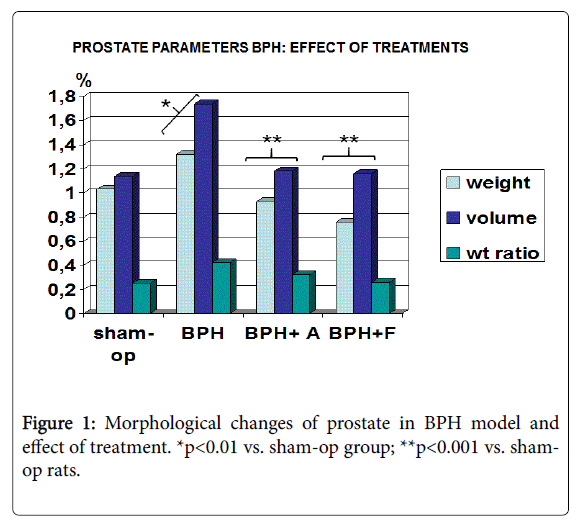
Body weight physiologically increased in sham-operated group and this was comparable to both untreated BPH model and both treatments groups without any significant difference although the Finasteride-group showed to have a trend towards lower weight (data not shown, p >0.05). As compared to sham-operated control, prostate weight, weight ratio and volume significantly increased in untreated BPH model (p<0.05). Both TR10/P3795-and finasteride-treated groups showed a significant and comparable reduction (Figure 1, p<0.05 vs. sham group).
Urinary output
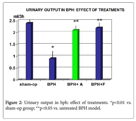
As compared to sham-op control group, rats with untreated BPH showed a significant contraction of urinary output of over 50% (p<0.01). Both TR10/P3795 and finasteride brought about a near to normal urinary volume (Figure 2, p<0.01 vs. untreated BPH). There was no significant difference between the two treatment groups.
Routine blood chemistry
Liver and renal functional blood parameters were within normal range throughout the study, irrespective of the group considered and no statistically significant difference appeared (data not shown).
Histological morphology of the prostate
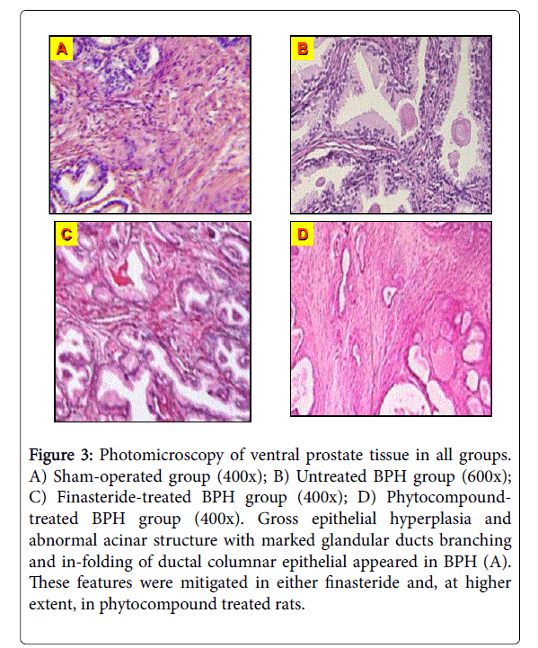
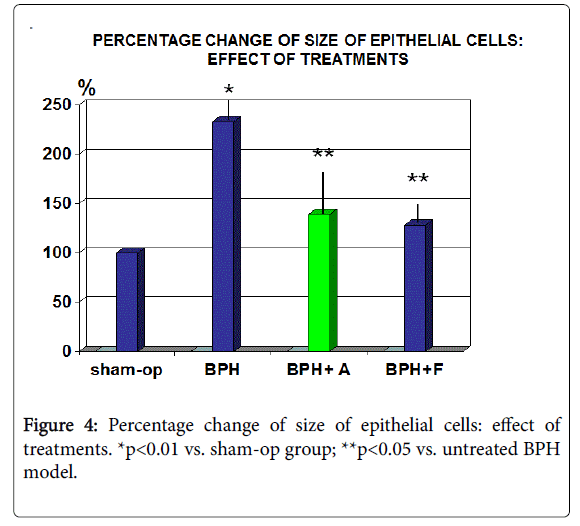
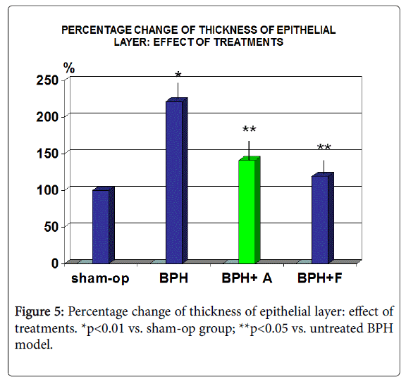
As expected, as compared to sham operated group, untreated BPH rats showed typical features such as epithelial hyperplasia with intraepithelial vacuoles. Moreover, an uneven acinar structure with evident increase of the glandular ducts branching and of in-folding of ductal columnar epithelial was recorded (Figure 3). When calculating these parameters, it appeared that there was a significant increase of epithelial size and thickness (Figures 4 and 5, p<0.01). Either TR10/ P3795 or Finasteride brought about a statistically significant protection of these features (p<0.05 vs. BPH model) whit an overall milder feature of BPH.
Figure 3: Photomicroscopy of ventral prostate tissue in all groups. A) Sham-operated group (400x); B) Untreated BPH group (600x); C) Finasteride-treated BPH group (400x); D) Phytocompound-treated BPH group (400x). Gross epithelial hyperplasia and abnormal acinar structure with marked glandular ducts branching and in-folding of ductal columnar epithelial appeared in BPH (A). These features were mitigated in either finasteride and, at higher extent, in phytocompound treated rats.
Assessment of serum dihydrotestosterone (DHT) and prostatic acid phosphatase (PAP)
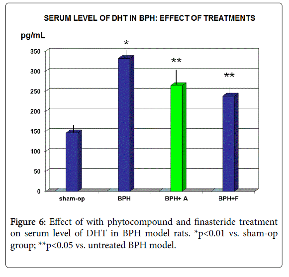
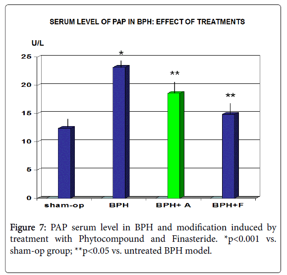
Rats with untreated BPH showed a significant increase of both DHT and PAP serum level (Figures 6 and 7, p<0.001 vs. sham-op group). Both TR10/P3795 and finasteride brought about a significant decrease of both parameters (p<0.05 vs. untreated BPH), finasteride showing a not significant better performance as for PAP serum level.
Assessment of oxidative and inflammatory status in BPH model
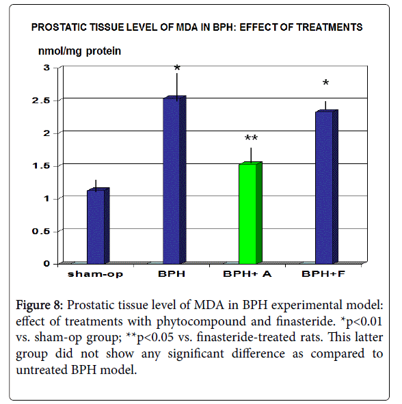
Oxidative stress, as measured by MDA concentration, was significantly elevated at a tissue level in BPH model when compared to same area in sham group (Figure 8, p<0.01). On the other hand, TR10/ P3795 significantly decreased MDA concentrations compared to the untreated BPH group (p<0.01).
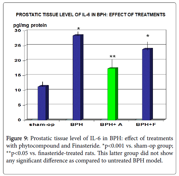
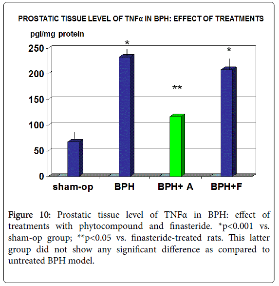
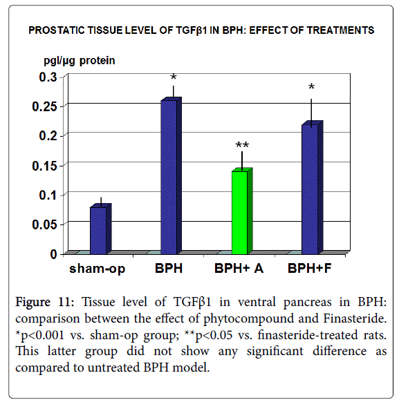
Accordingly, prostatic sample taken from untreated BPH rats showed a significantly abnormality of all tested inflammatory parameters [IL-6, TNF-α and TGF-β1 (Figures 9-11, p<0.001 vs. sham-op rats)].
Discussion
Natural compounds maintain a growing popularity in the treatment of Benign Prostatic Hyperplasia (BPH) and related Lower Urinary Tract Symptoms (LUTS) mainly due their overall general acceptance and reported lack of substantial side-effects. While the hormonal factor does represent a relevant pathophysiological variable of BPH occurrence and related drugs have been synthesized accordingly, several mechanisms have been advocated for its development. These include, among others, tissue and intracellular redox unbalance [30,31]. Indeed it is well known that human prostate tissue has a peculiar vulnerability to oxidative DNA damage due to more rapid cell turnover and also to the low activity of superoxide dismutase and catalase and increased endogenous levels of DNA base products, these two variables having being reported as to be inversely correlated in in BPH samples has received further recent confirmations [32].
In BPH, the exposure of prostate epithelia to ongoing oxidative stress molecules can trigger the otherwise inactive transcription factor NF-κB [33], a key inflammatory transcriptional regulator. As a consequence, by the TNF-α/AP-1 transduction pathway and the NF- κB-Inducing Kinase (NIK) transduction pathway, the local production of proinflammatory cytokines [34] is determined by triggering apoptotic pathway which limits uncontrolled cell proliferation. In this regard, TR10/P3795 exerted a significant reduction of MDA and inflammatory markers unlike finasteride which had already been demonstrated not to affect protein oxidation in the prostate [35]. This is likely to be advocated for by the total phenolic content of a selected extract from pumpkin in its composition. Indeed, it has been shown that they maintain an effective radical scavenging activity from as low as 0.16 mg/ml in vitro concentration [36]. Such anti-oxidant, anti-inflammatory and pro-apoptotic properties have been also demonstrated for two other main components of the present formula, i.e., pomegranate and serenoa [37-39]. In this setting, the triggered inflammatory cascade has not simply an ancillary meaning but a definite pathogenetic role of disease progression till potential cancerous transformation [40,41].
Thus, the proven anti-inflammatory properties of specific serenoa, pomegranate and pumpkin extracts [37-39,42] which are not directly shared by finasteride, come to be all the more relevant. This has been confirmed last month also in an obese mouse model [43] further paving the way to a wider systemic metabolic understanding of the disease [44-47]. In favour of such integrated phyto-pharmaceutic approach, it is worthwhile reporting that components such as specific pumking seed, red clover isoflavones, serenoa and pomegranate extracts have been shown to have antiandrogenic activity by dose-dependently blocking the binding of DHT besides am anti-aromatase and anti-5-α-reductase Type II action [38,48-51]. It goes without saying that inflammation per se exerts an endocrine disrupting effect at the prostate level by negatively affecting tissue estrogen metabolism [52], this being, together with antioxidant property, one of the suspected hormone receptor-independent anti-mutagenic mechanisms of some phytocompounds [53].
The very high dosage of arginine into pumpkin seed extract may be a further distinctive feature of the potency of the formula since it has been shown in rats that via a nitric oxide-mediated mechanism, this significantly improves in-bladder pressure and bladder volume adaptation [54]. While there is a growing evidence in clinics of the rationale use of phytocompounds for prostate health [55-57], the present experimental work using a formula combining a number of the most effective phytochemicals, offers new hints for its larger use either as sole therapeutic approach in prevention management, an alternative to chemicals when appropriate and as a potential adjuvant weapon during current chemical treatments.
Finally, although there are some assumptions on the role of fish oil in prostate health, there is a paucity of scientific evidence [58,59], moreover, it has been reported the possible deleterious role of -3 PUFAs alpha-linolenic acid in the development of prostate cancer [60]. Some of our group has tried in-house preliminary studies with a EPA-rich marine compound (Caviarlieri) with no results whatsoever and also have tested to no avail Celergen, a further fish-lipoprotein derivative with anti-inflammatory properties in vitro studies but which has indeed no published in vivo or clinical prove so far.
Conclusion
From this study, it appeared that either TR10/P3795- and finasteride-treated groups showed a significant comparable reduction of all typical morphometric and functional parameters seen in BPH, (volume, weight and weight/body weight ration and urinary output) together with a significant decrease of serum level of DHT and PAP. However, only TR10/P3795 brought about a partial but significant decrease of MDA, IL-6, TNFα and TGFβ1 (p<0.05 vs. sham and vs. finasteride). Taken overall and considering the multifactorial aetiology of BPH, the data from this experimental model show the promising larger spectrum of mechanisms of action of the tested poly-phytocompound.
References
- Bavendam TG, Norton JM, Kirkali Z, Mullins C, Kusek JW, et al. (2016) Advancing a Comprehensive Approach to the Study of Lower Urinary Tract Symptoms. J Urol 196: 1342-1349.
- Jung HB, Kim HJ, Cho ST (2015) A current perspective on geriatric lower urinary tract dysfunction. Korean J Urol 56: 266-275.
- McVary K (2006) BPH: Epidemiology and Comorbidities. Am J Manag Care 5: S122.
- Roehrborn CG, Rosen RC (2008) Medical therapy options for aging men with benign prostatic hyperplasia: focus on alfuzosin 10 mg once daily. Clin Interv Aging 3: 511-524.
- Lee C, Kozlowski J, Grayhack J (1997) Intrinsic and extrinsic factors controlling benign prostatic growth. Prostate 31: 131-136.
- Moss MC, Rezan T, Karaman UR, Gomelsky A (2017) Treatment of Concomitant OAB and BPH. Curr Urol Rep 18: 1.
- Kang M, Kim M, Choo MS, Paick JS, Oh SJ (2016) Urodynamic Features and Significant Predictors of Bladder Outlet Obstruction in Patients With Lower Urinary Tract Symptoms/Benign Prostatic Hyperplasia and Small Prostate Volume. Urology 89: 96-102.
- Ke QS, Jiang YH, Kuo HC (2015) Role of Bladder Neck and Urethral Sphincter Dysfunction in Men with Persistent Bothersome Lower Urinary Tract Symptoms after α-1 Blocker. Treatment. Low Urin Tract Symptoms 7: 143-148.
- Shao IH, Wu CC, Hsu HS, Chang SC, Wang HH, et al. (2016) The effect of nocturia on sleep quality and daytime function in patients with lower urinary tract symptoms: a cross-sectional study. Clin Interv Aging 11: 879-885.
- Abdul-Muhsin HM, Tyson MD, Andrews PE, Castle EP, Ferrigni RG, et al. (2016) Analysis of Benign Prostatic Hyperplasia Patients' Perspective Through a Third Party-administered Survey. Urology 88: 155-160.
- Song L, Shen W, Zhang H, Wang Q, Wang Y, et al. (2016) Differential expression of androgen, estrogen, and progesterone receptors in benign prostatic hyperplasia. Bosn J Basic Med Sci 16: 201-208.
- Husain I, Shukla S, Soni P, Husain N (2016) Role of androgen receptor in prostatic neoplasia versus hyperplasia. J Cancer Res Ther 12: 112-116.
- Wen S, Chang HC, Tian J, Shang Z, Niu Y, et al. (2015) Stromal androgen receptor roles in the development of normal prostate, benign prostate hyperplasia, and prostate cancer. Am J Pathol 185: 293-301.
- Mostaghel EA (2014) Beyond T and DHT-novel steroid derivatives capable of wild type androgen receptor activation. Int J Biol Sci 10: 602-613.
- Dahm P, Brasure M, MacDonald R, Olson CM, Nelson VA, et al. (2016) Comparative Effectivenes of Newer Medications for Lower Urinary Tract Symptoms Attributed to Benign Prostatic Hyperplasia: A Systematic Review and Meta-analysis. Eur Urol Oct 4. pii: S0302-2838(16)30667-4.
- Macey MR, Raynor MC (2016) Medical and Surgical Treatment Modalities for Lower Urinary Tract Symptoms in the Male Patient Secondary to Benign Prostatic Hyperplasia: A Review. Semin Intervent Radiol 33: 217-223.
- Berkseth KE, Thirumalai A, Amory JK (2016) Pharmacologic Therapy in Men's Health: Hypogonadism, Erectile Dysfunction, and Benign Prostatic Hyperplasia. Med Clin North Am 100: 791-805.
- Athanasopoulos A, Perimenis P (2005) Efficacy of the combination of an alpha1-blocker with an anticholinergic agent in the treatment of lower urinary tract symptoms associated with bladder outlet obstruction. Expert Opin Pharmacother 6: 2429-2433.
- Dimitropoulos K, Gravas S (2015) Solifenacin/tamsulosin fixed-dose combination therapy to treat lower urinary tract symptoms in patients with benign prostatic hyperplasia. Drug Des Devel Ther 9: 1707-1716.
- La Torre A, Giupponi G, Duffy D, Conca A, Cai T, et al. (2016) Sexual Dysfunction Related to Drugs: a Critical Review. Part V: α-Blocker and 5-ARI Drugs. Pharmacopsychiatry 49: 3-13.
- Traish AM, Melcangi RC, Bortolato M, Garcia-Segura LM, Zitzmann M (2015) Adverse effects of 5α-reductase inhibitors: What do we know, don't know, and need to know? Rev Endocr Metab Disord 16: 177-198.
- Gacci M, Ficarra V, Sebastianelli A, Corona G, Serni S, et al. (2014) Impact of medical treatments for male lower urinary tract symptoms due to benign prostatic hyperplasia on ejaculatory function: a systematic review and meta-analysis. J Sex Med 11: 1554-1566.
- Shen HN, Xu Y, Jiang ZZ, Huang X, Zhang LY, et al. (2015) Inhibitory effects of Tripterygium wilfordii multiglycoside on benign prostatic hyperplasia in rats. Chin J Nat Med 13: 421-427.
- Vidlar A, Student V Jr, Vostalova J, Fromentin E, Roller M, et al. (2016) Cranberry fruit powder (Flowens™) improves lower urinary tract symptoms in men: a double-blind, randomized, placebo-controlled study. World J Urol 3: 419-424.
- Allkanjari O, Vitalone A (2015) What do we know about phytotherapy of benign prostatic hyperplasia? Life Sci 126: 42-56.
- Pagano E, Laudato M, Griffo M, Capasso R (2014) Phytotherapy of benign prostatic hyperplasia. A minireview. Phytother Res 28: 949-955.
- Ørsted DD, Bojesen SE (2013) The link between benign prostatic hyperplasia and prostate cancer. Nat Rev Urol 10: 49-54.
- Gur S, Kadowitz PJ, Hellstrom WJ (2008) Guide to drug therapy for lower urinary tract symptoms in patients with benign prostatic obstruction : implications for sexual dysfunction. Drugs 68: 209-229.
- Bernichtein S, Pigat N, Capiod T, Boutillon F, Verkarre V, et al. (2015) High milk consumption does not affect prostate tumor progression in two mouse models of benign and neoplastic lesions. PLoS ONE 10: e0125423.
- Udensi UK, Tchounwou PB (2016) Oxidative stress in prostate hyperplasia and carcinogenesis. J Exp Clin Cancer Res 35: 139.
- Minciullo PL, Inferrera A, Navarra M, Calapai G, Magno C, et al. (2015) Oxidative stress in benign prostatic hyperplasia: a systematic review. Urol Int 94: 249-254.
- Pace G, Di Massimo C, De Amicis D, Corbacelli C, Di Renzo L, et al. (2010) Oxidative stress in benign prostatic hyperplasia and prostate cancer. Urol Int 85: 328-333.
- Shankar E, Bhaskaran N, Maclennan GT, Liu G, Daneshgari F, et al. (2015) Inflammatory Signaling Involved in High-Fat Diet Induced Prostate Diseases. J Urol Res 2: 2018.
- Popovics P, Schally AV, Salgueiro L, Kovacs K, Rick FG (2017) Antagonists of growth hormone-releasing hormone inhibit proliferation induced by inflammation in prostatic epithelial cells. Proc Natl Acad Sci U S A 114: 1359-1364.
- Hoque A, Ambrosone CB, Till C, Goodman PJ, Tangen C, et al. (2010) Serum oxidized protein and prostate cancer risk within the Prostate Cancer Prevention Trial. Cancer Prev Res (Phila) 3: 478-483.
- Xanthopoulou MN, Nomikos T, Fragopoulou E, Antonopoulou S (2009) Antioxidant and lipoxygenase inhibitory activities of pumpkin seed extracts. Food Res Int 42: 641-646.
- Ammar AE, Esmat A, Hassona MD, Tadros MG, Abdel-Naim AB, et al. (2015) The effect of pomegranate fruit extract on testosterone-induced BPH in rats. Prostate 75: 679-692.
- Latil A, Pétrissans MT, Rouquet J, Robert G, de la Taille A (2015) Effects of hexanic extract of Serenoa repens (Permixon® 160 mg) on inflammation biomarkers in the treatment of lower urinary tract symptoms related to benign prostatic hyperplasia. Prostate 75: 1857-1867.
- Iii Colado-Velázquez J, Mailloux-Salinas P, Medina-Contreras J, Cruz-Robles D, Bravo G (2015) Effect of Serenoa Repens on Oxidative Stress, Inflammatory and Growth Factors in Obese Wistar Rats with Benign Prostatic Hyperplasia. Phytother Res 9: 1525-1531.
- Norström MM, Rådestad E, Sundberg B, Mattsson J, Henningsohn L, et al. (2016) Progression of benign prostatic hyperplasia is associated with pro-inflammatory mediators and chronic activation of prostate-infiltrating lymphocytes. Oncotarget 7: 23581-23593.
- Torkko KC, Wilson RS, Smith EE, Kusek JW, van Bokhoven A, et al. (2015) Prostate Biopsy Markers of Inflammation are Associated with Risk of Clinical Progression of Benign Prostatic Hyperplasia: Findings from the MTOPS Study. J Urol 194: 454-461.
- Winkler C, Wirleitner B, Schroecknadel K, Shenmack H, Fuchs D (2005) Extracts of pumpkin (Cucurbita pepo L.) Seeds suppress stimulated peripheral blood mononuclear cells in vitro. Am J Immunol 1: 6-11.
- Fortis-Barrera Á, García-Macedo R, Almanza-Perez JC, Blancas-Flores G, Zamilpa-Alvarez A, et al. (2017) Cucurbita ficifolia (Cucurbitaceae) modulates inflammatory cytokines and IFN-γ in obese mice. Can J Physiol Pharmacol 95: 170-177.
- Castelli T, Russo GI, Reale G, Privitera S, Chisari M, et al. (2016) Metabolic syndrome and prostatic disease: potentially role of polyphenols in preventive strategies. A review. Int Braz J Urol 42: 422-430.
- Vignozzi L, Gacci M, Maggi M (2016) Lower urinary tract symptoms, benign prostatic hyperplasia and metabolic syndrome. Nat Rev Urol 13: 108-119.
- Yang TK, Woo P, Yang HJ, Chang HC, Hsieh JT, et al. (2016) Correlations of Metabolic Components with Prostate Volume in Middle-Aged Men Receiving Health Check-Up. PLoS One 11: e0145050.
- Chang SN, Han J, Abdelkader TS, Kim TH, Lee JM, et al. (2014) High animal fat intake enhances prostate cancer progression and reduces glutathione peroxidase 3 expression in early stages of TRAMP mice. Prostate 74: 1266-1271.
- Damiano R, Cai T, Fornara P, Franzese CA, Leonardi R, et al. (2016) The role of Cucurbita pepo in the management of patients affected by lower urinary tract symptoms due to benign prostatic hyperplasia: A narrative review. Arch Ital Urol Androl 88: 136-143.
- Kapoor R, Ronnenberg A, Puleo E, Chatterton RT Jr, Dorgan JF, et al. (2015) Effects of Pomegranate Juice on Hormonal Biomarkers of Breast Cancer Risk. Nutr Cancer 67: 1113-1119.
- Opoku-Acheampong AB, Penugonda K, Lindshield BL (2016) Effect of Saw Palmetto Supplements on Androgen-Sensitive LNCaP Human Prostate Cancer Cell Number and Syrian Hamster Flank Organ Growth. Evid Based Complement Alternat Med 2016: 8135135.
- Jarred RA, McPherson SJ, Jones ME, Simpson ER, Risbridger GP (2003) Anti-androgenic action by red clover-derived dietary isoflavones reduces non-malignant prostate enlargement in aromatase knockout (ArKo) mice. Prostate 56: 54-64.
- Mosli HA, Al-Abd AM, El-Shaer MA, Khedr A, Gazzaz FS, et al. (2012) Local inflammation influences oestrogen metabolism in prostatic tissue. BJU Int 110: 274-282.
- Medjakovic S, Hobiger S, Ardjomand-Woelkart K, Bucar F, Jungbauer A (2016) Pumpkin seed extract: Cell growth inhibition of hyperplastic and cancer cells, independent of steroid hormone receptors. Fitoterapia 110: 150-156.
- Hata, Tanahashi S, Wakida Y (2005) Effects of pumpkin seed extract on urinary bladder function in anesthetized rats. Med Sci Pharm Sci 54: 1-10.
- Kroeger N, Belldegrun AS, Pantuck AJ (2013) Pomegranate Extracts in the Management of Men's Urologic Health: Scientific Rationale and Preclinical and Clinical Data. Evid Based Compl Altern Med 2013: 701434.
- Shirvan MK, Mahboob MR, Masuminia M, Mohammadi S (2014) Pumpkin seed oil (prostafit) or prazosin? Which one is better in the treatment of symptomatic benign prostatic hyperplasia. J Pak Med Assoc 64: 683-685.
- Vahlensieck W, Theurer C, Pfitzer E, Patz B, Banik N, et al. (2015) Effects of pumpkin seed in men with lower urinary tract symptoms due to benign prostatic hyperplasia in the one-year, randomized, placebo-controlled GRANU study. Urol Int 94: 286-295.
- Comhaire F, Mahmoud A (2004) Preventing diseases of the prostate in the elderly using hormones and nutriceuticals. Aging Male 7: 155-169.
- Christensen JH, Fabrin K, Borup K, Barber N, Poulsen J (2006) Prostate tissue and leukocyte levels of n-3 polyunsaturated fatty acids in men with benign prostate hyperplasia or prostate cancer. BJU Int 97: 270-273.
- Krotkiewski M, Przybyszewska M, Janik P (2003) Cytostatic and cytotoxic effects of alkylglycerols (Ecomer). Med Sci Monit 9: PI131-PI15.
Relevant Topics
- Applied Biopharmaceutics
- Biomarker Discovery
- Biopharmaceuticals Manufacturing and Industry
- Biopharmaceuticals Process Validation
- Biopharmaceutics and Drug Disposition
- Clinical Drug Trials
- Clinical Pharmacists
- Clinical Pharmacology
- Clinical Research Studies
- Clinical Trials Databases
- DMPK (Drug Metabolism and Pharmacokinetics)
- Medical Trails/ Drug Medical Trails
- Methods in Clinical Pharmacology
- Pharmacoeconomics
- Pharmacogenomics
- Pharmacokinetic-Pharmacodynamic (PK-PD) Modeling
- Precision Medicine
- Preclinical safety evaluation of biopharmaceuticals
- Psychopharmacology
Recommended Journals
Article Tools
Article Usage
- Total views: 5384
- [From(publication date):
May-2017 - Apr 14, 2025] - Breakdown by view type
- HTML page views : 4452
- PDF downloads : 932
