Case Report Open Access
Atypical Carcinoid Tumor of the Larynx: A Challenging Diagnostic Case
Kastoer C1,2*, Vanderveken OM1,2, Lammens M2,3, Specenier P2,4, Carp L2,5, Vliet JV6, Verfaillie J1, Van de Heyning PH1,2 and Van Laer CG1,21Department of Otorhinolaryngology, Head and Neck Surgery, Antwerp University Hospital, Antwerp, Belgium
2Department of Medicine and Health Sciences, University of Antwerp, Antwerp, Belgium
3Department of Pathology, Wilrijkstraat, Antwerp University Hospital, Antwerp, Belgium
4Department of Oncology, Wilrijkstraat, Edegem, Antwerp University Hospital, Antwerp, Belgium
5Department of Nuclear Medicine, Wilrijkstraat, Antwerp University Hospital, Antwerp, Belgium
6Department of Otorhinolaryngology, Turnhout General Hospital, Turnhout, Antwerp, Belgium
- *Corresponding Author:
- Chloe Kastoer
Head and Neck Surgery
Department of Otorhinolaryngology
Antwerp University Hospital
Wilrijkstraat 10, 2650 Edegem
Antwerp, Belgium; Tel: +32038213436
Fax: +32038214271
E-mail: chloe.kastoer@uza.be
Received date: July 11, 2016; Accepted date: July 26, 2016; Published date: July 28, 2016
Citation: Kastoer C, Vanderveken OM, Lammens M, Specenier P, Carp L, et al. (2016) Atypical Carcinoid Tumor of the Larynx: A Challenging Diagnostic Case. J Clin Exp Pathol 6:287. doi:10.4172/2161-0681.1000287
Copyright: © 2016 Kastoer C, et al. This is an open-access article distributed under the terms of the Creative Commons Attribution License, which permits unrestricted use, distribution, and reproduction in any medium, provided the original author and source are credited.
Visit for more related articles at Journal of Clinical & Experimental Pathology
Abstract
Background: Atypical carcinoids are rare neuroendocrine tumors, but they are the most common type of nonsquamous cell laryngeal neoplasms. Recent literature addresses the importance of adequate diagnosis, but lacks consensus in treatment strategy.
Case report: A 69-year-old man presented with dysphagia. Diagnosis of supraglottic hemangioma was based on fiberoptic examination. After treatment with CO2 laser excision histopathological diagnosis stated paraganglioma. Dysphagia recurred after 6 months. (18)F-FDG PET/CT-scan revealed high FDG-uptake at the right aryepiglottic fold. After CO2 laser excision histopathology showed atypical carcinoid. Total laryngectomy and neck dissection was performed to secure tumor-free margins, followed by adjuvant radiotherapy due to lymph node metastases. A month after termination of radiotherapy no lesions were determined by somatostatin receptor scintigraphy.
Conclusion: Personalized management should be based mainly on mitotic rate and locoregional extension. Surgical treatment and lifelong multidisciplinary follow-up is mandatory, while the role of adjuvant therapy remains debatable.
Keywords
Hemangioma; Paraganglioma; Neuroendocrine tumors; Larynx
Introduction
Neuroendocrine tumors occur in all organs, but most often in the gastrointestinal or bronchial tract. They account for 0.5-2% of all malignancies and approximately 1% of laryngeal cancers [1]. Various classification systems and terms have been used to indicate neuroendocrine neoplasms of the larynx, without universal agreement. Precise diagnosis and definitive tumor subtyping is based on histomorphological features supported by immunohistochemistry. This can be challenging but crucial for prognosis, as neuroendocrine tumors vary from benign to very aggressive. A common classification in four groups: paraganglioma, typical carcinoid, atypical carcinoid and small cell carcinoma [2,3]. Atypical carcinoid tumors are also referred to as moderately differentiated neuroendocrine carcinoma, grade II or large cell neuroendocrine carcinoma [4].
Goldman et al. describe an atypical carcinoid tumor of the larynx for the first time in 1969. Atypical carcinoid tumors of the larynx are usually malignant, have an aggressive clinical prognosis and have been confused with other tumors frequently [5]. Paragangliomas of the larynx are first described by Blanchard and Saunders in 1955, less than 90 cases of paraganglioma of the larynx have been described since and are considered benign with an excellent prognosis [6]. Almost all reported cases of malignant paraganglioma and many cases of typical carcinoid are reclassified as atypical carcinoid on review [7]. Therefore, it is difficult to determine the exact number of atypical carcinoid cases.
In this paper, we present a challenging case of an atypical carcinoid tumor at the level of the right aryepiglottic fold.
Case Report
A 69-year-old man was referred to our tertiary center with the tentative endoscopic diagnosis of a laryngeal hemangioma. The patient reported dysphagia and globus sensation without accompanying symptoms. The patient had a 13 pack-year history of cigarette smoking and two units of alcohol were consumed daily.
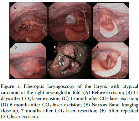
Endoscopic evaluation revealed a pedunculated red-brown submucosal mass (7 × 7 × 9 mm) at the level of the right aryepiglottic fold. No other relevant findings were present at clinical examination. Complete excision of the lesion at the level of the right arytenoid was performed using CO2 laser during direct laryngoscopy. The histopathology report suggested paraganglioma. The tumor was described as organoid and consisted of rounded nests of atypical cells without necrotic change. Four mitoses per 10 high-power fields (HPF) were found (Figure 1).
Figure 1: Fiberoptic laryngoscopy of the larynx with atypical carcinoid at the right aryepiglottic fold. (A) Before excision; (B) 11 days after CO2 laser excision; (C) 1 month after CO2 laser excision; (D) 6 months after CO2 laser excision; (E) Narrow Band Imaging close-up, 7 months after CO2 laser resection; (F) After repeated CO2 laser excision.
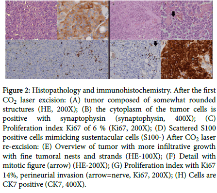
Immunohistochemical examination showed cytoplasm positive for synaptophysin and chromogranin and negative for cytokeratin (CK). Scattered S100 positive sustentacular cells were seen. The Ki67 index was up to 6% (Figure 2). Based on these results, clinical endoscopic follow-up was recommended.
Figure 2: Histopathology and immunohistochemistry. After the first CO2 laser excision: (A) tumor composed of somewhat rounded structures (HE, 200X); (B) the cytoplasm of the tumor cells is positive with synaptophysin (synaptophysin, 400X); (C) Proliferation index Ki67 of 6 % (Ki67, 200X); (D) Scattered S100 positive cells mimicking sustentacular cells (S100-) After CO2 laser re-excision: (E) Overview of tumor with more infiltrative growth with fine tumoral nests and strands (HE-100X); (F) Detail with mitotic figure (arrow) (HE-200X); (G) Proliferation index with Ki67 14%, perineurial invasion (arrow=nerve, Ki67, 200X); (H) Cells are CK7 positive (CK7, 400X).
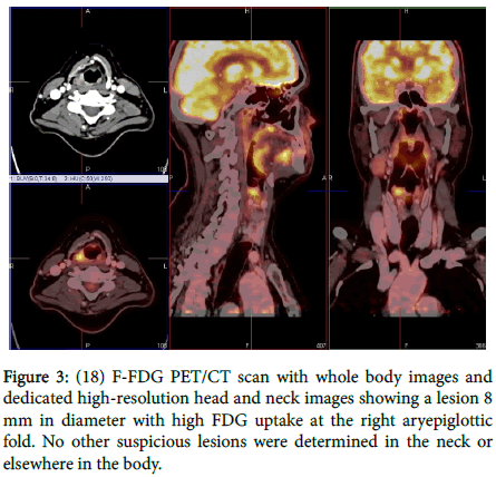
Symptoms of dysphagia recurred within 6 months, accompanied by a tingling sensation in the laryngeal region that worsened during ingestion of food. A pink-whitish lesion occurred on the right aryepiglottic fold (Figure 1). Narrow band imaging during fiberoptic laryngoscopy showed neovascularization suspect for malignancy (Figure 1). An (18) F-FDG PET/CT-scan revealed high FDG-uptake from a lesion at the right aryepiglottic fold with a diameter of 8 mm. No suspicious lesions were determined elsewhere in the body (Figure 3). Blood examination showed elevated levels of neuron-specific enolase (NSE, 17.3 μg/L, n<16.3 μg/L) and chromogranin A (CgA, 229 μg/L, n=40-170 μg/L).
CO2 laser excision of the lesion was repeated and histopathology revealed atypical carcinoid (Figure 2), surgical margins were not free of tumor cells. The lesion contained regions with infiltrative growth of fine tumoral strands and nests in addition to solid areas. Necrosis and perineural invasion were present. The mitotic index was 5 per 10 HPF. The cells were CK7 and synaptophysin positive; CK20 negative. The Ki-67 index was 14% (Figure 3).
Total laryngectomy and unilateral supraomohyoidal elective neck dissection of region II-III-IV at the right side was performed. Postoperative CgA blood levels remained elevated (219 μg/L) and NSE levels remained within normal limits. Final diagnosis was an atypical carcinoid tumor of the larynx, pT1N2bMx R0 (TNM 7th edition, UICC 2009). As metastatic lymph nodes were identified in the neck dissection specimen adjuvant postoperative radiotherapy (30 sessions of 2 Gy bilateral) was proposed.
Somatostatin receptor scintigraphy (SRS, OctreoScan) an (18) FFDG PET/CT-scan were performed a month and three months after termination of radiotherapy respectively. No lesions were determined. Every therapeutic decision was discussed with the local multidisciplinary head and neck tumor board.
Discussion
Atypical carcinoids have a 5-year disease-specific survival of about 50% and 30% after 10 years [4,7]. More than 60% of the patients have recurrence despite aggressive treatment. Delays can occur even after the conventional 5-year follow-up period. Despite adequate treatment, almost 70% of these patients develop distant metastasis [7]. Negative prognostic factors are a Ki67 index higher than 5% or the presence of regional lymphatic metastasis [8].
Overlap in histopathological features of atypical carcinoids and paragangliomas is presented (Table 1) [4,5,9].
| Atypical carcinoid tumor | Paraganglioma |
|---|---|
| Subepithelial lesion with or without ulceration of the mucosa | Subepithelial lesion without ulceration of the mucosa |
| Trabecular and/or organoid pattern | Organoid pattern with aggregation of chief cells in the Zellballen pattern characteristic but not pathognomonic of this tumor |
| Glandular and/or squamous differentiation | Sustentacular cells |
| Oncocytic and oncocytoid changes may be present | Prominent vascular lesion |
| Mucinous changes may be present | Mitoses and necrosis are infrequent |
| Squamous nest may be present | |
| Rosettes may be present | |
| 2 to 10 mitoses per 2 mm2 (10 high-power fields) | |
| Mild to moderate nuclear pleomorphism | |
| Sustencular cells may be present | |
| Necrosis (often punctuate) | |
| Tendency to grow in nests and cords of cells, often with peripheral palisading of nuclei | |
| Focally Zellballen pattern may be present | |
| Cysts with intracystic papillary like projections of tumor cells may be seen | |
| Large cells with abundant eosinophilic, finely granular cytoplasm with vesicular nuclei and prominent nucleoli |
Table 1: Histopathological criteria for diagnosis of atypical carcinoid tumor and paraganglioma.
Atypical carcinoids can have a distinct paraganglioma-like appearance, with cell nests resembling a Zellballen pattern [5]. Atypical carcinoids of the larynx show immunoreactivity for both epithelial and neuroendocrine markers.
In the reported case, findings on the first biopsy correlated with a tentative diagnosis of paraganglioma based on the cytokeratin (CK) negativity and presence of sustentacular cells with some positivity for S100. Detection of mitosis, however, is not common in cases of paraganglioma and might be more compatible with a diagnosis of atypical carcinoid (Table 1). Both paragangliomas and atypical carcinoids are immunohistochemically positive for chromogranin and synaptophysin [10,11]. In general atypical carcinoids are positive for low molecular weight CK as well as other epithelial markers, and CK is negative in cases of paraganglioma [4]. This makes CK is the most useful immunomarker for differential diagnosis. S-100 protein stains are strongly positive in the sustentacular cells of paragangliomas but they can also be positive in atypical carcinoids (Table 2) [4,5,9-11].
| Staining agent | Atypical carcinoid tumor | Paraganglioma |
|---|---|---|
| Cytokeratin (CK) | + (low molecular weight) | - |
| Epithelial membrane antigen | + | - |
| Carcinoembryonic antigen (CEA) | + | - |
| Chromogranin | + | + |
| Synaptophysin | + | + |
| CD56 | + | + |
| Neuron-specific enolase (NSE) | + | + |
| S-100 protein | +/- (negative in most cases) | + (in sustentacular cells) |
Table 2: Immunocytochemical findings in atypical carcinoid tumor and paraganglioma.
Mitosis (per 2 mm2, 10 HPF)<2 indicates typical carcinoid, 2-10 indicates atypical carcinoid and>11 indicates small cell neuroendocrine carcinoma; mitoses are rare in paraganglioma [4,12]. Mitotic rate may be the easiest way to differentiate between neuroendocrine tumors and their aggressiveness. In our case the mitotic rate of 4 in the first histopathological report may have pointed out the diagnosis of atypical carcinoid rather than paraganglioma.
X-ray computed tomography (CT) and magnetic resonance imaging are useful to demonstrate local extension of neuroendocrine tumors [13]. Small tumors may be visualized only by endoscopic assessment and not by conventional imaging. Multiple cases describe that somatostatin receptor scintigraphy (SRS, OctreoScan) can reveal lymph node metastases that are not clinically present [14]. SRS image fusion with low-dose CT is important for better anatomical localization of scintigraphic findings. The overall sensitivity of SRS is 89%. This is high compared to (123) I-MIBG scintigraphy (52%) and (18)F-FDG PET (58%) [15]. However, in tumors with a high proliferation rate (Ki67>15%), (18)F-FDG PET has the highest sensitivity (92%) compared to SRS (69%) and (123) I-MIBG scintigraphy (46%). SRS and (18) F-FDG PET provide complimentary diagnostic information and are both of value for neuroendocrine tumor patients [15]. In our case the histopathologic report states Ki67 14% and high uptake on (18)F-FDG PET before surgery. Follow-up by endoscopy and (18) F-FDG PET is provided.
Radical surgical removal is recommended in both paragangliomas and atypical carcinomas of the larynx [4,5,9]. In atypical carcinomas, selective neck dissection is recommended in addition to radical surgery of the primary tumor, patients who did not have neck dissection developed regional recurrence without local recurrence in almost 30% of cases [7]. Chemotherapy or radiation therapy does not improve the cure rate of atypical carcinoid tumors and does not result in a better 5-year disease-specific survival rate [4,7,9]. These results are challenged by small series and cases that state that atypical carcinoids respond well to combined therapy and that postoperative radiotherapy is useful in the case of substantial local extension, multiple node metastases and/or extracapsular disease [14].
Conclusion
The reported case indicates that differentiation between paraganglioma and atypical carcinoid is challenging. Expert analysis of histopathological and immunohistochemical findings is crucial to distinguish between neuroendocrine tumors. Personalized management should be based mainly on mitotic rate and locoregional extension. Surgical treatment and lifelong multidisciplinary follow-up is mandatory, while the role of adjuvant therapy remains debatable.
References
- Maggard MA, O'Connell JB, Ko CY (2004) Updated population-based review of carcinoid tumors. Ann surg 240: 117-122.
- Lewis JS, Jr., Ferlito A, Gnepp DR, Rinaldo A, Devaney KO, et al. (2011) Terminology and classification of neuroendocrine neoplasms of the larynx. Laryngoscope 121: 1187-1193.
- Vandist V, Deridder F, Waelput W, Parizel PM, Van de Heyning P, et al. (2010) A neuroendocrine tumour of the sphenoid sinus and nasopharynx: A case report. B-Ent 6: 147-151.
- Ferlito A, Silver CE, Bradford CR, Rinaldo A (2009) Neuroendocrine neoplasms of the larynx: An overview. Head Neck 31: 1634-1646.
- Mills SE (2002) Neuroectodermal neoplasms of the head and neck with emphasis on neuroendocrine carcinomas. Mod Pathol 15: 264-278.
- Smolarz JR, Hanna EY, Williams MD, Kupferman ME (2010) Paraganglioma of the endolarynx: A rare tumor in an uncommon location. Head Neck Oncol 2: 2.
- van der Laan TP, Plaat BE, van der Laan BF, Halmos GB (2014) Clinical recommendations on the treatment of neuroendocrine carcinoma of the larynx: A meta-analysis of 436 reported cases. Head Neck 37: 705-715.
- Oukabli M, Blechet C, Harket A, Gaillard F, Garand G, et al. (2008) Atypical carcinoid of the arytenoid: Report of six cases. Ann Pathol 28: 2-8.
- Mills SE, Johns ME (1984) Atypical carcinoid tumor of the larynx. A light microscopic and ultrastructural study. Arch Otolaryngol 110: 58-62.
- Milroy CM, Rode J, Moss E (1991) Laryngeal paragangliomas and neuroendocrine carcinomas. Histopathology 18: 201-209.
- Woodruff JM, Senie RT (1991) Atypical carcinoid tumor of the larynx. A critical review of the literature. ORL J Otorhinolaryngology Relat Spec 53: 194-209.
- Ferlito A, Barnes L, Rinaldo A, Gnepp DR, Milroy CM (1998) A review of neuroendocrine neoplasms of the larynx: Update on diagnosis and treatment. The J LaryngolOtol 112: 827-834.
- Subedi N, Prestwich R, Chowdhury F, Patel C, Scarsbrook A (2013) Neuroendocrine tumours of the head and neck: Anatomical, functional and molecular imaging and contemporary management. Cancer imaging 13: 407-422.
- Pein MK, Holzhausen H, Kosling S, Bartel-Friedrich S, Knipping S (2010) [Atypical carcinoid of the larynx. Case report and review of literature]. HNO 58: 812-817.
- Binderup T, Knigge U, Loft A, Mortensen J, Pfeifer A, et al. (2010) Functional imaging of neuroendocrine tumors: A head-to-head comparison of somatostatin receptor scintigraphy, 123i-mibg scintigraphy, and 18F-FDG PET. J Nucl Med 51: 704-712.
- Wenig BM, Hyams VJ, Heffner DK (1988) Moderately differentiated neuroendocrine carcinoma of the larynx. A clinicopathologic study of 54 cases. Cancer 62: 2658-2676.
Relevant Topics
Recommended Journals
Article Tools
Article Usage
- Total views: 11868
- [From(publication date):
August-2016 - Apr 03, 2025] - Breakdown by view type
- HTML page views : 11016
- PDF downloads : 852


