Assessment of Hematological and Biochemical Markers in the Early post-Burn Period for Predicting Recovery and Mortality in Animals with Burn Injuries
Received: 07-Apr-2018 / Accepted Date: 07-May-2018 / Published Date: 14-May-2018 DOI: 10.4172/2161-0681.1000343
Abstract
Here we investigate hematological and biochemical alterations in the burn animals. The values of these parameters are very important in allowing the veterinarians to know how the animal body is responding to the different therapies that being provided? And this will help the veterinary medical staff for proper management with less morbidity and mortality. Three burned animals were admitted to the University's Veterinary Hospital, Faculty of Veterinary Medicine, South Valley University. They were suffering from second to third degree burn injury. Blood samples were collected after 48 h post burn for hematological and biochemical analyses. For the hematological parameters, RBCs count, hemoglobin (Hb) concentration, packed cell volume (PCV%) and platelets count exhibited significant decreases in burnt animals in comparison to healthy control animals. Burns also induced increases in WBCs count, MCV and MCH in comparison to the controls. For the differential leukocytic count, lymphocytosis and neutrophilia were recorded in burned animals. For biochemical parameters, liver enzymes activities as well as serum creatinine and urea were significantly increased when compared with the control animals. The total serum protein and albumin of burn animals revealed significant decrease. The serum electrolytes, serum sodium showed a significant decrease while serum potassium showed significant increase.
Keywords: Burns; Biochemical; Hematological; Electrolytes; Health care team; Proper management
Introduction
The skin is one of the largest and most protective organs of the body. The animal skin has a surface area of 3.70 to 4 square meters and forms the major interface between the internal organs and the external environment. The skin is continuously exposed to many harmful materials, including liquids, gases, solid matters, sunlight, and microorganisms. Furthermore, skin acts as an immunological barrier where it contains the macrophage cells (langerhans cells) which detect harmful antigens, presenting protective effect in allergic dermatitis and skin graft rejection [1,2]. Burn is considered as one of the most common and highly destructive forms of skin trauma [3]. It causes damages to the skin by destroying the skin's tissues. Coming to the skin in contact with the source of injury such as flames, electricity, chemicals and radiation resulted in the burns. Thermal burns are caused by external sources of heat, such as flames, scalding liquids, or steams [1]. The severity of any burn injury is related to the burn's size and deepness and related also to which part of the body that has been burned. Expecting the burn-related mortality is depending on the specialized care, and the type and probability of complications of the burn, as well as the burn proportion to the animal's total body surface area (TBSA) [3]. Burns classification depends on the depth of the wound as in the first-degree burns the epidermal layers of the skin only are affected. In the second degree burns the skin's superficial and deep partial-thickness are included. Third-degree burns all layers of the skin are injured, while in the fourth-degree burns the full thicknesses of the skin with underlying structures are injured [4]. Decrease the morbidity and mortality from the major burn in the veterinary society was achieved by the good progress in trauma and burn management which resulted in improved the animal's survival. The improvement in the survived burnt animals caused by the advancements in the revival, well advanced surgical techniques, prevent and control the infection and nutritional support [1].
The most common effects of thermal injury may be both local and systemic. As response to the burn injury the body exhibited different responses includes hypermetabolism, prolonged catabolism, organ dysfunction, cellular protection mechanisms, inflammations and immunosuppression [5]. Burned animals commonly exhibit an inflammatory changes not only in the burn area but also involved in the entire organs (kidney, liver and other organs), which summarized in the term of systemic inflammatory response syndrome (SIRS). Burn sepses with SIRS are major indicators for detecting the morbidity and mortality rate in thermally injured animals [4]. Larger surface body burns are characterized by marked hypermetabolic response and marked losses in the total body mass. The hypermetabolic responses induced by burns are related to a progressive decrease in the host defenses mechanisms through the immunological changes [6]. The state of immunosuppression induced by thermal injuries induced predisposes burn animals to infectious complications and risks of sepsis [3]. Many veterinary surgeons feel uncomfortable conditions in managing the animals with major thermal trauma, although burn injuries are frequent in our veterinary society. These conditions are because of the high morbidity and mortality rates. In this study we estimate some hematological and biochemical alterations in the burn animals whom admitted to University Hospital. This will help the veterinary medical staff for proper management with less morbidity and mortality.
Materials and Methods
There 3 burned cattle were admitted to the University's Hospital, Faculty of Veterinary Medicine, South Valley University. The burnt animals were clinically investigated by specialist veterinarians and were classified between first and third degree burns. Blood samples were collected carefully after 48 h post burns. Five ml of blood was drawn from the three burnt animals and three other samples were collected from healthy control animals for hematological and biochemical study. The blood samples were divided in two portions; the first portion withdrawn on tubes contains EDTA as anti-coagulants to prevent clotting of blood to be used for hematological studies. The second portion was added in plain tubes without anti-coagulant, for obtaining serum for subsequent biochemical tests. The serum samples were frozen at -20°C for biochemical analysis [7].
Complete blood count
The whole blood samples were analyzed using an automatized blood analyzer (Urite, China) for red blood cell count (RBCs), hemoglobin (Hb) concentration, packed cell volume (PCV) percentage, white blood cell count (WBC), thrombocytes count and blood indices, mean corpuscular hemoglobin (MCH), mean corpuscular hemoglobin concentration (MCHC), and mean corpuscular volume (MCV). The Peripheral blood films smear was examined. Differential white cell counts were performed on blood films stained with Giemsa stain.
Serum biochemical analyses
Liver function test: Determination of alanine aminotransferase (ALT), Aspartate aminotransferase (AST), alkaline phosphatase (ALP), total bilirubin, total protein and albumin was performed according to the manufacturer’s protocol of reagent kits purchased from bio diagnostics, Egypt.
Kidney function test: Determination of serum creatinine, blood urea nitrogen (BUN) and uric acid were performed according to the manufacturer’s protocol of reagent kits purchased from Bio diagnostics, Egypt.
Electrolytes detection: Determination of serum sodium and potassium according to the manufacturer’s protocol of reagent kits purchased from Human Company, Germany.
Results
Hematological studies
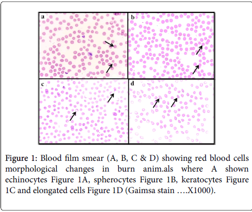
Peripheral blood film smear: Examined blood films exhibited paikilocytosis. The common cells observed were spherocytes, echinocytes, keratocytes and elongated cells. Also there were many cells with vesicle formation as well as the presence of fragmented erythrocytes as shown in Figure 1.
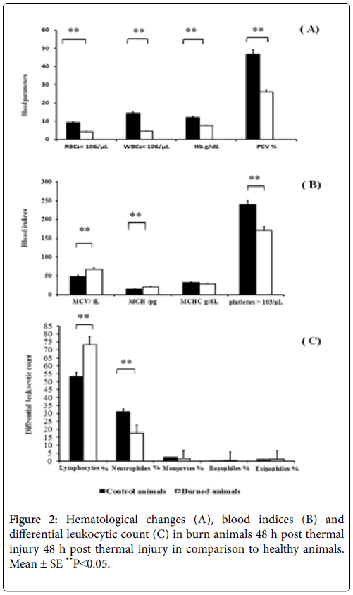
Blood parameters: Figures 1 and 2 exhibited the blood parameters of burned cattle 48 h after exposure to thermal injury. We noticed significance decrease in RBC's count, HB concentration and PCV% and thrombocytes count. Furthermore, burns induced significant increase in the WBC's count, MCV and MCH levels while the increase in MCHC level was proved non-significant in comparison to healthy controls. For differential leuckocytic count, thermal injury induced lymphocytosis and neutrophilia. On the other hand, no changes were seen in the count of both basophiles and Eosinophils in comparison with the control group according data in Figure 3.
Biochemical analyses
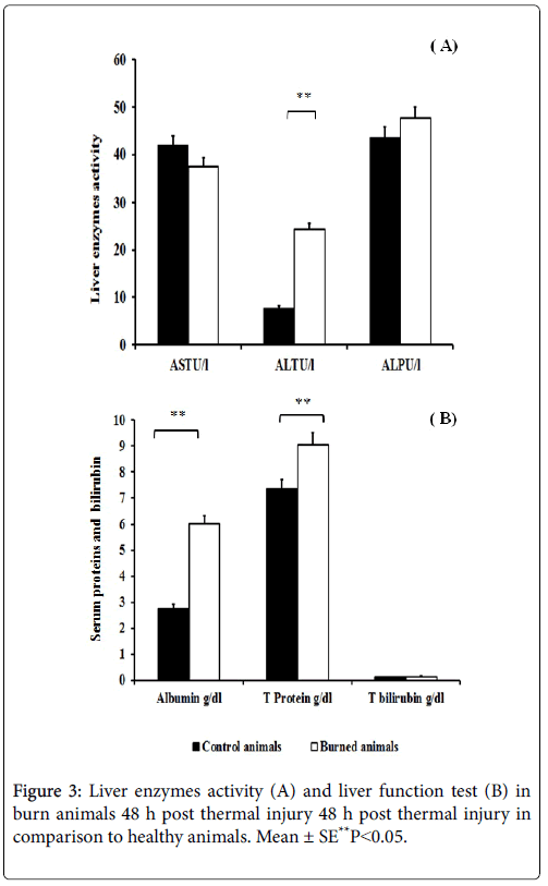
Liver function analysis: Liver enzymes activity were analyzed where there was increase in AST, ALT, ALP enzyme activities which proved highly significant in ALT enzyme when compared with control animals. In addition, burns induced significant decrease in serum total protein and albumin and total bilirubin which, proved non-significant when compared with normal control animals Figure 4.
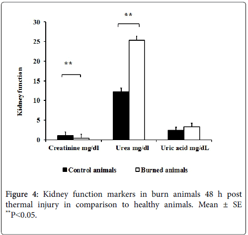
Kidney function analysis: As shown in Figure 5, burns induced significance increases in serum creatinine and blood urea nitrogen while non-significance increase was recorded in uric acid in comparison to control group.
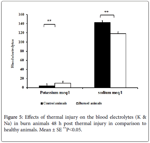
Serum electrolytes: Burns also lead to significant increase in potassium levels, while sodium level was significantly decreased.
Discussion
This study is aimed to estimate some hematological and biochemical changes in the burn animals, The levels of these parameters are very important in letting the veterinarian care team to know how the body is responding to the thermal injury and the response to the different therapies that being provided. For the hematological changes in the peripheral blood film smear many misshaping cells were observed. Echinocytes, Keratocytes and spherocytes were the dominant cells observed. The abnormal red cell morphology (paikilocytosis) resembles echinocytes, a form which is known to be due to depletion of ATP concentration in the erythrocytes [8]. RBC's count, HB concentration and PCV % values were significantly decreased in association with significantly increased in WBC's count as shown in Figure 1. Our results come in agree with EI-Sonbaty & EI-otiefy [8]. Anemia has been assumed in injured animals being due to the accelerated erythrocytes decomposition. The decreasing of hematocrit is to be expected with inadequate fluid resuscitation, and may also be a hallmark of occult bleeding [9].
Significant leukocytosis was noticed in burned animals in this study. These findings are consistent with many reports. Gruber and Farese [10] reported that a peripheral leukocytosis lasted for 35 days or more in murine granulopoiesis after inducement of a standardized sublethal third-degree burn.` A similar result was reported by D'Alesandro and Gruber [11] in an experimental study on rats, where after a 30% thermal injury leukocyte quantities were three to five times normal values. Burns induced significant increase in the MCV and MCH values. The increase in MCV and MCH values was attributed to hemolytic anemia and liver dysfunction [12]. In addition, there was significant decrease (thrombocytopenia) in PLT count when compared with control animals confirm data obtain by Fiebach et al. [13].
Thrombocytopenia was due to increase the platelet consumption for occult bleeding, dehydration and reduced thrombopoietin production due to liver failure. For differential leuckocytic count, the data in Figure 3 exhibited thermal injury induced lymphocytosis and neutrophilia. These findings are agreed with those of Marina and Lara [14]. The increase in the lymphocytes attributed to increase the histamine release, which play role in increase the vascular permeability post burning [15], while increase in the neutrophils % was due to sepsis occurred after burns [16]. To better understand, the alteration expected in the internal organs biochemical analysis was done for detection liver and kidney function markers. We have obtained elevated liver enzymes AST, ALT, ALP. Those findings were reported by previous studies [17,18]. Burns lead to an increase in edema formation that in turn leads to cell damage, with the release of hepatic enzymes [19]. The increase in the liver enzymes activities was due to the systemic inflammatory syndrome induced by thermal injury. Importantly, total protein and albumin decreased in burn animals. This study was supported by Demling [20] who recorded that marked decreased in plasma proteins occurs early post burn.
Other study also pointed out that burns injury results in dramatic changes in plasma proteins in which the concentration of protein was significantly lower in serum of burn animals than the control [21]. These results occur because local inflammatory cytokines enter the circulation and result in systemic inflammatory response. As burns approach 25% of TBSA, this will lead the microvascular escape to become generalized and permit the loss of fluid and protein from the intravascular to the extravascular spaces and finally they are lost through the wound [3].
The serum sodium of burnt animals was significantly decreased in comparison to healthy controls. This results agreed with Huang et al. [22] who states that during the first 3 days after burn, serum sodium levels were moderately elevated, as well as, these results were supported by Ge et al. [23] who explained that serum Na+ decreased post-burn and increased after resuscitation. The full explanations for these results are in the major burns, the intravascular volume is lost in burned and unburned tissues. Losing of the intravascular volume is due to increase the vascular permeability as well as the interstitial osmotic pressure in burn tissue and cellular edema developed.
The concentration of serum potassium is significantly increased in burnt animals. Our data are supported by other study, which states that in major burns, the initial resuscitation period (between 0 and 36 h) characterized by hyperkalemia because of the massive tissue destruction and necrosis [24]. As well as, Demling et al. [20] stated that potassium ions will increase if severe hemolysis has occurred or renal impairment is present. Our results reveled also increase in the level of urea and creatinine, the markers of kidney failure this come in agreement with Anandani [18]. Multiple conditions contribute to renal failure in the burn animals such as hypovolemia, cardiac dysfunction, release of inflammatory mediators and denatured proteins (from extensive tissue destruction), and nephrotoxic drugs [25,26]. Finally, the increase or decrease in the hematological and biochemical parameters may be due to the hypermetabolic state of the body which resulted from the increase in the adrenaline release, loss of fluid and electrolytes, hemolysis and sepsis.
References
- Abston S, Blakeney P, Desai M (2000) Acute Burn Management. Resident Orientation Manual. Galveston Shriners Burn Hospital and University of Texas Medical Branch Blocker Burn Unit.
- Porth CM (2007) Essential pathophysiology 2nd edition. Edited by. Lippincott Williams and Wilkins, pp 618-622.
- Church D, Elsayed S, Reid O, Winston B, Lindsay R (2006) Burn Wound Infections. Clinical Clin Microbiol Rev 19: 403-434.
- Brunicardi FC, Anderson DK, Billar TR, David LD, Hunter JG, et al. (2009) Principle of Surgery. 9th. ed., McGraw- Hill. USA, PP 189-221.
- Yang E, Maguire T, Yarmush ML, Berthiaume F, Androulakis IP (2007) Bioinformatics analysis of the early inflammatory response in a rat thermalinjury model. BMC Bioinformatics 8: 10.
- Marvaki C, Joannovich I, Kiritsi E, Iordanou P, Iconomo T (2001) The effecttiveness of early enteral nutrition in burn patients. Ann Burns Fire Disasters 16.
- Lewis SM, Bain BJ, Bates I (2006) Dacie and LEWIS Prctical haematology. 10th ed., Churchill Livingstone Elsevier. Germany.
- El-Sonbaty MA, El-Otiefy MA (1996) Haematological changes in severly Burned Patients . Ann Burns Fire Disasters 9(4).
- Stewart C (1998) Environmental Emergencies for Emergency Services. J .B. Diving medicine, 719: 265-1803.
- Gruber DF, Farese AM (1989) Bone marrow myelopoiesis in rats after 10%, 20% or 30% thermal injury. J Burn Care Rehabil 10: 410-417.
- D'Alesandro MM, Gruber DF (1990) Quantitative and functional alterations of peripheral blood neutrophils after 10% and 30% thermal injury. J Burn Care Rehabil 11: 295-300.
- Savage DG, Ogundipe A, Allen RH, Stabler SP, Lindenbaum J (2000) Etiology and diagnostic evaluation of macrocytosis. J Med Sci 319: 343-352.
- Fiebach NH, Barker LR, Burton JR, Zieve PD (2007) Principles of Ambulatory Medicine. Lippincott Williams & Wilkins. ISBN 9780781762274.
- Marina P, Lara M (2007) Platelet count monitoring in burn patients. Biochem Med 17: 212-219.
- Roszkowski W, Plaut N, Lichtenstein LM (1977) Selective display of histamine receptors on lymphocytes Science 195: pp.683-685.
- Tavares-Murta BM, Zaparoli M, Ferreira RB, Silva-Vergara ML, Oliveira CH, et.al. (2002) Failure of neutrophil chemotactic function in septic patients. Crit Care Med 30: 1056-1061.
- Hala MG, Mahfouz MH (2008) Biochemical Alterations of Amino Acids, Neurotransmitters and Hepatic Functions after Thermal Injury in Rats. Egypt J Biochem Mol Biol 26: 13-28.
- Anandani JH (2010) Impact of thermal injury on hematological and biochemical parameters in burnt patients.Biosc Biotech Res Comm 3: 97-100.
- Jeschke MG, Low JF, Spies M, Vita R, Hawkins HK, et.al. (2001) Cell proliferation, apoptosis, NF-kappaB expression, enzyme, protein, and weight changes in livers of burned rats. Am J Physiol Gastrointest Liver Physiol 280: 1314-1320.
- Demling RH, DeSanti LR, Orgill D (2004) Practical Approach To Treatment: Initial Management of the Burn Patient PART 2. Burn Surger Org.
- Vemula M, Berthiaume F, Jayaraman A , Yarmush ML (2004) Expression profiling analysis of the metabolic and inflammatory changes following burn injury in rats. Physiol Genomics 18: 87-98.
- Ge SD, Xhu SH, Liu SK, Chen YL (1996) Effects of hypertonic sodium lactate dextran 70 resuscitation severely burned dogs. Annals of Burns and Fire Disasters 9: 101.
- Huang PP, Stucky FS, Dimick AR, Treat RC, Bessey PQ, et al. (1995) Hypertonic sodium resuscitation is associated with renal failure and death. Ann Surg 221: 543-557.
- Ramos CG (2000) Management of fluid and electrolyte disturbances in the burn patient. Ann Burns Fire Disasters 13 (4).
- Davies DM, Pusey CD, Rainford DJ (1979) Acute renal failure in burns. Scand J Plast Reconstr Surg 13: 189-92.
- Schiavon M, Di Landro D, Baldo M (1988) A study of renal damage in seriously burned patients. Burns. 14: 107-12.
Citation: Khalil MA, Shanab OM, Zannoni GF, Heygazy EM (2018) Assessment of Hematological and Biochemical Markers in the Early post-Burn Period for Predicting Recovery and Mortality in Animals with Burn Injuries. J Clin Exp Pathol 8:343. DOI: 10.4172/2161-0681.1000343
Copyright: © 2018 Khalil AM, et al. This is an open-access article distributed under the terms of the Creative Commons Attribution License, which permits unrestricted use, distribution, and reproduction in any medium, provided the original author and source are credited.
Share This Article
Recommended Journals
Open Access Journals
Article Tools
Article Usage
- Total views: 6745
- [From(publication date): 0-2018 - Apr 04, 2025]
- Breakdown by view type
- HTML page views: 5839
- PDF downloads: 906

