Aretery of Percheron Stroke (About 3 Cases)
Received: 04-May-2023 / Manuscript No. roa-23-98748 / Editor assigned: 06-May-2023 / PreQC No. roa-23-98748 (PQ) / Reviewed: 20-May-2023 / QC No. roa-23-98748 / Revised: 23-May-2023 / Manuscript No. roa-23-98748 (R) / Published Date: 30-May-2023 DOI: 10.4172/2167-7964.1000444
Abstract
The Percheron artery is a rare thalamic vasculature variant and is a single dominant thalamoperforating artery supplying bilateral paramedian thalamic territories. Occlusion of the aretery of Percheron results in a characteristic pattern of bilateral paramedian thalamic infarcts and is estimated to represent between 0.1%-0.3% of all ischemic strokes and 4% to 35% of all thalamic strokes. Despite our knowledge of the characteristic radiologic features of an aretery of percheron stroke, the true incidence of aretery of percheron strokes is challenging to estimate due to nonspecific clinical symptoms and subtle findings on computed tomography (CT) and magnetic resonance imaging (MRI). We report a case series of three patients with aretery of percheron stroke confirmed by radiologic imaging.
Keywords
Percheron artery, MRI, Paramedian thalamus, Rostral midbrain
Introduction
Percheron artery is a rare variant of thalamic arteries characterized by a single dominant perforating artery, originating in the proximal segment of the posterior cerebral artery and supplying the bilateral paramedian thalamus and the rostral midbrain [1]. The incidence of Percheron arterial ischemia is estimated to be 0.1% to 0.3% [2]. MRI is a crucial examination to establish an accurate diagnosis and to ensure better therapeutic management. We report in our article three cases of arterial infarction of Percheron.
Case presentation
Case 1
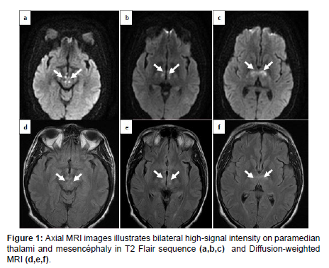
A 43 years old man with a past medical history of carotid surgery, presented to the emergency department with a fluctuant consciousness, followed by drowsiness episodes. On the neurological examination, the patient had right upper limb paraplegia. No other abnormalities were noted. A brain MRI showed bilateral thalamic and mesencephalic lesions in T2 Flair and hyper diffusion signal (Figure 1).
Case 2
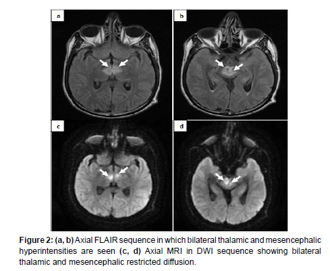
A 45-year-old patient with no significant past medical history, he was admitted to the emergency room for sudden onset of consciousness and diabetic ketoacidosis. On physical examination, he was unconscious with a Glasgow Coma Scale score of 7. He had neither sensitive nor motor focal signs. A brain MRI identified evidence of bilateral thalamic and mesencephalic infarction (Figure 2).
Case 3
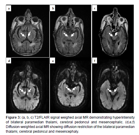
A 91-year-old patient was admitted to the emergency room for a brief loss of consciousness. On the neurological exam, the patient had a Glasgow Coma Scale (GCS) of 12 points (ocular: three points, verbal: five points, motor: four points) with bilateral symmetrical sluggish pupils, however, he had neither sensitive nor motor focal signs. Magnetic resonance imaging (MRI) of the brain was performed, revealing bilateral thalamic mesencephalic, and peduncles lesions in T2 Flair and diffusion hyper signal (Figure 3).
Discussion
The artery of Percheron is a rare anatomical variant of paramedian thalamic vessels [3]. It is present in 7-10% of the general population [4]. Bilateral perforating thalamic arteries branch from a single arterial trunk originating from the proximal segment (P1) of the posterior cerebral artery [3]. It provides arterial supply to the paramedian territories of the thalamus and rostral zones of the midbrain [3].
Infarction caused by occlusion of the Percheron artery is a subtype of stroke that is often under-diagnosed [5] due to the functional anatomy of the thalamus; occlusion of the Percheron artery causes variable and non-specific clinical manifestations [1].
Four distinct ischemic patterns of Percheron artery infarction have been identified: a bilateral thalamic region with mesencephalic involvement (43%), bilateral paramedian thalamic without
Mesencephalic involvement (38%), bilateral paramedian thalamic and anterior thalamus with mesencephalic participation (14%), and bilateral paramedian thalamic and anterior thalamus without mesencephalic involvement (5%) [2].
Infarction of this artery is often characterized by a triad of symptoms, including anemia, supranuclear and vertical gaze palsy, and altered consciousness [6]; other symptoms have been reported: severe bradycardia, seizures, behavioral disturbances, slurred speech, motor deficits [6, 7].
Early diagnosis of Percheron arterial infarction can be difficult because it is uncommon, and early CT or MRI may be harmful [8]. Cerebral CT and angioscan usually show no abnormalities [9]. On CT, Percheron arterial infarction may be manifested by hypodensity in the bilateral paramedian thalamic regions concerning cytotoxic edema [8].
MRI is the examination of choice for early diagnosis, and diffusionweighted imaging (DWI) and Flair sequence are the key sequences. The infarct appears as a T2 Flair and DWI hyper signal [3], a V-shaped hyper signal on Flair and DWI sequence along the pial surface of the midbrain in the interpeduncular fossa [10]. The sensitivity of this V-sign is 67% in case of associated midbrain involvement [10].
There may be other types of infarcts that can lead to bilateral thalamic lesions apart from Percheron arterial infarction; these include deep cerebral venous thrombosis by occlusion of the internal cerebral veins as well as basilar artery apex syndrome, in which case territories supplied by the posterior cerebral, superior cerebellar and pontine arteries are also involved [10].
Wernicke’s encephalopathy is also included in the differential diagnosis as it involves bilateral lesions of the thalami, periaqueductal gray tectal plate, dorsal medulla, and mammillary bodies [10]. Infections and osmotic myelinosis may also be included in the differential diagnosis [10].
Treatment of Percheron arterial infarction includes thrombolysis and intravenous heparin therapy combined with long-term anticoagulants. However, as there is often a delay in diagnosing Percheron arterial infarction, thrombolysis cannot be performed due to its narrow therapeutic window. This highlights the importance of early diagnosis so that therapy can be initiated [8].
Conclusion
Percheron’s artery is a rare anatomical variant. Percheron’s arterial infarction leads to a characteristic ischemic, bilateral paramedian thalamic injury with or without midbrain involvement. Early neuroimaging and a precise diagnosis are necessary for better patient management.
References
- Kichloo A, Jamal SM, Zain EA, Wani F, Vipparala N (2019) Artery of Percheron Infarction: A Short Review. J Investig Med High Impact Case Rep 7: 2324709619867355.
- Sandvig A, Lundberg S, Neuwirth J (2017) Artery of Percheron infarction: a case report. J Med Case Rep. 11: 221.
- Ranasinghe KMIU, Herath HMMTB, Dissanayake D, Seneviratne M (2020) Artery of Percheron infarction presenting as nuclear third nerve palsy and transient loss of consciousness: a case report. BMC Neurol 20: 320.
- Snyder HE, Ali S, Sue J, Unsal A, Fong C, et al. (2020) Artery of Percheron infarction with persistent amnesia: a case report of bilateral paramedian thalamic syndrome. BMC Neurol. 20: 370.
- Lazzaro NA, Wright B, Castillo M, Fischbein NJ, Glastonbury CM, et al. (2010) Artery of percheron infarction: imaging patterns and clinical spectrum. AJNR Am J Neuroradiol 31: 1283-1289.
- Flowers J, Gandhi S, Guduguntla L, Yang A, Moudgil S (2022) Artery of Percheron Strokes: Three Cases in Three Months. Cureus. 14: e21688.
- Khanni JL, Casale JA, Koek AY, Espinosa Del Pozo PH, Espinosa PS (2018) Artery of Percheron Infarct: An Acute Diagnostic Challenge with a Spectrum of Clinical Presentations. Cureus 10: e3276.
- Macedo M, Reis D, Cerullo G, Florêncio A, Frias C, et al. (2022) Stroke due to Percheron Artery Occlusion: Description of a Consecutive Case Series from Southern Portugal. J Neurosci Rural Pract 13: 151-154.
- Sharma A, Bande D, Matta A (2021) A Case of Diagnostic Difficulty: Transient Loss of Consciousness in Artery of Percheron Infarct. Cureus 13: e12918.
- Kheiralla OAM (2020) Artery of Percheron infarction a rare anatomical variant and a diagnostic challenge: Case report. Radiol Case Rep 16: 22-29.
Indexed at, Google Scholar, Crossref
Indexed at, Google Scholar, Crossref
Indexed at, Google Scholar, Crossref
Indexed at, Google Scholar, Crossref
Indexed at, Google Scholar, Crossref
Indexed at, Google Scholar, Crossref
Indexed at, Google Scholar, Crossref
Indexed at, Google Scholar, Crossref
Indexed at, Google Scholar, Crossref
Citation: Houssni JE, Dehayni F, Neftah I, Nouali H, Edderai M, et al. (2023) Aretery of Percheron Satroke (About 3 Cases). OMICS J Radiol 12: 444. DOI: 10.4172/2167-7964.1000444
Copyright: © 2023 Houssni JE, et al. This is an open-access article distributed under the terms of the Creative Commons Attribution License, which permits unrestricted use, distribution, and reproduction in any medium, provided the original author and source are credited.
Select your language of interest to view the total content in your interested language
Share This Article
Open Access Journals
Article Tools
Article Usage
- Total views: 2401
- [From(publication date): 0-2023 - Nov 23, 2025]
- Breakdown by view type
- HTML page views: 2057
- PDF downloads: 344


