Case Report Open Access
Apical Thrombus Mimicking Cardiac Myxoma: Application of Cardiovascular Magnetic Resonance
| Rimsha Hasan, John Saia, Patrick O’Beirne, Kenneth Khaw, Gerald Ukrainski, Edward Wrobleski, Lannae Ewing, Chad Bousanti, Wehner LJ and Jingsheng Zheng* | |
| Division of Cardiology, AtlantiCare Regional Medical Center, Pomona, New Jersey | |
| Corresponding Author : | Jingsheng Zheng Division of Cardiology AtlantiCare Regional Medical Center 65 Jimmie Leeds Road Pomona, NJ 08205,USA Tel: +609-561-8500 Fax: +609-567-0432 E-mail: jszheng@comcast.net |
| Received February 10, 2015; Accepted March 23, 2015; Published March 26, 2015 | |
| Citation: Hasan R, Saia J, O’Beirne P, Khaw K, Ukrainski G, et al. (2015) Apical Thrombus Mimicking Cardiac Myxoma: Application of Cardiovascular Magnetic Resonance. OMICS J Radiol 4:181. doi: 10.4172/2167-7964.1000181 | |
| Copyright: © 2015 Rimsha Hasan, et al. This is an open-access article distributed under the terms of the Creative Commons Attribution License, which permits unrestricted use, distribution, and reproduction in any medium, provided the original author and source are credited. | |
Visit for more related articles at Journal of Radiology
Abstract
Echocardiography is the most common imaging modality to visualize cardiac masses. However, sometimes it is difficult to distinguish between thrombus and cardiac tumors. Other imaging modalities should be used to delineate detailed anatomy of the cardiac masses. A 63-year-old white male with past medical history of coronary artery disease, myocardial infarct and coronary artery bypass graft surgery, was found to have a “cardiac mass” by a routine 2-dimensional echocardiogram. Echocardiogram revealed a large mass in left ventricle attached with a long narrow stalk to the apex. Contrast echocardiogram with DEFINITY revealed a non-opacified contrast defect in the left ventricular apex. It is very rare that apical thrombus has a long narrow stalk. Other possibilities such as cardiac myxoma or other cardiac tumors cannot be excluded. Cardiac magnetic resonance with and without contrast was therefore performed. It revealed a nonenhancing rounded thrombus within the apex. There was left apical thinning/ aneurysm with dyskinesis. Patient was treated with Coumadin for anticoagulation. He was doing well with current regimen.
| Keywords |
| Cardiac Mass; Echocardiogram; Cardiovascular Magnetic Resonance |
| Introduction |
| Cardiac Masses are broadly classified into tumors, vegetation, thrombi, iatrogenic and normal variants. 2-dimensional echocardiogram is the primary modality commonly used for evaluation of these masses. However this modality has its limitations. Recent study by Weinsaft et al. showed that sensitivity and specificity for evaluation of LV thrombus by 2D echocardiogram is 33 and 91%, respectively. Intra-observer variability also limits the utilization of conventional echocardiogram to accurately diagnose thrombi. The recent advances in cardiac MRI have made it possible to delineate subtle differentiation points between cardiac masses. Present study reveals a rare case of cardiac thrombus mimicking atrial myxoma. |
| Case Report |
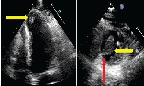
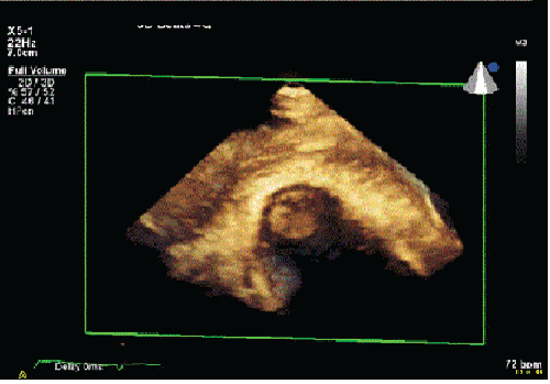
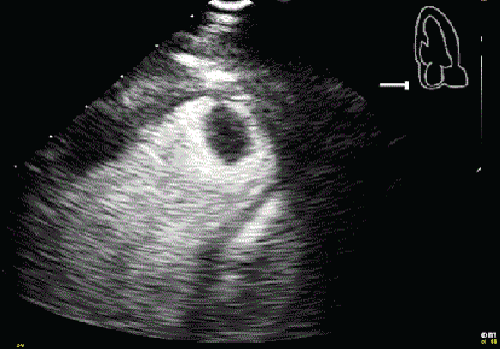
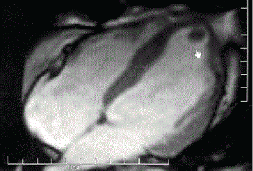
| A 63 year old male with history of coronary artery disease, post myocardial infarction in 2008 with four-vessel coronary artery bypass graft surgery (CABGx4) underwent a follow up echocardiogram. Patient was in his usual state of health and did not have any complaints of dyspnea, palpitations, decreased functional capacity, chest pain or syncope. Electrocardiogram was consistent with sinus tachycardia at 100 bpm with prior anteroseptal infarction. 2- dimensional transthoracic echocardiogram (TTE) revealed a large 1.4 cm × 0.9 cm, ovoid and heterogeneous mass (yellow arrow) in left ventricle (LV) attached with a long narrow stalk (red arrow) to apex (Figure 1). 3-dimensional echocardiogram revealed a large apical mass (Figure 2). Contrast echocardiogram with DEFINITY revealed apical akinesis and severe hypokinesis of the inferoapical and anterior wall with LV ejection fraction of 45%. There was a non-opacified contrast defect in the LV apex (Figure 3). Patient was started on anticoagulation with heparin with suspicion of possible apical thrombus however possibility of different etiology of cardiac mass was not entirely excluded. Cardiac MRI was subsequently done on 1.5 Tesla magnet system without and with intravenous contrast. 23 ml of Magnevist was administered intravenously. EKG synchronization was utilized for cardiac gating. Delayed myocardial enhancement compatible with infarct within anterior wall extending from base to the apex and into the lateral wall was noted. A non-enhancing rounded thrombus within the apex was noted measuring 10 mm along with left apical thinning/aneurysm with dyskinesis (Figure 4). Right ventricle was found to be normal in size with normal right ventricular wall motion. No pericardial effusion or abnormal pericardial enhancement was seen. Patient was treated with coumadin for anticoagulation. He is currently doing well. |
| Discussion |
| Left ventricular thrombus formation is a frequent complication in the patient with history of anterior wall myocardial infarct and in those patients with systolic left ventricular dysfunction. The incidence of LV thrombus was reported as high as 30-40% in patients with anterior wall myocardial infarct2. Cardiac thrombi are at risk for thromboembolic events. Cardiac tumors, both primary and secondary, occur with an estimated prevalence of 0.002-0.3% at autopsy and 0.15% in echocardiographic series3. Cardiac myxomas are the most common benign primary cardiac tumor. Cardiac myxomas occur mostly in the left atrium and are rarely seen in the LV (< 5%). There are multiple reports of LV myxomas attaching to the LV wall via a long stalk4,5,6. Prompt and accurate diagnosis of LV thrombus and cardiac tumors (such as myxoma) is very important since treatment for cardiac thrombus and cardiac myxomas are significant different. Systemic anticoagulation is generally recommended for LV thrombus to prevent potential thromboembolic event, while surgical resection may be the choice for cardiac tumors. |
| TTE is the most common imaging modality for detecting intracardiac masses. However, it has several limitations, including operator dependence and restrictive field of view (especially in the patient with a large body habitus)7. It has been reported that significant interobserver variability exists in diagnosing LV thrombus. One recent study1 showed that sensitivity and specificity for using TTE for detection of apical thrombus is 33% and 91%, respectively, with positive and negative predicted value of 29% and 93%. |
| In the present study, a patient with prior myocardial infarct, S/P CABG, and LV dysfunction, was found to have a LV mass attaching to the apex via a long thin stalk during a routine TTE. This finding was confirmed with contrast echocardiography using DEFINITY and 3- dimensional echocardiogram. It is very rare to have a long thin stalk with apical thrombus. Other possibilities include cardiac myxoma or other cardiac tumors |
| Cardiovascular magnetic resonance (CMR) has become a mainstream imaging modality in the workup of intracardiac masses. CMR provides an unrestricted field of view, and superior soft-tissue depiction without ionizing radiation. CMR can also provide optimal tumor characterization. Contrast-enhanced CMR using gadolinium contrast has been shown to distinguish viable and infarcted myocardium on the basis of contrast uptake pattern. Recently, this technique was shown to be a useful tool to detect LV thrombus. Since thrombus is avascular, it does not have contrast uptake 8,9. The patient in this study underwent CMR with contrast, which confirmed LV thrombus. Patient was doing well with anticoagulation. |
| Summary |
| TTE is the most common initial imaging modality to evaluate LV mass. Contrast echocardiogram and 3-dimensional echocardiogram may provide detailed anatomical appearance of the apical mass. Contrast-enhancement CMR can identify thrombus based on tissue characteristics rather than anatomic appearance. CMR enables thrombus identification irrespective of location and morphology. |
References |
|
Figures at a glance
 |
 |
 |
 |
| Figure 1 | Figure 2 | Figure 3 | Figure 4 |
Relevant Topics
- Abdominal Radiology
- AI in Radiology
- Breast Imaging
- Cardiovascular Radiology
- Chest Radiology
- Clinical Radiology
- CT Imaging
- Diagnostic Radiology
- Emergency Radiology
- Fluoroscopy Radiology
- General Radiology
- Genitourinary Radiology
- Interventional Radiology Techniques
- Mammography
- Minimal Invasive surgery
- Musculoskeletal Radiology
- Neuroradiology
- Neuroradiology Advances
- Oral and Maxillofacial Radiology
- Radiography
- Radiology Imaging
- Surgical Radiology
- Tele Radiology
- Therapeutic Radiology
Recommended Journals
Article Tools
Article Usage
- Total views: 14608
- [From(publication date):
April-2015 - Jul 06, 2025] - Breakdown by view type
- HTML page views : 9990
- PDF downloads : 4618
