Research Article Open Access
Anterolateral Ankle Pain: Comparison of Two Areas of Clinical Anterolateral Pain Using Imaging and Arthroscopic Findings
Kentaro Amaha1*, Taiki Nozaki2, Sachiko Ohde3 and Atsushi Tasaki1
1Department of Orthopedic Surgery, St. Luke’s International Hospital, Tokyo, Japan
2Department of Radiology, St. Luke’s International Hospital, Tokyo, Japan
3Life Science Institute, St. Luke’s International Hospital, Tokyo, Japan
- *Corresponding Author:
- Amaha K
Department of Orthopedic Surgery
St. Luke’s International Hospital, Tokyo, Japan
Tel: 81-335415151
Fax: 81-335440649
E-mail: amaken@luke.ac.jp
Received Date: July 21, 2016; Accepted Date: June 9, 2016; Published Date: June 13, 2016
Citation: Amaha K, Nozaki T, Ohde S, Tasaki A (2016) Anterolateral Ankle Pain: Comparison of Two Areas of Clinical Anterolateral Pain Using Imaging and Arthroscopic Findings. Clin Res Foot Ankle 4:187. doi:10.4172/2329-910X.1000187
Copyright: © 2016 Amaha K, et al. This is an open-access article distributed under the terms of the Creative Commons Attribution License, which permits unrestricted use, distribution, and reproduction in any medium, provided the original author and source are credited.
Visit for more related articles at Clinical Research on Foot & Ankle
Abstract
Keywords
Anterolateral ankle pain; Ankle sprain; Ankle instability; CT findings; MRI findings; Arthroscopic findings; Scar tissue
Abbreviations
MRI: Magnetic Resonance Imaging; CT: Computed Tomography; ATFL: Anterior Talofibular Ligament; SD: Standard Deviation
Introduction
Lateral ligament injuries of the ankle are common in athletes. The majority of these patients can be treated conservatively [1,2]. However, patients with continuous pain and instability affecting their ability to participate in sports can be treated surgically [3]. Chronic anterolateral ankle pain is a common complaint following ankle sprain in athletes [4].
Although the exact definition of the anterolateral aspect of the ankle is ambiguous, the area typically referred to as anterolateral is composed of the capsule and ligament anteriorly, fibula laterally, talus medially and inferiorly, and tibia medially and superiorly. To date, arthroscopic examination and imaging studies have demonstrated a variety of anterolateral ankle pathologies [4-13].
With respect to the anterolateral region of the ankle, several pathologic conditions reportedly cause “anterolateral ankle impingement”, including osteophytes, synovitis, thickened scar tissue, an accessory anterior inferior tibiofibular (Bassett’s) ligament, and loose bodies. Anterolateral impingement is defined by the presence of the interposition of abnormal soft tissue [4,7,11].
Currently, MRI is a validated, noninvasive diagnostic tool to detect anterolateral impingement that is strongly associated with abnormality at arthroscopy [8]. Although anterolateral ankle impingement is now better understood, the exact relationship between structural abnormality and physical anterolateral ankle pain remains unclear.
In the clinical setting, there are two primary areas of pain in the anterolateral region: the lateral edge of the plafond (lateral shoulder) and the area over the anterior talofibular ligament (ATFL). Only a few studies have described the relationship between findings on clinical examination and the presence of physical pain in these specific regions [8,13].
Furthermore, there are no published studies assessing the anterolateral ankle region divided into these two separate areas. The aim of this study was to assess the relationship between imaging and arthroscopic intra-articular abnormalities in these two distinct areas of anterolateral ankle pain.
Materials and Method
Between 2011 and 2013, the electronic medical records of consecutive patients who underwent ankle joint arthroscopy were reviewed. The study was a retrospective review of the medical records and hence did not require review by the Local Research Ethics Committee.
Patients with chronic ankle pain or laxity of the ankle joint after two months of conservative treatment, consisting of anti-inflammatory medication and physical therapy, were included in this retrospective study. An ankle specialist (KA) assessed ankle laxity by performing the talar tilt test and anterior drawer test.
These tests were considered positive if there was soft end feel to the translation of the talus. Patients with septic ankle arthritis, crystal ankle arthritis, osteoarthritis of the subtalar joint, those who had undergone previous ankle surgery, and those with a history of a foot or ankle fracture were excluded.
A total of 32 patients (21 males, 11 females) with an average age of 36.5 (±15.9 standard deviation [14]) years were included in this analysis. The average duration between the onset of symptoms and the day of surgery was 9.3 (±7.2 SD) months and the average duration of follow-up was 25.1 (±11.7 SD) months.
The foot and ankle specialist (KA) assessed all patients preoperatively. Physical examination was performed for each ankle to determine if patients had tenderness over the anterolateral area.
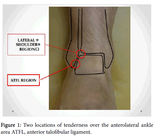
To assess the point of tenderness precisely, we divided the anterolateral area into two regions: the lateral shoulder region and the ATFL region (Figure 1). The presence of tenderness was recorded in all patients. On clinical examination, there was no foot deformity or systemic hyperlaxity in any patient.
Imaging assessment
All patients were examined preoperatively by non-enhanced MRI and non-enhanced CT. CT studies were performed on a 16-slice CT (Bright Speed Elite, GE Healthcare, Milwaukee, USA) with a 0.625 × 16 mm detector or a 64-slice CT (Discovery CT750 HD, GE Healthcare,Milwaukee, USA) with a 0.625 × 64 mm detector.
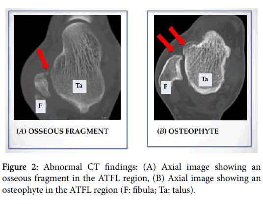
CT findings, including osteophytes and osseous fragments in the lateral edge and ATFL regions, were evaluated. MRI studies were performed on a 3.0T-MRI unit (Verio, Siemens AG, Erlangen, Germany) or a 1.5T-MRI unit (Optima 450w, GE Healthcare, Milwaukee, USA). For the Verio, MR images included coronal and axial short tau inversion recovery (STIR) images (TR/TE/TI/FA = 5000/77/220/150) and T1WI images (TR/TE/FA = 600/10/150).
Each slice was 4 mm thick, and the slice gap was 0.5 mm. The field of view was 16 cm, and the matrix size was 320 × 256 mm in all images. With the Optima 450w, MR images included coronal and axial STIR images (TR/TE/TI/FA = 6000/52/150/160) and PDWI images (TR/TE/FA = 2500/22/160).
Each slice was 4 mm thick, and the slice gap, 0.5 mm. The field of
view was 16 cm, and the matrix size was 320 × 224 mm in all images.
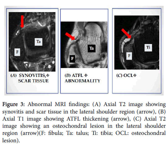
MRI findings, including synovitis, scar tissue, and abnormal ATFL and
osteochondral lesions in the lateral shoulder and ATFL regions, were
evaluated.
A board-certified radiologist experienced in musculoskeletal radiology (13 years of experience) retrospectively reviewed MR and CT images while blinded to clinical and arthroscopic findings (Figures 2 and 3).
Figure 3: Abnormal MRI findings: (A) Axial T2 image showing synovitis and scar tissue in the lateral shoulder region (arrow), (B) Axial T1 image showing ATFL thickening (arrow), (C) Axial T2 image showing an osteochondral lesion in the lateral shoulder region (arrow)(F: fibula; Ta: talus; Ti: tibia; OCL: osteochondral lesion).
Arthroscopy method
Ankle arthroscopy was performed in all patients under general
anesthesia by a single surgeon (KA). The limb was prepared in the
usual fashion. Topographic landmarks of the anterior aspect of the
ankle were identified. A non-invasive distraction device and an
automated inflow pump system administering isotonic saline were
used in all patients. A tourniquet was used only when scope visibility
was impaired because of bleeding. A 2.7-mm 30-degree arthroscope,
2.9-mm shaver, and burr were used. Anteromedial and anterolateral
portals were performed using blunt dissection with straight mosquito
forceps to avoid injury to the superficial peroneal nerve, and
penetration of the joint capsule was accomplished using a trocar with a
rounded tip. Exploration of the ankle joint was performed in a
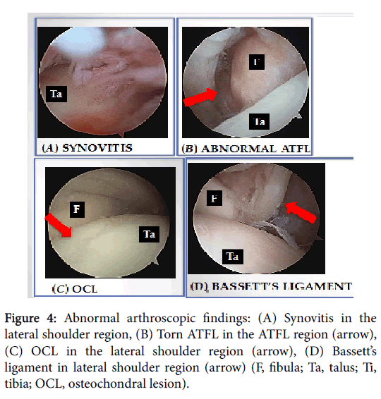
systematic manner (Figure 4). All surgeries were recorded in DVD
format from the beginning to the end of the procedure. The recordings
were reviewed by the author (KA). The assessment of anterolateral
lesions was made based on the presence of intra-articular pathologies
including synovitis, scar tissue, Bassett’s ligament, osseous fragment, abnormal ATFL, and osteochondral lesions in the lateral edge and
ATFL regions. Appropriate postoperative care tailored to each patient
was provided.
Figure 4: Abnormal arthroscopic findings: (A) Synovitis in the lateral shoulder region, (B) Torn ATFL in the ATFL region (arrow), (C) OCL in the lateral shoulder region (arrow), (D) Bassett√ʬ?¬?s ligament in lateral shoulder region (arrow) (F, fibula; Ta, talus; Ti, tibia; OCL, osteochondral lesion).
Statistics analysis
Chi-squared tests were used to compare the results of CT and MRI versus arthroscopy. A level of p<0.05 was considered statistically significant; all statistical tests were two-tailed. Data were analyzed using IBM SPSS statistics software version 22.0 J (IBM, Tokyo, Japan).
Results
Individual patient findings are listed in Table 1. Almost all patients showed some abnormalities on both imaging and arthroscopy, regardless of physical tenderness.
| No. | age | √?¬†gender | tenderness | tenderness | CT | CT | MRI | MRI | arthroscopy | arthroscopy |
|---|---|---|---|---|---|---|---|---|---|---|
| (LS) | (ATFL) | (LS) | (ATFL) | (LS) | (ATFL) | (LS) | (ATFL) | |||
| 1 | 53 | male | 1 | 0 | 1 | 2 | 1,2 | 1,2 | 2,4 | 1,4 |
| 2 | 28 | female | 0 | 0 | 0 | 2 | 0 | 0 | 1 | 4 |
| 3 | 30 | male | 0 | 0 | 1 | 0 | 4 | 0 | 3 | 1 |
| 4 | 30 | male | 1 | 1 | 1 | 1,2 | 1,2 | 1 | 1,2 | 1,3 |
| 5 | 32 | male | 1 | 0 | 1 | 1,2 | 1,2 | 1 | 1,2 | 1,3 |
| 6 | 27 | male | 1 | 1 | 1 | 1,2 | 1,2 | 1 | 1,2 | 1,3 |
| 7 | 19 | male | 1 | 1 | 1 | 1,2 | 1,2 | 1 | 1,2,4 | 1,3 |
| 8 | 19 | male | 0 | 1 | 0 | 0 | 1 | 0 | 1 | 1 |
| 9 | 44 | female | 0 | 1 | 0 | 0 | 0 | 0 | 0 | 0 |
| 10 | 49 | male | 1 | 0 | 1 | 1 | 1,2 | 1,2 | 1,4 | 1,4 |
| 11 | 21 | male | 1 | 1 | 1 | 2 | 0 | 0 | 1,2 | 1,3 |
| 12 | 29 | male | 0 | 0 | 1 | 0 | 2 | 1,2,3 | 0 | 0 |
| 13 | 20 | male | 1 | 1 | 1 | 0 | 0 | 0 | 1,2 | 0 |
| 14 | 24 | female | 0 | 1 | 1 | 2 | 0 | 0 | 1,3 | 0 |
| 15 | 44 | male | 0 | 1 | 0 | 2 | 0 | 0 | 1,4 | 1 |
| 16 | 30 | female | 1 | 0 | 1 | 1 | 1,2 | 0 | 1 | 1 |
| 17 | 49 | male | 1 | 1 | 1 | 1 | 1 | 1,2 | 1,2 | 3 |
| 18 | 40 | male | 0 | 1 | 1,2 | 1 | 1,2 | 1,2,3 | 1 | 1,3 |
| 19 | 29 | male | 1 | 1 | 2 | 1 | 0 | 1,2 | 1 | 1,3 |
| 20 | 64 | female | 1 | 1 | 1 | 1 | 1,2 | 1,2,3 | 1,4 | 1,2,3,4 |
| 21 | 21 | male | 1 | 1 | 2 | 0 | 1 | 1,2 | 1 | 1 |
| 22 | 21 | male | 0 | 0 | 0 | 0 | 1 | 0 | 2 | 1 |
| 23 | 27 | female | 1 | 1 | 0 | 0 | 1 | 1 | 1 | 3 |
| 24 | 18 | female | 0 | 0 | 0 | 1 | 1 | 1,2 | 1 | 3 |
| 25 | 36 | female | 1 | 0 | 0 | 0 | 0 | 1 | 1,2 | 3 |
| 26 | 40 | male | 1 | 0 | 1,2 | 1,2 | 1 | 1 | 1,3 | 1,2,4 |
| 27 | 64 | male | 1 | 0 | 1 | 1,2 | 1,2 | 1 | 1,3 | 2,4 |
| 28 | 57 | female | 1 | 0 | 0 | 0 | 1 | 1 | 2 | 1,4 |
| 29 | 67 | female | 0 | 0 | 1 | 1 | 1,2 | 1 | 1,3,4 | 1,2,4 |
| 30 | 73 | male | 1 | 0 | 1,2 | 1,2 | 1,2 | 1,3 | 1 | 1,3 |
| 31 | 20 | female | 1 | 1 | 0 | 0 | 1 | 0 | 1 | 0 |
| 32 | 43 | male | 0 | 0 | 1 | 0 | 0 | 0 | 1 | 1 |
Table 1: Patient abnormalities on clinical, radiographic, and arthroscopic evaluation.TENDERNESS: 0: No pain 1: Pain
CT (LS): 0: Normal 1: Osteophyte 2: Osseous fragment
CT (ATFL): 0: Normal 1: Osteophyte 2: Osseous fragment
MRI (LS): 0: Normal 1: Synovitis, Scar tissue 2: Osteochondral lesion
MRI (ATFL): 0: Normal 1: Synovitis, Scar tissue 2: Abnormal ATFL 3: OCL
ARTHOSCOPIC FINDING (LS) 0: Normal 1: Synovitis, Scar tissue 2: Bassett's ligament
3: Osseous fragment 4: OCL
ARTHOSCOPIC FINDING (ATFL) 0: Normal 1: Synovitis, Scar tissue 2: Osseous fragment
3: Abnormal ATFL 4: OCL
Physical examination
Twenty-six patients (81.3%) showed tenderness over the anterolateral aspect: 20 (62.5%) had a lateral shoulder lesion, 17 (54.1%) had an ATFL lesion, and 11 (34.4%) had lesions in both regions.
Preoperative CT revealed bony abnormalities on the anterolateral aspect in 28 patients (87.5%). Concerning lateral shoulder lesions, 20 patients (62.5%) had bony spurs and 5 patients (15.6%) had osseous fragments. With regard to ATFL lesions, 15 patients (46.9%) had bony spurs and 12 patients (37.5%) had osseous fragments.
Preoperative MRI revealed abnormalities on the anterolateral aspect in a total of 25 patients (78.1%). Twenty-three patients (71.9%) had abnormalities of the lateral shoulder, including synovitis/scar tissue and osteochondral lesions, in descending order of frequency. Twenty patients (62.5%) had abnormalities of the ATFL region, including synovitis/scar tissue, abnormal ATFL, and osteochondral lesions, in descending order of frequency.
On arthroscopy, 30 patients (97.0%) showed abnormalities of the entire anterolateral aspect. Thirty patients (97.0%) showed abnormalities of the lateral shoulder region, including synovitis/scar tissue, Bassett’s ligament, osteochondral lesions, and osseous fragments, in descending order of frequency. Twenty-seven patients (84.4%) showed abnormalities of the ATFL region, including synovitis/ scar tissue, abnormal ATFL, osteochondral lesions, and osseous fragments, in descending order of frequency.
Statistical analysis showed that abnormal radiographic and arthroscopic findings correlated with clinical pain. In the lateral shoulder region, synovitis, soft tissue scarring, and Bassett’s ligament correlated with clinical pain. In the ATFL region, abnormal ATFL and osteochondral lesions correlated with clinical pain (Table 2).
| Findings | LSpain (n=20) | ATFL pain (n=17) |
|---|---|---|
| CT osteophyte | 0.29 | 0.63 |
| CT ooseous fragment | 0.37 | 0.54 |
| MRI synovitis, scar tissue | 0.03* | 0.46 |
| MRI abnormal ATFL | - | 0.29 |
| MRI OCL | 0.15 | 0.65 |
| A-S synovitis, scar tissue | 0.12 | 0.31 |
| A-S Bassett ligament | 0.018* | - |
| A-S osseous fragment | 0.261 | 0.25 |
| A-S Abnormal ATFL | - | 0.03* |
| A-S OCL | 0.6 | 0.01* |
A-S: arthroscopy; ATFL: anterior talofibula ligament; OCL:osteochondral lesion
Table 2: Correlation between clinical pain and abnormal findings.
Discussion
A number of studies have reported on anterolateral ankle pain [4-13]. Wolin [15] first described anterolateral soft tissue impingement as early as 1950. Bosien et al. [16] reported that one-third of acute ankle sprain patients develop chronic pain. To determine the exactcause of anterolateral ankle pain, various intra-articular abnormalities have been confirmed by imaging and arthroscopic examination [13]. Symptoms from the anterolateral area of the ankle occur when there is a build-up of scar tissue that is not physiologically present. Instability subsequent to a sprained ankle produces synovitis and a prominent soft tissue mass, ultimately resulting in anterolateral ankle impingement [4]. To date, anterolateral ankle impingement is thought to be the result of pathological soft tissue growth [4-6,13].
The current study shows that there are various abnormalities of the
anterolateral ankle region. Six of our patients showed no tenderness
over the anterolateral aspect, despite the presence of abnormalities on
imaging and arthroscopy. In the lateral shoulder region, our findings
showed that the presence of synovitis and scar tissue on MRI
correlated with clinical anterolateral pain. In terms of scar tissue, the etiology and mechanism of its formation remains poorly understood.
However, recent studies have revealed the mechanism of pathologic
scarring [17,18]. Ishise et al. [17] concluded that repetitive mechanical
stretching activates TRPC3 channels in fibroblasts, leading to
increased production of fibronectin, a regulator of wound scarring. In
the light of this data, the fact that a scar tissue mass can produce
anterolateral ankle pain highlights the importance of immobilization
and rest after ankle injury. Although early initiation of range of motion
exercise is currently felt to improve outcomes, immobilization after
ankle injury should be reconsidered in an effort to prevent scar tissue
formation and subsequent disability. The same can be said regarding
management following arthroscopic debridement procedures. Early
range of motion training after arthroscopic debridement could lead to
the formation of secondary scar tissue, as previously reported [19].
Bassett’s ligament is another well-known finding associated with lateral ankle pain [20]. Despite arguments over its prevalence as a normal variant or its association with ankle instability, Bassett’s ligament is an accepted possible cause of anterolateral pain [20,21]. Considering the lack of knowledge concerning functional alteration following surgical removal of Bassett's ligament, careful surgical procedures are needed in order to avoid producing damage to the main body of the anterior inferior tibiofibular ligament.
In the present study, we found that an abnormal ATFL and
osteochondral lesions on arthroscopy correlated with clinical pain in
the ATFL region. A previous study showed that, despite no
demonstrable abnormal lateral laxity, morphologic ATFL abnormality
can be observed on arthroscopic evaluation [13]. Morphologic ATFL
abnormality causes microinstability that can lead to osteochondral
lesions and soft tissue impingement in the ATFL region. In terms of
osteochondral pathology, previous reports refer only to talar dome
osteochondral lesions [22,23]. Other studies have shown that ATFL
insufficiency leads to a significant increase in internal rotational
instability in the transverse plane [24-26]. This instability results in
microtrauma, causing osteochondral lesions of the lateral and medial
ankle joint. It is important to realize that osteochondral lesions cause
not only trauma but also instability. The locations of osteochondral
lesions caused by instability were varied in the current study.
Evaluation for osteochondral lesions requires careful inspection due to
the potential to miss them at the tip of the lateral malleolus (within the
ATFL region) because of narrowing, proliferative synovitis, and scar
tissue.
There are several types of impingement that can occur in the ankle
joint, including anterior, anterolateral, anteromedial, posterior, and
posteromedial impingement [12]. Anterolateral ankle impingement is
often considered to be a result of abnormal soft tissue. The current
study revealed that osteocartilaginous tissues are potentially associated
with clinical pain. As with anterior impingement, bony injury and
cartilage damage result in anterolateral osteophytes with associated
synovitis and scarring of the soft tissue. To better treat the underlying
pathologic condition, anterolateral impingement should be considered
a result of both soft tissue and osteocartilaginous growth. When
evaluating these lesions, one must consider the fact that these soft
tissue and osteochondral abnormalities may be old and asymptomatic.
It is crucial to carefully assess ankle status by evaluating changes in
physical activity and time course following acute injury.
This study has several limitations. All abnormal findings are poorly defined, and the best method of assessing these abnormalities remains controversial. In fact, in some cases, the same pathologic findings, such as an abnormal ATFL, showed no correlation with clinical pain when diagnosed by MRI but did correlate when diagnosed arthroscopically. Another limitation is that only a small number of patients were reviewed, and this study was retrospective in design. Future prospective clinical trials enrolling a larger population, and taking into account the patient’s duration of symptoms in relation to injury and activity level, are warranted.
Conclusion
In conclusion, patients affected by anterolateral ankle pain have intra-articular pathologic findings that may include both soft tissue and osteocartilaginous tissue abnormalities. In the anterolateral ankle, pain in the lateral shoulder region and the ATFL region showed different clinical findings. Although abnormal soft tissue was thought to be the cause of clinical pain in the lateral shoulder region, abnormal osteocartilaginous tissues were more common in the ATFL region. Inappropriate care resulting in scar tissue formation and the presence of microinstability are possible origins of abnormal findings. Future studies assessing the most beneficial treatment modalities for ankle injury are warranted.
Conflict of Interest
The authors declare no potential conflicts of interest with respect to the research, authorship, and/or publication of this study.
Funding
The authors received no financial support for the research, authorship, and/or publication of this study.
References
- Akseki D, Pinar H, Yaldiz K, Akseki NG, Arman C (2002) The anterior inferior tibiofibular ligament and talar impingement: a cadaveric study. Knee Surg Sports TraumatolArthrosc 10: 321-326.
- Pihlajam√?¬§ki H, Hietaniemi K, Paavola M, Visuri T, Mattila VM (2010) Surgical versus functional treatment for acute ruptures of the lateral ligament complex of the ankle in young men: a randomized controlled trial. J Bone Joint Surg Am 92: 2367-2374.
- Maffulli N,Ferran NA (2008) Management of acute and chronic ankle instability. J Am AcadOrthopSurg 16: 608-615.
- Ferkel RD, Karzel RP, Del Pizzo W, Friedman MJ, Fischer SP (1991) Arthroscopic treatment of anterolateral impingement of the ankle. Am J Sports Med 19: 440√ʬ?¬?446.
- Russo A, Zappia M, Reginelli A, Carfora M, D'Agosto GF, et al. (2013) Ankle impingement: a review of multimodality imaging approach. MusculoskeletSurg 97 Suppl 2: S161-168.
- Robinson P, White LM (2002) Soft-tissue and osseous impingement syndromes of the ankle: role of imaging in diagnosis and management. Radiographics 22: 1457-1469.
- Duncan D,Mologne T, Hildebrand H, Stanley M, Schreckengaust R, et al. (2006) The usefulness of magnetic resonance imaging in the diagnosis of anterolateral impingement of the ankle. J Foot Ankle Surg 45: 304-307.
- Haller J, Bernt R, Seeger T, Weissenb√?¬§ck A, T√?¬ľchler H, et al. (2006) MR-imaging of anterior tibiotalar impingement syndrome: agreement, sensitivity and specificity of MR-imaging and indirect MR-arthrography. Eur J Radiol 58: 450-460.
- Odak S, Ahluwalia R, Shivarathre DG, Mahmood A, Blucher N, et al. (2015) Arthroscopic Evaluation of Impingement and Osteochondral Lesions in Chronic Lateral Ankle Instability. Foot Ankle Int 36: 1045-1049.
- Hauger O, Moinard M, Lasalarie JC, Chauveaux D, Diard F (1999) Anterolateral compartment of the ankle in the lateral impingement syndrome: appearance on CT arthrography. AJR Am J Roentgenol 173: 685-690.
- Cochet H, Pel√?¬© E, Amoretti N, Brunot S, Lafen√?¬™tre O, et al. (2010) Anterolateral ankle impingement: diagnostic performance of MDCT arthrography and sonography. AJR Am J Roentgenol 194: 1575-1580.
- Cha SD, Kim HS, Chung ST, Yoo JH, Park JH, et al. (2012) Intra-articular lesions in chronic lateral ankle instability: comparison of arthroscopy with magnetic resonance imaging findings. ClinOrthopSurg 4: 293-299.
- Vega J, Pe√?¬Īa F, Golan√?¬≥ P (2014) Minor or occult ankle instability as a cause of anterolateral pain after ankle sprain.Knee Surg Sports TraumatolArthrosc 24: 1116-1123.
- Wolin I, Glassman F, Sideman S, Levinthal DH (1950) Internal derangement of the talofibular component of the ankle. SurgGynecolObstet 91: 193-200.
- Bosien WR, Staples OS, Russell SW (1955) Residual disability following acute ankle sprains. J Bone Joint Surg Am 37-37A: 1237-1243.
- Sarrazy V, Billet F, Micallef L, Coulomb B, Desmouli√?¬®re A (2011) Mechanisms of pathological scarring: role of myofibroblasts and current developments. Wound Repair Regen 19 Suppl 1: S10-S15.
- Ishise H, Larson B, Hirata Y, Fujiwara T, Nishimoto S, et al. (2015) Hypertrophic scar contracture is mediated by the TRPC3 mechanical force transducer via NFkB activation. Sci Rep 5: 11620.
- Ferkel RD, Chams RN (2007) Chronic lateral instability: arthroscopic findings and long-term results. Foot Ankle Int 28: 24-31.
- Bassett FH , Gates HS , Billys JB, Morris HB, Nikolaou PK (1990) Talar impingement by the anteroinferiortibiofibular ligament. A cause of chronic pain in the ankle after inversion sprain. J Bone Joint Surg Am 72: 55-59.
- Akseki D, Pinar H, Yaldiz K, Akseki NG, Arman C (2002) The anterior inferior tibiofibular ligament and talar impingement: a cadaveric study. Knee Surg Sports TraumatolArthrosc 10: 321-326.
- Orr JD, Dutton JR, Fowler JT (2012) Anatomic location and morphology of symptomatic, operatively treated osteochondral lesions of the talus. Foot Ankle Int 33: 1051-1057.
- Elias I,Zoga AC, Morrison WB, Besser MP, Schweitzer ME, et al. (2007) Osteochondral lesions of the talus: localization and morphologic data from 424 patients using a novel anatomical grid scheme. Foot Ankle Int 28: 154-161.
- Ringleb SI, Dhakal A, Anderson CD, Bawab S, Paranjape R (2011) Effects of lateral ligament sectioning on the stability of the ankle and subtalar joint. J Orthop Res 29: 1459-1464.
- Hollis JM, Blasier RD, Flahiff CM (1995) Simulated lateral ankle ligamentous injury. Change in ankle stability. Am J Sports Med 23: 672-677.
- Kerkhoffs GM, Blankevoort L, Van Poll D, Marti RK, Van Dijk CN (2001) Anterior lateral ankle ligament damage and anterior talocrural-joint laxity: an overview of the in vitro reports in literature. ClinBiomech 16: 635-643.
- Lee M, Kwon JW, Choi WJ, Lee JW (2015) Comparison of Outcomes for Osteochondral Lesions of the Talus With and Without Chronic Lateral Ankle Instability. Foot Ankle Int 36: 1050-1057.
Relevant Topics
Recommended Journals
Article Tools
Article Usage
- Total views: 16440
- [From(publication date):
July-2016 - Apr 04, 2025] - Breakdown by view type
- HTML page views : 15544
- PDF downloads : 896


