Another Unusual Anatomical Variant of the Celiac Trunk: A Case Report
Received: 03-May-2024 / Manuscript No. roa-24-136575 / Editor assigned: 06-May-2024 / PreQC No. roa-24-136575 / Reviewed: 20-May-2024 / QC No. roa-24-136575 / Revised: 27-May-2024 / Manuscript No. roa-24-136575 / Published Date: 31-May-2024
Abstract
Anatomical variations of the celiac trunk are numerous, and a thorough understanding of these anatomical variations is essential to avoid potentially life-threatening complications. We report the case of a patient with hepato-renal polycystic disease, who is a candidate for liver transplantation, presenting with an unusual anatomical variant of the celiac trunk. In this variant, there is quadrifurcation of the celiac trunk, giving rise separately to four branches: the left gastric artery, which further gave rise to the left hepatic artery, the splenic artery, the gastroduodenal artery, and the right hepatic artery
Introduction
The celiac trunk arises from the ventral aspect of the abdominal aorta, emerging at a nearly fixed location at the level of the first lumbar vertebra. The celiac trunk typically trifurcates, giving rise to the splenic artery, the common hepatic artery, and the left gastric artery. The common hepatic artery further divides into the proper hepatic artery and the gastroduodenal artery; the proper hepatic artery then bifurcates at the hepatic hilum into the right hepatic branch and the left hepatic branch [1]. Approximately 15% of the population exhibits variations in the branches of the celiac trunk [2]. The frequency of iatrogenic hepatic vascular injuries increases in the presence of anatomical variations [3]. A thorough understanding of these anatomical variations is crucial to avoid potentially life-threatening complications that can occur, especially in hepatopancreatic surgery, liver transplantation, and chemoembolization [1,2]. Here, we report an unusual anatomical variant involving a quadrifurcation of the celiac trunk, giving rise to a left gastric artery that supplies the left hepatic artery, followed by a right hepatic artery, and then bifurcating into the splenic artery and a gastroduodenal artery, in a patient with hepato-renal polycystic disease who is a candidate for liver transplantation.
Case Presentation
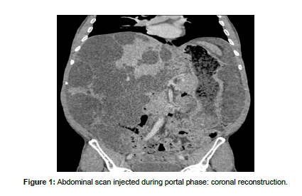
The patient was a 47-year-old male with hepato-renal polycystic disease, who was a candidate for liver transplantation. An abdominal angioscan revealed multiple bilateral hepatic and renal cystic lesions of varying sizes, some of which exhibited wall calcifications, leading to hepatomegaly and bilateral nephromegaly (Figure 1). Additionally, an anatomical variant was observed, characterized by a quadrifurcation of the celiac trunk, giving rise separately to four branches: the left gastric artery, which further gave rise to the left hepatic artery, the splenic artery, the gastroduodenal artery, and the right hepatic artery (Figure 2). It is also noteworthy that the common hepatic artery and the proper hepatic artery were absent.
Discussion
The celiac trunk arises from the anterior surface of the abdominal aorta and typically gives rise to three branches in 72% to 90% of the population: the left gastric artery, the splenic artery, and the common hepatic artery [1,3]. The common hepatic artery then bifurcates into the proper hepatic artery and the gastroduodenal artery, with the proper hepatic artery further dividing at the hepatic hilum into the right and left hepatic branches [1,2]. However, this typical vascularization pattern, known as the modal pattern, is only present in 52% to 80% of cases [2]. Various anatomical variations of the celiac trunk and the hepatic arterial system have been described in several studies and classifications, with the Uflacker and Michels classifications being the most well-known and widely used. The former describes eight anatomical variants of the celiac trunk, while the latter describes ten anatomical variants of the hepatic arterial system. The most common variant reported is the origin of the right hepatic artery from the superior mesenteric artery [2,3].
Our observation presents an unusual anatomical variant that has not been previously classified or reported to our knowledge. In this variant, the celiac trunk separately gives rise to four branches: the left gastric artery, which in turn gives rise to the left hepatic artery, the splenic artery, the gastroduodenal artery, and the right hepatic artery, with the latter two branches arising directly from the celiac trunk. Another unique aspect of our observation is its presence in a patient with hepato-renal polycystic disease who is a candidate for liver transplantation.
Knowledge of these anatomical variations during surgery is crucial for planning surgical techniques to avoid potentially life-threatening complications such as hepatic ischemia, hemorrhage, and liver abscesses [2]. During liver transplantation, precise determination of the extrahepatic arterial schemes of both the donor and recipient livers is essential [4]. Segment IV of the liver is critically important in transplantation surgery, and therefore, understanding its blood supply origin is crucial [3]. Erroneous ligation due to ignorance of these variations could result in necrosis of a liver segment or lobe. Thus, all variations must be defined and managed appropriately to ensure adequate vascular supply during liver transplantation [4].
Conclusion
Anatomical variations of the celiac trunk and the hepatic arterial system are common. Preoperative knowledge of these variations is essential to reduce morbidity and mortality rates. Therefore, collaboration between surgeons and radiologists is crucial before any surgical intervention to ensure effective and safe patient management.
References
- Miora Lovatiana, Randrianalison (2023) Morphometric and anatomical variations of digestive arteries view in Computer Tomography. Ann Afr Med 16.
- Ferjaoui Wael (2019) Une autre variante anatomique du tronc cœliaque. JMR 2: 13-16.
- Ugurel MS (2010) Anatomical variations of hepatic arterial system, coeliac trunk and renal arteries: an analysis with multidetector CT angiography. Brit J Radiol 83: 661-667.
- Bao-Gui Wang, Rosemarie Frober (2009) Accessory extrahepatic arteries: Blood supply of a human liver by three arteries A case report with brief literature review. Ann Anat 191: 477-484.
Indexed at, Google Scholar, Crossref
Citation: Khadija E (2024) Another Unusual Anatomical Variant of the CeliacTrunk: A Case Report. OMICS J Radiol 13: 567.
Copyright: © 2024 Khadija E. This is an open-access article distributed under theterms of the Creative Commons Attribution License, which permits unrestricteduse, distribution, and reproduction in any medium, provided the original author andsource are credited.
Select your language of interest to view the total content in your interested language
Share This Article
Open Access Journals
Article Usage
- Total views: 1197
- [From(publication date): 0-2024 - Nov 22, 2025]
- Breakdown by view type
- HTML page views: 904
- PDF downloads: 293


