Make the best use of Scientific Research and information from our 700+ peer reviewed, Open Access Journals that operates with the help of 50,000+ Editorial Board Members and esteemed reviewers and 1000+ Scientific associations in Medical, Clinical, Pharmaceutical, Engineering, Technology and Management Fields.
Meet Inspiring Speakers and Experts at our 3000+ Global Conferenceseries Events with over 600+ Conferences, 1200+ Symposiums and 1200+ Workshops on Medical, Pharma, Engineering, Science, Technology and Business
Case Report Open Access
An Unusual Case of Arachnoid Cyst
| Reem Zakzouk* and Ali Alhaidey | |
| Department of Radiology, Prince Sultan Medical Complex, Riyadh, KSA | |
| Corresponding Author : | Reem Zakzouk Department of Radiology Prince Sultan Medical Complex Riyadh, Kingdom of Saudi Arabia E-mail: reem365@hotmail.com |
| Received July 30, 2013; Accepted September 25, 2013; Published September 28, 2013 | |
| Citation: Zakzouk R, Alhaidey A (2013) An Unusual Case of Arachnoid Cyst. OMICS J Radiology 2:144 doi: 10.4172/2167-7964.1000144 | |
| Copyright: © 2013 Zakzouk R, et al. This is an open-access article distributed under the terms of the Creative Commons Attribution License, which permits unrestricted use, distribution, and reproduction in any medium, provided the original author and source are credited. | |
Visit for more related articles at Journal of Radiology
Abstract
Arachnoid cysts are benign, congenital, space-occupying lesions that are filled with CSF (Cerebrospinal fluid). We report an unusual case of a multi-compartmental arachnoid cyst of 26 years old male who complained of gradually increasing headache. Computed tomography and magnetic resonance imaging revealed a large arachnoid cyst involving the left middle and anterior cranial fossae with sub-falcine extension into the right anterior cranial fossa.
| Keywords |
| Arachnoid cyst; Computed tomography; Magnetic resonance imaging |
| Case Report |
| A 26-year-old male presented to the Emergency Department complaining of a gradually worsening headache and recent history of trauma. On noncontrast Computed Tomography examination of the head performed in an outside hospital, the patient was diagnosed with an intracranial neoplasm and was referred to our institution for further assessment. |
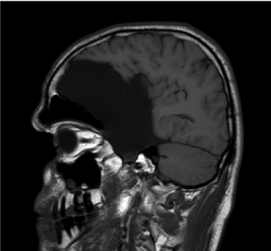
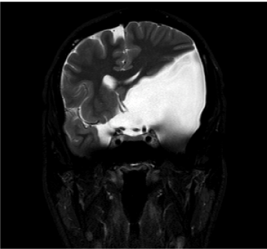
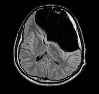
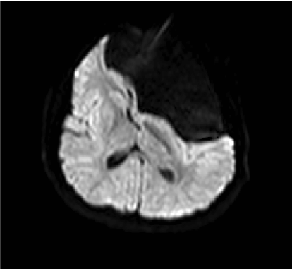
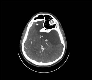
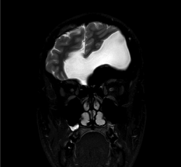
| Magnetic Resonance Imaging of the brain showed a large, extraaxial, well-defined multicompartmental lesion involving the left middle and anterior cranial fossae with sub-falcine extension into the right anterior cranial fossa. The lesion follows CSF signal intensity on all sequences, demonstrating T1 hypointensity (Figure 1a) and T2 hyperintensity (Figure 1b), suppression of signal on Fluid Attenuated Inversion Recovery (FLAIR) (Figure 1c) sequences and facilitation of diffusion on DWI sequences (Figure 1d). It displaces the overlying brain parenchyma superiolaterally and results in effacement of the frontal horn of the left lateral ventricle with no hydrocephalous (Figures 1b and 2). It does not communicate with the ventricular system. It results in mild mass effect on the circle of Willis, the left middle cerebral artery and bilateral anterior cerebral arteries as well as on the pituitary stalk. The Computed Tomography demonstrates characteristic bony Scalloping and remodeling of the inner table of the adjacent calvarium, indicating the slow growth of the lesion. The left frontal sinus is much larger than the right frontal sinus, which is also unusual (Figure 3). |
| Discussion |
| Despite the imaging findings of compressed pituitary stalk, cranial vessels and brain parenchyma by the large lesion the patient had normal pituitary and cognitive functions. The findings may be explained by malformation of the menix primitiva associated with a component of dural dysplasia. |
| Arachnoid cysts are benign, congenital, space-occupying lesions that are filled with CSF. They do not communicate with the ventricular system. The cysts tend to be unilocular, smoothly marginated expansile lesions [1-3]. The incidence is somewhat higher in men [4]. They represent 1% of non-traumatic intra-cranial mass lesions and are often discovered incidentally on cranial imaging examinations. They are congenital lesions, probably arising due to maldevelopment of the meninx primitiva in early embryogenesis. More than 50% of arachnoid cysts occur in the middle cranial fossa and larger lesions are often associated with temporal lobe hypoplasia. Symptoms ranging from headaches and seizures to cognitive impairment and focal neurological [5]. Other locations include the suprasellar cistern and posterior fossa (10%), where they occur most commonly in the cerebellopontine angle cistern (Figure 4). Less common locations are within the interhemispheric fissure; over the cerebral convexity; or in the choroidal fissure, cisterna magna, quadrigeminal cistern, and the vermian fissures [2-6]. |
| The precise mechanism for the formation of arachnoid cysts is not known. It is possible that they are secondary to “splitting” or a diverticulum of the developing arachnoid. The cause has also been attributed to trauma, mastoiditis, meningitis, and subarachnoid hemorrhage [2-7]. Arachnoid cysts are generally stable over time, although cases of sudden or progressive enlargement, as well as spontaneous resolution, have been reported [8,9]. |
| The best diagnostic clue is a sharply demarcated extra axial cyst that can displace or deform adjacent brain. Scalloping of the adjacent calvarium is often seen. The classic arachnoid cyst has no identifiable internal architecture and does not enhance. The cyst typically has the same signal intensity as CSF at all sequences. Occasionally, however, hemorrhage, high protein content, or lack of flow within the cyst may complicate the MR appearance [2-5]. Hemorrhage may occur not only in the cyst but also in the subdural or extradural spaces. Arachnoid cysts have an increased prevalence of coexisting subdural hematomas, especially when they occur in the middle cranial fossa [10,11]. Regarding the differential Diagnosis, the most difficult lesion to distinguish from the arachnoid cyst is an epidermoid cyst. Chronic subdural hematoma and porencephalic cyst can also be confused for an arachnoid cyst [12]. |
| Teaching Point |
| Being a multicompartmental lesion does not necessarily indicate malignancy. |
References |
|
Figures at a glance
 |
 |
 |
 |
| Figure 1a | Figure 1b | Figure 1c | Figure 1d |
 |
 |
 |
|
| Figure 2 | Figure 3 | Figure 4 |
Post your comment
Relevant Topics
- Abdominal Radiology
- AI in Radiology
- Breast Imaging
- Cardiovascular Radiology
- Chest Radiology
- Clinical Radiology
- CT Imaging
- Diagnostic Radiology
- Emergency Radiology
- Fluoroscopy Radiology
- General Radiology
- Genitourinary Radiology
- Interventional Radiology Techniques
- Mammography
- Minimal Invasive surgery
- Musculoskeletal Radiology
- Neuroradiology
- Neuroradiology Advances
- Oral and Maxillofacial Radiology
- Radiography
- Radiology Imaging
- Surgical Radiology
- Tele Radiology
- Therapeutic Radiology
Recommended Journals
Article Tools
Article Usage
- Total views: 17642
- [From(publication date):
October-2013 - Apr 17, 2025] - Breakdown by view type
- HTML page views : 13012
- PDF downloads : 4630
