An Ultra-rare Hereditary Disease of 28–year Old Pregnant Patient withMalignant Alteration
Received: 23-Nov-2018 / Accepted Date: 06-Dec-2018 / Published Date: 12-Dec-2018 DOI: 10.4172/2332-0877.1000388
Abstract
Familial adenomatous polyposis (FAP) is an autosomal dominant disorder characterized by the development of hundreds to thousands of colorectal adenomatous polyps and the inevitable occurrence of colorectal carcinoma, if the colon is not removed. In a 28-year old patient, following diagnostic criteria, have been established more than 500 colorectal adenomas and family history of FAP.
Keywords: Colorectal; Carcinoma; Adenocarcinoma; Rectal carcinoma; Antibodies
Introduction
Having in the mind that the colorectal carcinoma is the most frequent carcinoma of the digestive system, and that often arises from adenoma by Marson’s “adenoma-carcinoma sequence” theory, we have undertaken the following study: morphological, histochemical and immunohistochemical examinations of one hundred adenomas of various size and macroscopically pattern in FAP, complicated by rectal mucinous adenocarcinoma.
Material and Methods
Total colectomy was induced by FAP, associated with rectal carcinoma. Numerous hundreds of tubulars, tubulo-villous and villous adenomas, size from 5 to 35 mm, as well as 10 biopsies of the cancerous rectal tissue, together with 10 regional lymph nodes, were fixed in 10% formaldehyde and processed in auto technicon. Paraffin sections of 2 microns were stained with; classic H&E method, histochemical PAS, HID-AB, pH2.5 methods as well as immunohistochemical ABC method, by using antibodies to; Ki-67 (mitotic activity), p53 suppressor activity and CEA (marker for colonic adenocarcinoma) [1-5].
Results
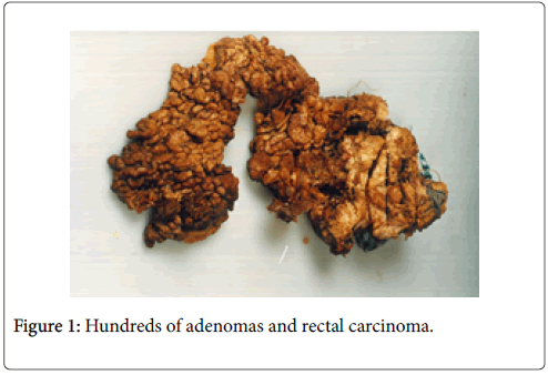
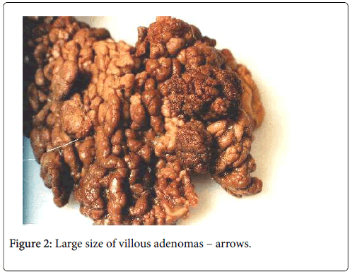
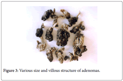
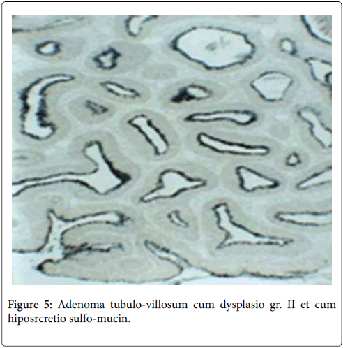
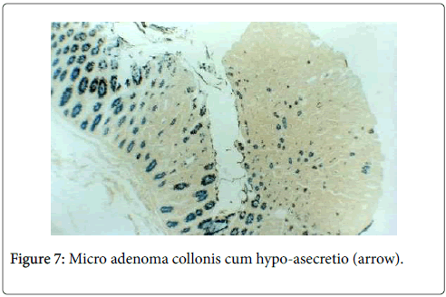
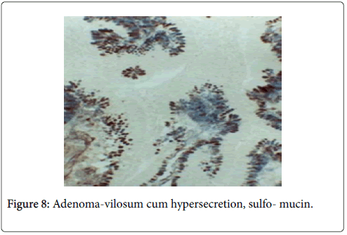
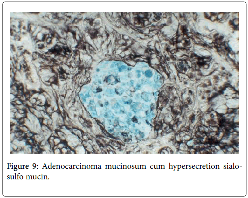
Most polyps were small, sessile and lobulated, “carpeting” the lining of the whole large bowel and numbering hundreds (Figures 1 and 2). Scattered larger sessile and pedunculated polyps were much less common, but the largest was surrounded by mucinous infiltrative rectal carcinoma (Figure 2). Histopathological, wide spectrum of size, structure and dysplasia of adenomas was obsreved (Figures 3-5). Malignant alteration of tubular adenoma with mucinous secretion was also seen (Figures 6 and 7). Strong mucinous secretion was found in both villous adenoma and mucinous adenocarcinoma (Figures 8 and 9).
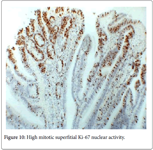
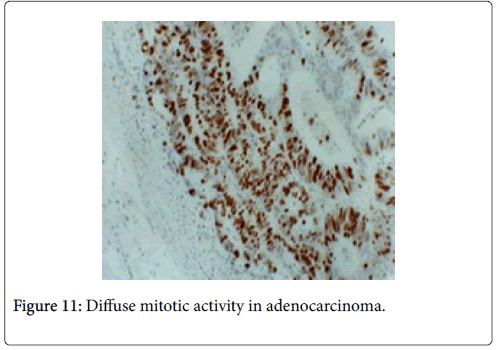
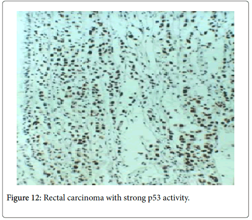
High expression of proliferative nuclear activity (Ki-67 antigen) was also observed (Figures 10 and 11). Strong p53 nuclear activity of the suppressor gene in adenocarcinoma has also been found (Figure 12) [5-7].
Conclusion
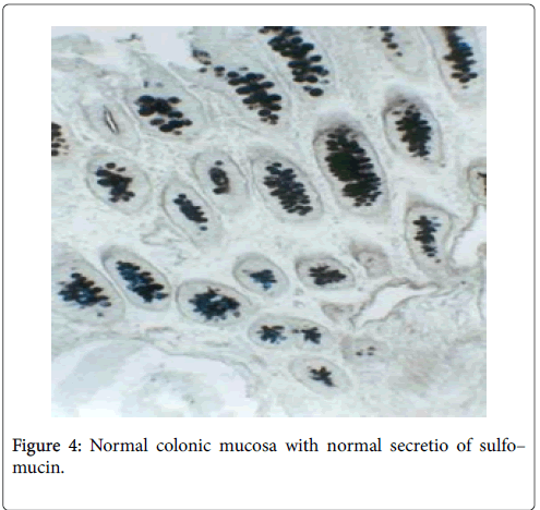
We have confirmed the theory “adenoma-carcinoma sequence”, showing that size, villous structure, severe dysplasia, as well as their number, represent the risk factors in colorectal carcinogenesis [7-9]. On the basis of the expression of normal HID and coexpression of small intestinal AB-mucin in both villous adenoma and surrounding mucinous adenocarcinoma, we have pointed out the direct connection between villous adenoma and mucinous colorectal adenocarcinoma. The discovery of the malignant alteration of the FAP in very young (28-years old) patient, could be explained by her pregnant state with decreased host immunity [10-14].
References
- Fenoglio CM, Lane N (1975) The Anatomial precursor of colorectal carcinoma. JAMA 23: 819-823.
- Chetty R (2003) Morson and Dawson’s Gastrointestinal Pathology. J Clin Pathol 56: 399.
- Potet F, Brousse J, Brousse NS (1975) Polyps of the colon: a study of 356 cases discovered during 2,067 routine necropsies. Archives françaises des maladies de l'appareil digestif  64: 201-208.
- Morson BC (1974) Evolution of cancer of the colon and rectum. Cancer 35: 2251-2270.
- Rotterdam H, Sommers C, Waye D (1981) Biopsy Diagnosis of the Digestive Tract. Raven Press, New York: 398-427.
- Katic V, Vukasinovic R, Stamenkovic I, Kutlesic C, Karanikolic D (1983) Relationship between benignant and malignant epithelial tumours of the large intestine. Serbian Archive 3: 377-386
- Katic V (1998) Pathology in the diseases of both small and large intestinum. Gastroenterology Part II: 480-558.
- Nagorni A, Tasic T, Stamenkovic I, Katic V (1996) Multiple Synchronous Carcinomas of the colon: A case report. Arch Gastroentero- hepatology 15: 70-72.
- Heinimann K, Mullhaup B, Weber B, Attenhofer M, Scott RJ, et al. (1998) Phenotipic differences in familial adenomatous polyposis based on APC gene. GUT 43: 675-679.
- Bussey HJR (1975) Familial polyposis coli. Johns Hopkins University Press, Baltimore.
- Powell SM, Petersen GM, Krush AJ, Booker S, Jen J, et al. (1993) Molecular diagnosis of familial adenomatous polyposis. N Eng J Med 329: 1982-1987.
- Pauli RM, Pauli Me, Hall JG (1980) Gardner syndrome and periampullary malignancy. Am J Med Genet 6: 205-219.
Citation: Katic V, Radovanovi Z, Karadzic A, Rankovic G, Nagorni I, et al. (2018) An Ultra-rare Hereditary Disease of 28–year Old Pregnant Patient with Malignant Alteration. J Infect Dis Ther 7:388. DOI: 10.4172/2332-0877.1000388
Copyright: © 2018 Katic V, et al. This is an open-access article distributed under the terms of the Creative Commons Attribution License, which permits unrestricted use, distribution, and reproduction in any medium, provided the original author and source are credited.
Share This Article
Recommended Journals
Open Access Journals
Article Tools
Article Usage
- Total views: 3714
- [From(publication date): 0-2019 - Apr 28, 2025]
- Breakdown by view type
- HTML page views: 2920
- PDF downloads: 794

