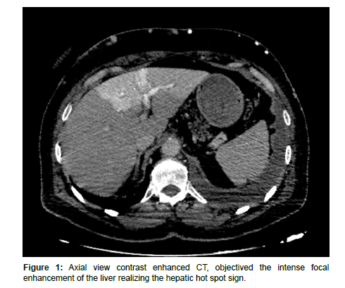
An Image of the Hepatic Hot Spot Sign
Received: 02-Nov-2023 / Manuscript No. roa-23-119389 / Editor assigned: 06-Nov-2023 / PreQC No. roa-23-119389 / Reviewed: 20-Nov-2023 / QC No. roa-23-119389 / Revised: 24-Nov-2023 / Manuscript No. roa-23-119389 / Published Date: 30-Nov-2023
Keywords
Hepatic hot spot sign, Superior vena cava occlusion
Image Article
A 30 year old women consulted for intense chest pain with cyanosis and dyspnea in whom a CT-chest angiography was performed in emergency looking for a superior cave syndrome: We observed a thrombosis of the superior vena cava with at the limit of the abdominal cuts performed of the CT chest a hyper dense focal hepatic opacification realizing the classic hepatic hot spot sign.
This sign was first described by Ishikawa in 1983 and it manifests as an area of intense focal enhancement of the quadrate lobe in the arterial phase [1].
In patient with obstruction of the superior vena cava, collateral veins return blood to the left hepatic lobe via the internal mammary and left umbilical veins, creating a hot spot in the area of insertion of the left umbilical vein and left main branches of the portal vein [2].
The hepatic hot spot sign has been reported in Budd-Chiari syndrome, the causes of the superior vena cava syndrome (vascular Behcet disease, lung carcinoma and lymphoma, fibrosing mediastinitis and luetic aneurysm), and masses of the liver (CHC, focal nodular hyperplasia, and abscess haemangioma) [3] (Figure 1).
References
- Virmani V, Lai, Ahuja CK, Khandelwal N (2010). The CT Quadrate lobe hot spot sign. Ann Hepatol 9: 296-298.
- Dickson AM (2005) The focal hepatic hot spot sign. Radiology 237: 647-648
- Vo NQ, Trinh CT, Nguyen HQ, Chansomphou V, Le TB (2020). The focal hepatic hot spot sign with lung cancer in computed tomography. Respirol Case Rep 8: e00671-e00671.
Indexed at, Google Scholar, Crossref
Indexed at, Google Scholar, Crossref
Citation: Adjou N, Sfar K, Bahlouli N, Jaddour M, Jroundi L, et al. (2023) An Imageof the Hepatic Hot Spot Sign. OMICS J Radiol 12: 508.
Copyright: © 2023 Adjou N, et al. This is an open-access article distributed underthe terms of the Creative Commons Attribution License, which permits unrestricteduse, distribution, and reproduction in any medium, provided the original author andsource are credited.
Share This Article
Open Access Journals
Article Usage
- Total views: 922
- [From(publication date): 0-2023 - Apr 20, 2025]
- Breakdown by view type
- HTML page views: 705
- PDF downloads: 217

