Case Report Open Access
An Exceptional Foreign Body in the Stomach
Makhoul E* and Zaarour A
Department of Gastroenterology University Hospital Notre Dame de Secours, Byblos, Lebanon
- *Corresponding Author:
- Elias Makhoul
Department of Gastroenterology
Faculty of Medicine and sciences Holy Spirit
University Hospital Notre Dame De Secours Byblos
POB-3 Byblos, Lebanon
Tel: +961 3711787
Fax: +961 9944483
E-mail: eliemakhoul@hotmail.com
Received date: May 26, 2015; Accepted date: June 15, 2015; Published date: June 24, 2015
Citation: Makhoul E, Zaarour A (2015) An Exceptional Foreign Body in the Stomach. J Gastrointest Dig Syst 5:302. doi:10.4172/2161-069X.1000302
Copyright: © 2015 Makhoul E, et al. This is an open-access article distributed under the terms of the Creative Commons Attribution License; which permits unrestricted use; distribution; and reproduction in any medium; provided the original author and source are credited.
Visit for more related articles at Journal of Gastrointestinal & Digestive System
Abstract
Foreign body ingestion is a common event that occurs most often in children who account for 75 to 85% of patients; and in psychiatric patients. In adults it is uncommon and usually happens accidentally where these bodies are ingested together with food. The most common foreign bodies accidentally ingested by adults are bones; especially fish bones 9-45% and less commonly dentures 4-18% whereas Children commonly ingest toys and small coins. Most of such foreign bodies pass through gastrointestinal tract uneventfully and rarely cause obstruction or perforation unless a context of pathological change of the digestive tract exists. These changes include strictures; malignancy; esophageal rings and achalasia. We hereby present an unusual and interesting case of a 51-year-old male patient in whom endoscopic removal from the stomach; of an endotracheal tube; was carried out.
Keywords
Foreign body; Endotracheal tube; Weight loss; Endoscopic removal; Stomach
Introduction
Foreign body ingestion is a common event that occurs most often in children who account for 75 to 85% of patients; and in psychiatric patients. In adults it is uncommon and usually happens accidentally where these bodies are ingested together with food. The most common foreign bodies accidentally ingested by adults are bones; especially fish bones 9-45% and less commonly dentures 4-18% whereas Children commonly ingest toys and small coins.
Most of such foreign bodies pass through gastrointestinal tract uneventfully and rarely cause obstruction or perforation unless a context of pathological change of the digestive tract exists. These changes include strictures; malignancy; esophageal rings and achalasia.
We hereby present an unusual and interesting case of a 51-year-old male patient in whom endoscopic removal from the stomach; of an endotracheal tube; was carried out.
Case Report
A 51-year-old male patient with unnoticed past medical history; who underwent cholecystectomy 2 years ago at another hospital; presented to the emergency department of our institution with his chief complaint being an epigastric pain; associated with repeated vomiting. The patient claimed that he began to suffer from abdominal pain about 1 year ago but he used to take pain reliever medications and antiemetic to get well. Two weeks ago the situation deteriorated as he suffered from repeated greenish vomiting occurring after meals; his pain increased in intensity and wasn’t relieved by medications anymore; but passage of flatus and stools was normal [1-6].
On examination; the patient was afebrile; but mildly agitated because of his pain. His abdomen was soft with a severe pain in the epigastric region; no palpable masses were noticed. The rest of the exam was otherwise unremarkable.
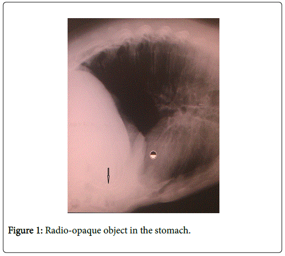
The patient was admitted; treated by intra venous antiemetics; antispasmodics and PPI. We noted normal routine blood tests. The chest x-ray showed an unknown radio-opaque object in the stomach (Figure 1).
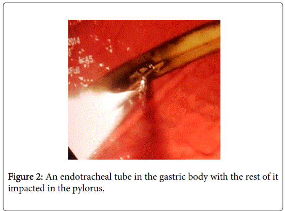
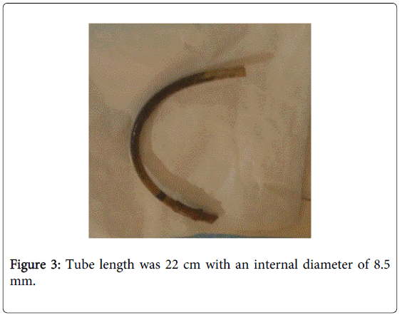
The patient underwent an upper GI endoscopy under intravenous sedation; which showed an endotracheal tube in the gastric body with the rest of it impacted in the pylorus (Figure 2). First; an attempt was made to remove the tube with a foreign body forceps; but it continued to slide. The second alternative was the polypectomy snare which was useful to dislodge the tube from the pylorus and pulling it to the proximal esophagus without any injury to the stomach or esophagus. At the cricopharyngeus muscle level; travel of the foreign body was again impeded; thereby the situation required laryngoscopy with a Magill forceps which helped in the removal of the tube from the pharynx with minor bleeding in the upper esophagus. The tube length was 22 cm with an internal diameter of 8.5 mm (Figure 3).
Discussion
Naturally; 80-90% of ingested foreign bodies that reach the stomach pass through the gastrointestinal tract without intervention. 10-20% must be removed endoscopically; whereas surgery is required in approximately 1% of cases [7].
Clinical management is based on knowledge of the chemical properties; the size; the location and the sharpness of the foreign body [3,5,8].
The passage through the duodenum depends on the length as well as the diameter of the foreign body. A length greater than 6 cm with a diameter of more than 2.5 cm makes the passage more difficult through the duodenum due to its fixed retroperitoneal position [8]. Conservative management is the best approach for short <6 cm and narrow <2.5 cm objects where spontaneous passage is expected without any intervention within one week. Endoscopic removal is indicated if the foreign body is longer than 6 cm; proximal to the first portion of the duodenum andor wider than 2.5 cm. Surgical removal should be considered if objects remain in the same location distal to the duodenum for more than 1 week [3,5,8].
In case of coins; endoscopic removal is indicated if they remain longer than 12-24 hours in the esophagus; or 3-4 weeks in the stomach in an asymptomatic patient [1,8].
The pointed foreign body is always more difficult to pass spontaneously than the rounded one. Sharp and sharp-pointed objects in the esophagus; stomach or duodenum; require urgent endoscopic removal. Surgical intervention should be considered when endoscopic treatment fails; and if the sharp body beyond the duodenum ceases to progress radiographically for 3 consecutive days [8].
Emergent endoscopic removal is indicated for a suspected disk battery discovered in the upper gastrointestinal tract. Laparotomy should be considered if it appears that the passage of the battery in the bowel has been arrested [7].
The presence of Bezoars needs always endoscopic disruption and removal of the mass. However many bezoars require surgical treatment [6,7,9-12].
Concerning narcotic packets; endoscopic removal is relatively contraindicated due to the risk of rupture and contents leakage. Surgery is only indicated when drug packets fail to progress or if there is an intestinal obstruction [7].
Unusual foreign bodies such as an ingested toothbrush [10], speaking valve [8] and endoscopic capsule [5]; removed endoscopically or by surgical intervention were reported in the literature.
Our case is an uncommon complication. The circumstances leading to this medical malpractice are not clear and whether the intubation was done by an inexperienced medical staff or not; is not exactly known. The tube could have been removed immediately but this didn’t happen; and during these two years; the patient went seeing several specialists who didn’t even think to do an abdominal imaging.
The difficulty to pass through the duodenum is due to the length of the endotracheal tube (more than 6 cm) and to the rigidity of duodenal intestinal part. This kind of foreign body carries a high risk of perforation. Weight loss is due to the pain and to the blockage of the endotracheal tube into the pylorus; thus reducing the intake of nutrients. The tube removal was carried out with a difficulty to pass through the cricopharyngeus muscle; the condition then required using a laryngoscope and a Magill forceps.
A few cases were reported in the literature on the treatment for removal of an endotracheal tube in the esophagus following intubation [2,4,13], and in one case, the tube was impacted in the stomach; therefore requiring surgery after unsuccessful endoscopic removal.
We report this case; to raise awareness of the danger of accidental esophageal intubation in the operating rooms as it is still a major cause of morbidity and even mortality.
References
- Webb WA (1995) Management of foreign bodies of the upper gastrointestinal tract: update.GastrointestEndosc 41: 39-51.
- Block EF, Cheatham ML, Parrish GA, Nelson LD, Beam N (1999) Ingested endotracheal tube in an adult following intubation attempt for head injury.Am Surg 65: 1134-1136.
- Eisen GM, Baron TH, Dominitz JA, Faigel DO, Goldstein JL, et al. (2002) Guideline for the management of ingested foreign bodies.GastrointestEndosc 55: 802-806.
- Er M (2005) An unusual foreign body of the esophagus.Asian CardiovascThorac Ann 13: 70-71.
- Cave D, Legnani P, de Franchis R, Lewis BS; ICCE (2005) ICCE consensus for capsule retention.Endoscopy 37: 1065-1067.
- Hewitt AN, Levine MS, Rubesin SE, Laufer I (2009) Gastric bezoars: reassessment of clinical and radiographic findings in 19 patients.Br J Radiol 82: 901-907.
- Gorter RR, Kneepkens CM, Mattens EC, Aronson DC, Heij HA (2010) Management of trichobezoar: case report and literature review.PediatrSurgInt 26: 457-463.
- ASGE Standards of Practice Committee, Ikenberry SO, Jue TL, Anderson MA, Appalaneni V, et al. (2011) Management of ingested foreign bodies and food impactions.GastrointestEndosc 73: 1085-1091.
- Ambe P, Weber SA, Schauer M, Knoefel WT (2012) Swallowed foreign bodies in adults.DtschArzteblInt 109: 869-875.
- Gomes CA Jr (2013) Endoscopic removal of an ingested toothbrush.Endoscopy 45 Suppl 2 UCTN: E129.
- Guelfguat M, Kaplinskiy V, Reddy SH, DiPoce J (2014) Clinical guidelines for imaging and reporting ingested foreign bodies.AJR Am J Roentgenol 203: 37-53.
- Osborne MS, Saunders T, Fuerstenberg F, Costello D, Dhesi B (2014) A man with absolute dysphagia after eating a steak.BMJ 349: g5462.
- Sing RF, Huynh TT, Gibbs MA, Perron AD (2001) The "swallowed" endotracheal tube.Am J Emerg Med 19: 606-607.
Relevant Topics
- Constipation
- Digestive Enzymes
- Endoscopy
- Epigastric Pain
- Gall Bladder
- Gastric Cancer
- Gastrointestinal Bleeding
- Gastrointestinal Hormones
- Gastrointestinal Infections
- Gastrointestinal Inflammation
- Gastrointestinal Pathology
- Gastrointestinal Pharmacology
- Gastrointestinal Radiology
- Gastrointestinal Surgery
- Gastrointestinal Tuberculosis
- GIST Sarcoma
- Intestinal Blockage
- Pancreas
- Salivary Glands
- Stomach Bloating
- Stomach Cramps
- Stomach Disorders
- Stomach Ulcer
Recommended Journals
Article Tools
Article Usage
- Total views: 15212
- [From(publication date):
June-2015 - Apr 05, 2025] - Breakdown by view type
- HTML page views : 10648
- PDF downloads : 4564


