Make the best use of Scientific Research and information from our 700+ peer reviewed, Open Access Journals that operates with the help of 50,000+ Editorial Board Members and esteemed reviewers and 1000+ Scientific associations in Medical, Clinical, Pharmaceutical, Engineering, Technology and Management Fields.
Meet Inspiring Speakers and Experts at our 3000+ Global Conferenceseries Events with over 600+ Conferences, 1200+ Symposiums and 1200+ Workshops on Medical, Pharma, Engineering, Science, Technology and Business
Case Report Open Access
Altered Coiling with Stent Assistance for an Iatrogenic Traumatic Aneurysm of the Internal Carotid Artery
| Akira Kurata1*, Sachio Suzuki1, Madoka Inukai1, Makoto Sasaki1, Kazuhisa Iwamoto1, Kuniaki Nakahara1, Shingo Konno2, Takao Kitahara2, Kiyotaka Fujii1 and Shinichi Kan3 | |
| 1Departments of Neurosurgery, Kitasato University School of Medicine, Kanagawa, Japan | |
| 2Critical Care Medicine, Kitasato University School of Medicine, Kanagawa, Japan | |
| 3Radiology, Kitasato University School of Medicine, Kanagawa, Japan | |
| Corresponding Author : | Akira Kurata Department of Neurosurgery Kitasato University School of Medicine 1-15-1 Kitasato, Minamik Sagamihara Kanagawa 228-8555, Japan E-mail: akirak@med.kitasato-u.ac.jp |
| Received February 02, 2011; Accepted February 17, 2012; Published February 20, 2012 | |
| Citation: Kurata A, Suzuki S, Inukai M, Sasaki M, Iwamoto K, et al. (2012) Altered Coiling with Stent Assistance for an Iatrogenic Traumatic Aneurysm of the Internal Carotid Artery. OMICS J Radiology. 1:102. doi: 10.4172/2167-7964.1000102 | |
| Copyright: © 2012 Kurata A et al. This is an open-access article distributed under the terms of the Creative Commons Attribution License, which permits unrestricted use, distribution, and reproduction in any medium, provided the original author and source are credited. | |
Visit for more related articles at Journal of Radiology
Abstract
We report a rare case of a traumatic aneurysm which developed after clipping surgery with review of the relevant literature. Endovascular treatment using coiling and stent assistance taking into account technical considerations is described. Monitoring of the aneurysmal pressure showed marked decrease immediately after the stent emplacement.
| Case Report |
| A 57-year old woman suffering from subarachnoid hemorrhage from a large internal carotid artery (ICA) aneurysm with a broad neck defined by computed tomography (CT) followed by three dimensional CT angiography in the local hospital, who was emergency transferred to our institute. Neurological status on admission was grade I on the H&H scale. This patient had had a past history of subarachnoid hemorrhage 14 years previously, undergoing surgery for a basilar artery ruptured aneurysm arising from the superior cerebellar artery (BA-SCA aneurysm) in other hospital. During the clipping surgery, the right internal carotid artery was injured, but fortunately it was repaired by further clipping. She and her family were Jehovahs Witness, rejecting the blood transfusion during the operation on grounds of religion. |
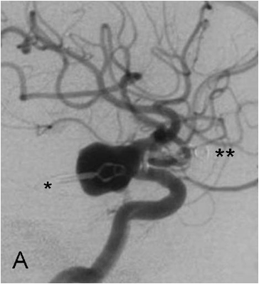
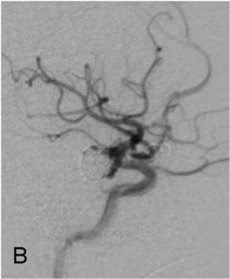
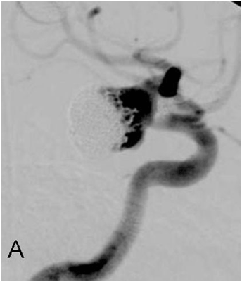
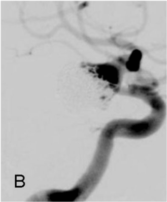
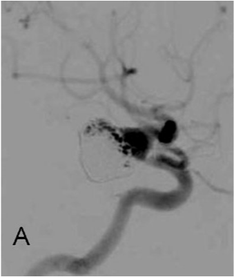
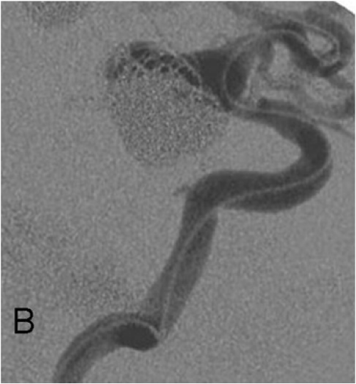
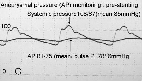
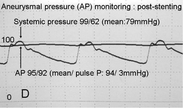
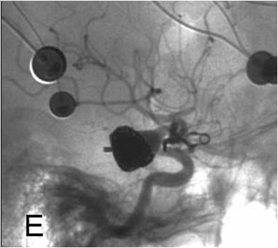
| At our institute, endovascular surgery was selected for the large traumatic aneurysm. Two clips were made, one completely dislocated from the internal carotid artery and the other for BA-SCA aneurysm, which disturbed the therapeutic window (Figure 1A) but endovascular surgery could be successfully performed (Figure 1B) and the immediate clinical course was uneventful. Follow-up angiography one month after the treatment showed opening of the aneurysm by coil compaction (Figure 2A) so additional coil embolization was performed (Figure 2B). However, follow-up angiography 4 months thereafter again showed reopening of the aneurysm (Figure 3A). Therefore, endovascular surgery for adding coils with stent emplacement was advocated. A 7F sheath was inserted into the right femoral artery and thereafter a 7F catheter (Brite tip guiding catheter, Cordis, J & J, USA ) preceded by a 5F catheter (Cathex, Japan) with a coaxial system was introduced into the right internal carotid artery. Initially, an attempt was made to advance a microcatheter (Prowler Select Plus, Codman, J & J, USA) was advanced into the middle cerebral artery (MCA) beyond the aneurysm but this failed and a soft microcatheter (Excelsior SL 10, Striker, Boston) was therefore introduced into the MCA. For the guide wire technique a 300cm long-guide wire (Accelerator, Covidien, USA) was applied. The microcatheter for the stent was successfully advanced into the MCA through this long guide wire (Figure 3B). Thereafter another microcatheter (Excelsior SL10, Stryker, USA) for coil embolization was introduced into the upper part of the residual lumen, which was inflow-zone. A self-expanding nitinol stent (Enterprise, Codman, J & J, USA) 37mm length was put in place from the M1 portion to a site distal to the carotid cavernous ICA to cover the broad neck of the aneurysm adequately. Thereafter coil embolization using jailing technique was successfully performed with monitoring of the aneurysmal internal pressure. After emplacement of the stent, the aneurysmal pressure showed marked decrease, pulse pressure in particular falling from 6 mmHg to 3mm Hg (Figure 3C & D). Final angiography showed no flow into the aneurysmal fundus (Figure 3E) and the clinical course was uneventful. Two days after the treatment, the patient was discharged without neurological deficit. Follow-up angiography 6 months after the final treatment showed persistent complete occlusion of the aneurysm. |
| Discussion |
| Traumatic intracranial aneurysms caused by iatrogenic injury are a very rare. Bank et al. [1] described a traumatic occlusion of the basilar artery, and their review of intracranial traumatic aneurysms summarized only 41 cases in the world literature. Although the iatrogenic source was not always clear, the aneurysm were usually associated with contiguous skull fractures or penetrating head injuries. Regarding iatrogenic intracranial aneurysms after aneurysmal surgery, to our knowledge there have only 10 cases in the literature, including present case (Table 1) [2-10]. |
| Cerebral angiography is instrumental in the diagnosis of traumatic aneurysms. Certain features on angiography suggest a traumatic etiology [11] such as a poorly defined neck, unusual sites or projections of the aneurysm, irregularly shape and delayed filling and emptying. Our case was an irregularly shaped aneurysm with a poorly defined neck was seen in. |
| Traumatic aneurysms rarely regress and because the wall is often only an organized clot have a high incidence of rupture (as high as 67%) [12]. Six of the 10 reported iatrogenic aneurysms after aneurysmal surgery were ruptured, the time being after less than 1 month in all except present case. Yatsuzuka et al. [7] emphasized the importance of early diagnosis and treatment. |
| Thus, treatment should be instituted once a diagnosis is made and in traumatic cases an encircling clip is very useful. However, when the aneurysm cannot be obliterated by a clip, it may needed to be trapped with an accompanying vascular bypass. Since this patient and her family were Jehovahs Witness, and did not accept such an operation on the grounds of religion because they rejected the blood transfusion, endovascular surgery was selected. The problem is that endovascular procedures also carry a significant risk given friable aneurysms with poorly defined necks. In the case presented, stenting proved a useful technique to prevent the coil migration and disturbance of the aneurysmal inflow. The monitoring of the aneurysmal pressure during endovascular surgery showed marked decrease immediately after the emplacement of the stent, this resulting in disturbance of the water-hammer effect, influenced by diversion of the blood inflow through the stent mesh (the diameter is about 3mm). Tremmel et al. [13] reported flow diversion effect using Enterprise stents. Evaluation of stagnation time of contrast material is an experimental model after emplacement of stents, showed increasing time delay associated with their number with one the delay was 114-117% compared with control, with two it was 127-128% and with three was 141%. Flowdiversion therapy for aneurysms has become available in Europe, with the Silk device (Balt, Montmorency, France) and the Pipeline Embolic Device (ev3, Irvine, California) and Lylyk et al. [14] and Szikora et al.[15] reported flow-diversion treatment of 63 and 19 aneurysms, respectively, with no hemorrhages. Such flow-diversion therapy might be particularly recommended for high risk aneurysms that are amenable to coil therapy or surgical clipping but are likely to recur as with the present case. |
| Technical considerations for enterprise stenting |
| Initially, attempts to advance a microcather (Prowler Select Plus, Codman, J & J, USA) was advanced into the middle cerebral artery (MCA) beyond the aneurysm failed. Prowler Select Plus (2.3F/3.0F) is stiff and its poor trackability resulted in protrusion into the aneurysm lumen. Therefore a soft microcatheter (Excelsior SL 10, Striker, Boston) was introduced into the MCA. Application of a exchanging guide wire technique with a 300cm long-guide wire (Accelerator, Covidien, USA) and a microcatheter (Prowler Select Plus, Codman, J & J, USA) for stenting should be recommended. |
References |
|
Tables and Figures at a glance
| Table 1 |
Figures at a glance
 |
 |
 |
 |
 |
| Figure 1a | Figure 1b | Figure 2a | Figure 2b | Figure 3a |
 |
 |
 |
 |
|
| Figure 3b | Figure 3c | Figure 3d | Figure 3e |
Post your comment
Relevant Topics
- Abdominal Radiology
- AI in Radiology
- Breast Imaging
- Cardiovascular Radiology
- Chest Radiology
- Clinical Radiology
- CT Imaging
- Diagnostic Radiology
- Emergency Radiology
- Fluoroscopy Radiology
- General Radiology
- Genitourinary Radiology
- Interventional Radiology Techniques
- Mammography
- Minimal Invasive surgery
- Musculoskeletal Radiology
- Neuroradiology
- Neuroradiology Advances
- Oral and Maxillofacial Radiology
- Radiography
- Radiology Imaging
- Surgical Radiology
- Tele Radiology
- Therapeutic Radiology
Recommended Journals
Article Tools
Article Usage
- Total views: 6304
- [From(publication date):
April-2012 - Mar 29, 2025] - Breakdown by view type
- HTML page views : 1705
- PDF downloads : 4599
