Research Article Open Access
Age-related Changes of White Matter in the Elderly Population Measured by Diffusion Tensor Imaging
| Sheng-Heng Tsai1, Jachih Fu2, Jyh-Wen Chai1,3,4, Yi-Ying Wu1 and Clayton Chi-Chang Chen1,5,6,7* | |
| 1Department of Radiology, Taichung Veterans General Hospital, Taiwan | |
| 2Computer Aided Measurement and Diagnostic Systems Laboratory, Department of Industrial Engineering and Management, National Yunlin University of Science and Technology, Yunlin, Taiwan | |
| 3Division of Radiology, College of Medicine, China Medical University, Taichung, Taiwan | |
| 4Department of Biomedical Engineering, HungKuang University, Taichung, Taiwan | |
| 5Department of Radiological Technology and Graduate Institute of Radiological Science, Central Taiwan University of Science and Technology, Taichung, Taiwan | |
| 6Department of Physical Therapy, Hungkuang University of Technology, Taichung, Taiwan | |
| 7Department of Physical Therapy and Assistive Technology, National Yang Ming University, Taipei, Taiwan | |
| Corresponding Author : | Dr. Clayton Chi-Chang Chen No 160, Sec 3, Taichung Harbor oad Department of Radiology Taichung Veterans General Hospital Taichung, Taiwan Tel: 04-23592525 ext 3701 Fax: 04-23592639 E-mail: ccc@vghtc.gov.tw |
| Received July 23, 2012; Accepted August 19, 2012; Published August 22, 2012 | |
| Citation: Tsai SH, Fu J, Chai JW, Wu YY, Chen CCC (2012) Age-related Changes of White Matter in the Elderly Population Measured by Diffusion Tensor Imaging. OMICS J Radiology. 1:107. doi: 10.4172/2167-7964.1000107 | |
| Copyright: © 2012 Tsai SH. This is an open-access article distributed under the terms of the Creative Commons Attribution License, which permits unrestricted use, distribution, and reproduction in any medium, provided the original author and source are credited. | |
Visit for more related articles at Journal of Radiology
Abstract
Background: A decline in cerebral White Matter (WM) integrity occurs across the adult lifespan, with more rapid degradation later in life. Diffusion Tensor Imaging (DTI) is a non-invasive method to evaluate the microscopic changes of WM in vivo. Linear regression models have been applied to depict the relationship between diffusion properties, such as Fractional Anisotropy (FA) and Mean Diffusivity (MD), derived from DTI and age. However, the fit of these linear regression models is unsatisfactory.
Objectives: The purpose of our study was to investigate the age-related changes of WM in an elderly population.
Methods: We performed statistical parametric mapping with DTI to evaluate the patterns of age-related microscopic WM changes with voxel-based analysis in the elderly population (>55 years old). A linear regression model was used to examine the associations between DTI parameters and age.
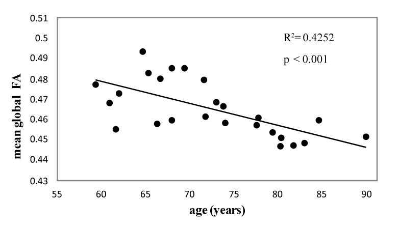
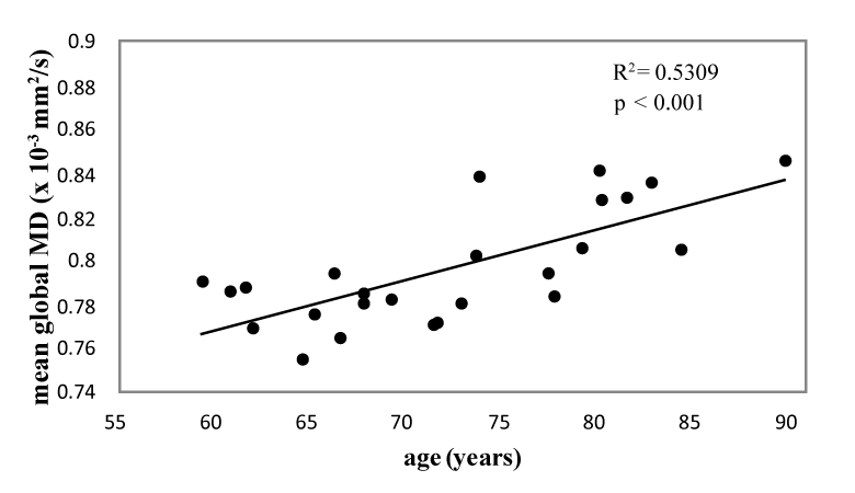
Results: The fit of the linear regression model depicting the association between mean global FA, MD values (FA, MD derived from global WM), and age was better than that reported in prior studies using DTI (R2=0.4252, p<0.001 for global FA and age; R2=0.5309, p<0.001 for global MD and age). Moreover, comparable to previous studies, the mean global FA showed a significant negative correlation with mean global MD, indicating a true age-related phenomenon of increased interstitial or intracellular fluid accumulation in cerebral WM. The mean frontal FA value was significantly lower than the mean global FA value. However, there was no significant difference between the mean frontal MD value and the mean global MD value.
Conclusions: Focusing on the elderly population, we can improve the fit of linear regression models to investigate the age-related changes of WM by DTI. These results suggest that age-related microscopic changes of WM might be occurring more consistently in later life.
| Keywords |
| Diffusion Tensor Imaging (DTI); White Matter (WM); Age-related change; Fractional Anisotropy (FA); Mean Diffusivity (MD); Voxel-based analysis |
| Introduction |
| Age-related changes of the brain, such as brain atrophy or degradation of fiber tracks integrity, are increasingly being quantified owing to advances in Magnetic Resonance Imaging (MRI). Macroscopic age-related changes of brain volume have been explored using a variety of methods. Voxel-based analysis is a fully automated, objective method that overcomes the problems associated with the manual placement of Region(s) of Interest (ROI) in examining Gray Matter (GM) and white matter (WM) differences across the entire brain [1]. Studies of the relationship between regional cerebral volume and aging using voxel-based analysis have been reported [2,3]. Diffusion tensor imaging (DTI) is an MRI technique that enables the measurement of the diffusion of water molecules within the brain [4]. In normal WM, the motion of water molecules is restricted by the parallel-oriented fibers and thus diffusion is highly anisotropic. The Fractional Anisotropy (FA) is a quantitative measure of the degree of anisotropy [5], and the Mean Diffusivity (MD) is a quantitative measure of the mean motion of water considered in all directions [6]. Age-related degradation of WM connections, being related to the decline in cognitive function [7], is usually demonstrated by the correlation between DTI parameters and age. In healthy subjects, the most common findings using DTI are age-related increases in MD and decreases in FA. However, the fit of linear regression models being used to investigate the relationship between FA, MD, and age are not satisfactory [2,8-12]. |
| Manual or semi-automated ROI-guided measurements have been utilized in the previous studies [10-12], and the fit of the linear regression models in these 3 studies was variable. Bias can be introduced as a result of a small number of regions that are insensitive to differences elsewhere in the brain. In the recent studies, a voxelbased approach was used owing to its advantages over manual ROI analysis when searching for abnormalities throughout the entire brain [2,8,9]. However, the R2 values of the linear regression models in these studies were low. Of note, younger subjects were included in these studies. Nevertheless, age-related changes in WM are thought to be late-onset in comparison to age-related changes in GM [13,14]. Allen et al. [13] proposed that WM volume increases until the mid-50s, after which it declines at an accelerated rate. Furthermore, it is interesting to note that normal myelination follows a posterior-to-anterior progression. The WM over the frontal lobe is late-myelinating, and it is thought to be vulnerable during the aging process [15]. |
| In the present study, we investigated 25 healthy elderly individuals (>55 years old) using DTI and voxel-based analysis to determine if the fit of linear regression models for demonstrating the relationship between DTI parameters and age would be better than those reported in the previous studies. The association between FA and MD values derived from global WM was also investigated. Lastly, we investigated the diffusion properties of frontal WM by DTI. |
| Materials and Methods |
| Subjects |
| A total of 25 subjects (mean age=72.3 ± 8.1 years; age range=59.3-89.8 years) meeting the following criterion were enrolled: 1) A Mini-Mental Status Examination (MMSE) score of no less than 28; 2) A Clinical Dementia Rating (CDR) scale equal to 0; and 3) Age>55 years. Table 1 lists the age distribution of the subjects. All subjects underwent a structured clinical interview to exclude any present or past neuropsychiatric illnesses, abuse of alcohol or drugs, and a history of serious head trauma. The cut-off for the MMSE score was set at >27 to exclude dementia and mild cognitive impairment with higher accuracy [16]. Brain WM properties, as observed on T2- weighted images, were also rated using the age-related WM changes (ARWMC) score [17]. The ARWMC score was 1 in most of the subjects, except for 4 subjects in which the ARWMC score was 2. |
| Image acquisition |
| The MRI was performed with a Siemens Symphony 1.5T MR scanner, and DTI, axial T1-weighted, axial T2-weighted, and 3-dimensional T1-weighted magnetization prepared rapid gradient echo (3D MPRAGE) images were obtained. The DTI images were obtained by using the single-shot echo planar imaging sequence (9600/98 msec/6 [TR/TE/NEX], 280 mm field of view, number of slices 65, matrix 128, voxel size 2.2×2.2×2.2 mm, b-value 1000 s/ mm2) with diffusion gradients in 13 directions. Axial T1-weighted images were obtained by spin echo sequence (500/8.1 msec/1 [TR/ TE/NEX], section thickness 6.0 mm, number of slices 20). Axial T2- weighted images were obtained by fast spin echo sequence (4320/114 msec/1 [TR/TE/NEX], section thickness 6.0 mm, number of slices 20). The 3-dimensional T1-weighted MP-RAGE images were obtained with parameters of 2800/4.19/1000 msec/1 [TR/TE/TI/NEX], section thickness 1.2 mm, and number of slices 176. |
| Image processing and quantification |
| The tissue probability maps for global WM were created from the 3D MPRAGE T1-weighted images using Statistical Parametric Mapping 8 (SPM 8) software (Members & Collaborators of the Wellcome Trust Centre for Neuroimaging; http://www.fil.ion.ucl. ac.uk/spm/software/spm8) and the East Asian brain template of the software. Since histograms of WM tissue probability maps show the WM clearly separated from the background, the histogram thresholding technique was employed for WM segmentation. The WM boundary in MR T1-weighted images can be visualized by mapping the segmented WM border to the original MR input images. The region of the frontal lobe, i.e. the anterior portion of each cerebral hemisphere, situated in front of the central sulcus and sylvian sulcus, were manually delineated on MR T1-weighted images fused with the WM border. |
| The segmented WM was then used as the mask to define the ROI in DTI. Since the tensor information is kept in DTI, and the WM location is segmented from MPRAGE T1-weighted images, image registration is required for motion compensation. Affine transformation was applied to register MPRAGE images with the b0 diffusion weighted images (DWI) by using mutual information metric combining with the quasi-Newton optimizer. |
| The eigen vectors and eigen values (λ1, λ2, λ3) of the diffusion tensor in the DTI were obtained by using multivariate linear fitting from the DWI. Two measurements, the FA and the MD, were calculated in the DTI on a voxel-by-voxel basis as follows [18]: |
 |
| The quantification of the FA and MD for each subject was acquired by averaging the voxel-based FA and MD values within the region of interest (which is the areas of segmented WM in this study). The output analysis for the global WM was arranged in 3 types of diagrams, showing the relationships between: 1) age and FA; 2) age and MD; and 3) FA and MD. Furthermore, the difference of DTI parameters from global and frontal WM was also assessed. |
| Statistical analyses |
| Statistical analysis was performed using SPSS version 14.0 for Windows (SPSS, Inc., Chicago, Illinois, USA). Linear regression models were applied to determine the correlations between FA, MD values of global WM, and age, as well as between FA and MD values of global WM. The MD and FA values of global WM were used as the dependent variables in the linear regression model in each voxel as a function of age. Moreover, we examined the mean global FA value using a linear regression model, and provided the model p-value and the slope estimate of mean global MD value. The differences between FA, MD values of frontal WM, and global WM were assessed using the Wilcoxon signed rank test. A p-value<0.001 was considered to indicate statistical significance. |
| Results |
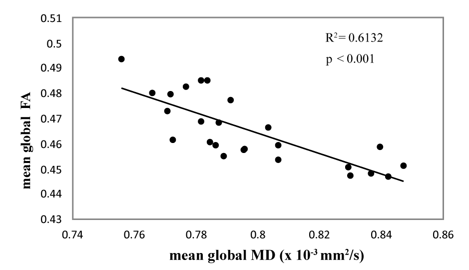
| Scatter plots for the association between the mean global FA, MD, and age are depicted in figures 1 and 2. Simple regression analysis showed a significant negative linear correlation between the FA value of global WM and age (R2=0.4252, p<0.001, FA=0.5426– 0.0011×age [y]) (Figure 1). A significant positive linear correlation was seen between the MD value of global WM and age (R2=0.5309, p<0.001, MD=0.6279-0.0023×age [y]) (Figure 2). Scatter plots for the association between mean global FA and mean global MD are shown in Figure 3. A significant negative linear correlation was seen between the mean FA and mean MD value of global WM (R2=0.6132, p<0.001, FA=0.7875–0.4043×age [y]) (Figure 3). |
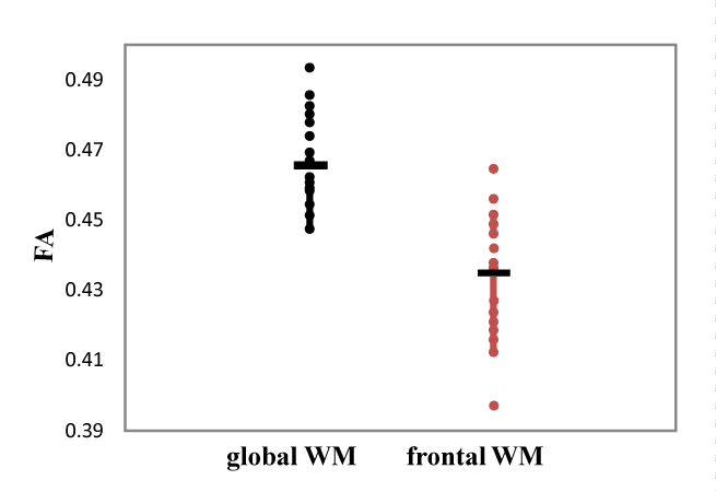
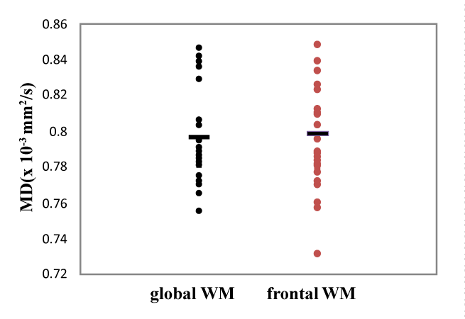
| There was statistically significant difference between the mean frontal FA and mean global FA (p<0.001) (Figure 4), but no significant difference between the mean frontal MD and mean global MD (p=0.946) (Figure 5). |
| Discussion |
| Changes in the structural organization of WM can be measured in vivo with DTI. The current study focused on changes in WM in the elderly population (>55 years old), and the age-related change of global WM was investigated by DTI parameters (FA and MD). We found a significant negative correlation between mean global FA and age, as well as a significant positive correlation between mean global MD and age. Moreover, a significant negative correlation between mean global FA and MD was noted. To the best of our knowledge, the linear regression models used in this study to investigate the association between FA, MD, and age are the best fitting models developed in DTI aging studies. In addition, the difference in age-related changes between frontal WM and global WM was also tested by DTI. The FA value of frontal WM was significantly lower than the FA value of global WM; however, there was no significant difference between the MD value of frontal WM and that of global WM. |
| Global effects of age |
| In the past, the relationship between age and brain volume has been explored by examining changes of regional cerebral volume and tissue concentration differences using structural MR images [19]. In addition to macroscopic changes, intact WM connections in the brain are important for the processing and integration of information generated by neural networks. The “disconnection hypothesis” postulates that a loss of the structural integrity of WM connections will lead to a loss of integration of these networks, and consequently a decline in performance on cognitive tasks [7]. There is evidence from postmortem studies that WM breakdown, such as myelin loss, is more widespread in persons with dementia [20]. Moreover, microscopic WM changes may begin before brain atrophy can be detected macroscopically [3]. Measures reflecting the microstructural integrity of WM may provide additional information on the relationship between WM and cognition. |
| DTI is an MRI technique that enables the measurement of the diffusion of water molecules within the brain [4]. In normal WM, the motion of water molecules is restricted by the parallel-oriented fibers, and diffusion is therefore highly anisotropic. Degradation of the microstructural organization of WM is accompanied by changes in measurable DTI parameters. The FA, a quantitative measure of the degree of anisotropy, can be used to probe the integrity of brain tissue [5]. A lower FA, as measured by DTI, signifies less anisotropic diffusion and thus lowers microstructural integrity [6]. The MD is a quantitative measure of the mean motion of water considered in all directions, and is used to identify pathological changes in cerebral tissue [6]. DTI therefore provides a noninvasive assessment of WM microstructural integrity. The 2 parameters are used to investigate microstructural changes in patients with Alzheimer’s disease [21]. |
| Assessing changes of the brain occurring with aging using DTI is currently a topic of many researchers. Previous studies have used age as an independent variable, and DTI parameters as dependent variables, to find regions with a significant linear correlation [2,8-12]. Sullivan et al. [12] published the first report regarding the linear correlation between FA and age in some regions of WM (R2<0.4). Salat et al. [11] improved the fit of linear regression models to depict the relationship between the FA value of global WM and age (R2=0.56). However, in that study the FA value of global WM was calculated from 9 ROIs in each hemisphere, and in the genu and splenium of the corpus callosum. |
| Voxel-based analysis has recently been used to overcome problems associated with the manual placement of ROIs across the entire brain [1]. Abe et al. [2] investigated the relationship between the FA value of the whole brain and age, as well as the relationship between the MD value of the whole brain and age with voxel-based analysis; however, the fit of the linear regression model was not satisfactory (R2=0.228 for FA and age, R2=0.309 for MD and age). Inano et al. [8] also reported an unsatisfactory result (R2=0.18). Hsu et al. [9] attempted to demonstrate the age-related changes of WM by DTI with higher-order polynomial models, and the quadratic regression model showed a higher R2 than the linear model in the analysis of the relationship between MD and age (R2=0.45). Nevertheless, the linear regression model was still the best-fitting model in the analysis of the relationship between FA and age (R2=0.11). The low and variable fit of the linear regression models could be due to methodological differences in approach to analysis or acquisition, and differences in the study populations. In past studies, younger aged populations were included. However, age-related volume changes in WM are thought to be late-onset in comparison to age-related changes in GM [13,14]. The current study examined age-related changes of global WM in an elderly population by DTI with voxel-based analysis, and the results showed higher R2 values of the linear regression models for the association between FA and age (R2=0.4252, p<0.001) and between MD and age (R2=0.5309, p<0.001) than those reported in prior studies. This result suggests that age-related microscopic changes of WM might be occurring more consistently in later life. |
| Relationship between anisotropy and diffusivity |
| An examination of the relationship between anisotropy and diffusivity provides information about the nature of the observed agerelated microstructural changes. Age-related degradation, as reflected by decreased anisotropy, is potentially associated with increased diffusivity if there is tissue loss that is replaced by free water, or decreased diffusivity if tissue loss is replaced by gliosis or compaction [22]. In agreement with prior study, our results showed the FA value of global WM was significantly negatively correlated with the MD value of global WM, suggesting a true age-related phenomenon of increased interstitial or intracellular fluid accumulation in cerebral WM. |
| Diffusion properties of frontal white matter |
| The constellation of frontal lobe processes called executive functions is especially vulnerable to undesirable effects of advancing age. Age-associated volume reductions have been reported to be more pronounced in the frontal lobe compared to other brain regions [14,23-25]. An age-related FA decline over frontal region had also been reported [15]. The FA value of frontal WM was found to be significantly lower than the FA value of global WM in this study, which is compatible with the suggestion that age-related WM microstructural alterations are more prominent in the frontal region. However, we found no significant difference between the mean frontal MD and mean global MD. There are 2 possible reasons for this finding. First, MD and FA are complementary markers of the effects of aging, and a correlation between MD value and age may be observed in regions other than the frontal lobe [2]. In addition, FA and MD might be associated with different neuropsychological functions in the aging process [26]. Second, there may be a better DTI parameter than MD value, such as radial diffusivity, that can be used to investigate the effect of age on microscopic changes of frontal WM integrity [8,27]. |
| Limitations |
| The principal limitation of the current study is the small sample size, thus the results need to be interpreted with caution. Our results only reflect differences among subjects of different age, rather than changes associated with aging in each individual. Therefore, a longitudinal study of these changes would be a better way to demonstrate the correlation between regional FA changes and aging. The effect of volume reduction and small WM lesions, even though we excluded the subjects with ARWMC scores >2, on aging WM were not taken into consideration in this study. These might influence the evaluation of microscopic changes of WM with aging by DTI [28]. Furthermore, global WM was tested in our study; however, there might be regional differences associated with aging in WM. In image processing, WM segmentation solely relies on the WM tissue probability maps generated by the SPM8 software. In addition to WM tissue probability maps, the SPM8 software was used to generate tissue probability maps of gray matter and cerebrospinal fluid. The future direction of this research is to develop an automatic WM segmentation algorithm by integrating the aforementioned 3 types of tissues. Despite these limitations, strength of this study was that it provided a better fit linear regression model to investigate the association between DTI parameters and age than reported in prior studies. Further study with a larger sample size, taking volume reduction and the white matter lesions as covariates, is needed to further explore microscopic changes of WM associated with aging. |
| Conclusion |
| We applied linear regression model to investigate age-related WM changes in the elderly population with DTI. Our results show significant linear correlations between diffusion properties (FA and MD) and age. To the best of our knowledge, the model proposed is the best fit of any linear regression model for demonstrating the association between diffusion properties and age. These results suggest that age-related microscopic changes of WM might be occurring more consistently in later life. Moreover, a significant negative correlation between mean global FA and mean global MD indicates a true age-related phenomenon of increased interstitial or intracellular fluid accumulation in cerebral WM. The mean frontal FA value was significantly lower than the mean global FA value. However, there was no significant difference between the mean frontal MD value and the mean global MD value. |
| Acknowledgements |
| This research was supported by the Wong Vung-Hau Radiology Foundation. The authors are sincerely and heartily grateful to Professor Fu-Chou Cheng for offering his insightful comments on this research. |
References |
|
Tables and Figures at a glance
| Table 1 |
Figures at a glance
 |
 |
 |
 |
 |
| Figure 1 | Figure 2 | Figure 3 | Figure 4 | Figure 5 |
Relevant Topics
- Abdominal Radiology
- AI in Radiology
- Breast Imaging
- Cardiovascular Radiology
- Chest Radiology
- Clinical Radiology
- CT Imaging
- Diagnostic Radiology
- Emergency Radiology
- Fluoroscopy Radiology
- General Radiology
- Genitourinary Radiology
- Interventional Radiology Techniques
- Mammography
- Minimal Invasive surgery
- Musculoskeletal Radiology
- Neuroradiology
- Neuroradiology Advances
- Oral and Maxillofacial Radiology
- Radiography
- Radiology Imaging
- Surgical Radiology
- Tele Radiology
- Therapeutic Radiology
Recommended Journals
Article Tools
Article Usage
- Total views: 6845
- [From(publication date):
October-2012 - Jul 01, 2025] - Breakdown by view type
- HTML page views : 2209
- PDF downloads : 4636
