Affirmation on Gastrointestinal Radiology: Illuminating Insights into the Inner Landscape
Received: 06-Jul-2023 / Manuscript No. roa-23-107665 / Editor assigned: 08-Jul-2023 / PreQC No. roa-23-107665 (PQ) / Reviewed: 22-Jul-2023 / QC No. roa-23-107665 / Revised: 24-Jul-2023 / Manuscript No. roa-23-107665 (R) / Published Date: 31-Jul-2023 DOI: 10.4172/2167-7964.1000468
Image Article
Gastrointestinal Radiology, a captivating branch of medical imaging, has empowered healthcare professionals to delve into the intricacies of the human digestive system. Employing various imaging techniques, this field has played a pivotal role in diagnosing, treating, and monitoring a myriad of gastrointestinal disorders. Through this image article, we embark on a journey to explore the fascinating world of gastrointestinal radiology, celebrating its invaluable contributions to modern medicine.
Gastrointestinal Radiology owes its profound advancements to cutting-edge technology. Today, medical professionals have an array of powerful imaging modalities at their disposal. X-rays, computed tomography (CT), magnetic resonance imaging (MRI), and fluoroscopy are just a few examples of the impressive tools used to visualize the gastrointestinal tract with remarkable precision.
The barium swallow study is a classic radiological examination performed to evaluate the upper gastrointestinal tract. Patients ingest a barium-based contrast material that coats the esophagus, stomach, and small intestine, allowing radiologists to observe their structure and function in real-time using fluoroscopy. This test aids in diagnosing conditions like gastroesophageal reflux disease (GERD), hiatal hernias, and swallowing disorders.
CT enterography is a sophisticated imaging technique that focuses on the small bowel. By combining CT scanning with oral contrast agents and intravenous contrast, radiologists obtain detailed images of the intestinal walls and surrounding structures [1]. This non-invasive approach is particularly valuable for diagnosing inflammatory bowel disease (IBD), small bowel tumors, and obscure gastrointestinal bleeding.
MRI enterography complements CT imaging, providing a radiation-free alternative to investigate gastrointestinal issues. This technique uses magnetic fields and radio waves to generate highresolution images of the abdomen. Radiologists employ this method to assess Crohn’s disease, ulcerative colitis, and other gastrointestinal disorders affecting the soft tissues [2].
Endoscopic ultrasound (EUS)-merging endoscopy and radiology
Endoscopic ultrasound (EUS) stands at the intersection of endoscopy and radiology, revolutionizing gastrointestinal diagnostics. During EUS, a specialized endoscope equipped with an ultrasound probe is introduced into the body, allowing simultaneous visualization of the digestive tract’s inner layers and adjacent structures. EUS is instrumental in staging gastrointestinal cancers, detecting pancreatic lesions, and evaluating submucosal tumors.
The virtual colonoscopy
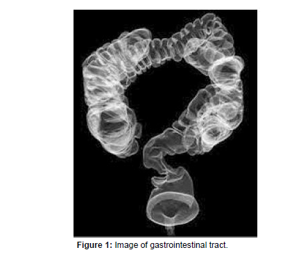
The virtual colonoscopy, also known as CT colonography is a less invasive alternative to traditional colonoscopy for colorectal cancer screening. Patients undergo CT scanning after their colon is gently inflated with air or carbon dioxide. Radiologists then reconstruct 3D images of the colon, allowing for the detection of polyps or other abnormalities without the need for an endoscope (Figure 1).
Gastrointestinal Radiology has undeniably revolutionized the diagnosis and management of gastrointestinal diseases. The amalgamation of state-of-the-art technology with medical expertise has allowed healthcare professionals to navigate the complexities of the digestive system, providing patients with timely and accurate diagnoses. As technology continues to evolve, the future of gastrointestinal radiology holds even greater promise, promising brighter prospects for patients and practitioners alike.
Acknowledgement
None
Conflict of Interest
None
References
- Mandell GA, Teplick SK (1982) Glucagon-its application to childhood gastrointestinal radiology. Gastrointest Radiol 7: 7-13.
- Schulman A, Bornman P, Kaplan C, Morton P, Rose A (1979) Gastrointestinal mucormycosis. Gastrointest Radiol 4: 385-388.
Indexed at, Google Scholar, Crossref
Citation: Akirha T (2023) Affirmation on Gastrointestinal Radiology: Illuminating Insights into the Inner Landscape. OMICS J Radiol 12: 468. DOI: 10.4172/2167-7964.1000468
Copyright: © 2023 Akirha T. This is an open-access article distributed under the terms of the Creative Commons Attribution License, which permits unrestricted use, distribution, and reproduction in any medium, provided the original author and source are credited.

