Affirmation on Gastrointestinal Radiology
Received: 25-Nov-2021 / Accepted Date: 29-Nov-2021 / Published Date: 07-Dec-2021 DOI: 10.4172/2167-7964.1000353
Image Article
Gastrointestinal (GI) Radiology is the subspecialty dedicated to the imaging of issues of the gastrointestinal system (the stomach and digestion tracts, the liver, the biliary tree, and the pancreas) utilizing a wide assortment of modalities, including Digital Radiography (X-RAY), Computed Tomography (CT), Ultrasound and Magnetic Resonance Imaging (MRI). Our radiologists work intimately with gastroenterologists, oncologists, and specialists to analyse and support the treatment of these sicknesses.
Pathologies of gastrointestinal Track are different and influence patients of various ages. Both of these conditions impact the imaging modalities of gastrointestinal parcel that went through pertinent changes during late years. Attractive Resonance (MR) and Computed Tomography (CT) procedures, enhanced for gastrointestinal imaging, are assuming today an expanding part in the assessment of gastrointestinal issues, and a few investigations have shown the upside of these methods over custom barium fluoroscopic assessments optional to upgrades in spatial and worldly goal joined with further developed gut distending specialists. In light of late writing and rules, there is a difference in ideal models in regards to the finding of throat and gastrointestinal malignant growth towards CT, while for little gut imaging in incendiary sickness MRI with another attention on Diffusion Weighted Imaging (DWI) are the main imaging modalities, on the grounds that DWI can be handily carried out in standard MRI for routine use to additional upgrade the analytic exactness in illness evaluation CT and MRI assume a significant part likewise in utilitarian issues. Specifically, the new advancement of quicker MRI beat successions gives fast, on-going imaging of the gastrointestinal lot, pinpointing spaces of injury and giving significant data on motility.
What is upper gastrointestinal tract (UGI?)
Upper gastrointestinal parcel radiography, likewise called an upper GI, is a x-beam assessment of the throat, stomach and initial segment of the small digestive tract (otherwise called the duodenum). Pictures are created utilizing an exceptional type of x-beam called fluoroscopy and an orally ingested contrast material like barium. A x-beam test assists specialists with diagnosing and treat ailments. It opens you to a little portion of ionizing radiation to deliver photos of within the body. X-beams are the most seasoned and regularly utilized type of clinical imaging.
Fluoroscopy makes it possible to see internal organs in motion. When the upper GI tract is coated with barium, the radiologist is able to view and assess the anatomy and function of the oesophagus, stomach and duodenum.
A x-beam assessment that assesses just the pharynx and throat is known as a barium swallow.
As well as drinking barium, a few patients are additionally given baking-soft drink gems (like Alka-Seltzer) to additionally working on the pictures. This strategy is called an air-differentiation or twofold difference upper GI.
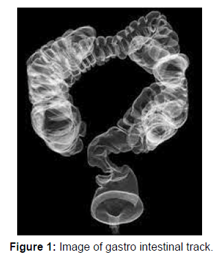
Once in a while, a few patients are given different types of orally ingested contrast, normally containing iodine. These elective difference materials might be utilized assuming that the patient has as of late gone through a medical procedure on the GI parcel, or has hypersensitivities to other differentiation materials. The radiologist will figure out which kind of difference material will be utilized Figure 1.
Citation: Thomas J (2021) Affirmation on Gastrointestinal Radiology. OMICS J Radiol 10: 353. DOI: 10.4172/2167-7964.1000353
Copyright: © 2021 Thomas J. This is an open-access article distributed under the terms of the Creative Commons Attribution License, which permits unrestricted use, distribution, and reproduction in any medium, provided the original author and source are credited.
Select your language of interest to view the total content in your interested language
Share This Article
Open Access Journals
Article Tools
Article Usage
- Total views: 2116
- [From(publication date): 0-2021 - Nov 03, 2025]
- Breakdown by view type
- HTML page views: 1494
- PDF downloads: 622

