Adult Cystic Lymphangioma in Posterior Omentum Cavity
Received: 02-Jan-2024 / Manuscript No. roa-24-125503 / Editor assigned: 05-Jan-2024 / PreQC No. roa-24-125503 / Reviewed: 19-Jan-2024 / QC No. roa-24-125503 / Revised: 26-Jan-2024 / Manuscript No. roa-24-125503 / Published Date: 31-Jan-2024
Abstract
A cystic lymphangioma is a non-malignant tumor that arises from the lymphatic vessels, we report the case of a 60-year-old female patient, presenting with chronic abdominal pain, An abdominal computed tomography was performed, revealing a cystic lymphangioma located in the posterior cavity of the omentum.
Keywords
Cystic; Lymphangioma; Imaging; Omentum
Case Report
A cystic lymphangioma is a non-malignant tumor that arises from the lymphatic vessels [1]. The suggested cause is an embryological anomaly, where primary lymphatic cysts do not properly connect with the main lymphatic system [2].
We report the case of a 60-year-old female patient with no significant medical history, presenting with chronic abdominal pain, evolving in the context of afebrile condition, and maintaining general well-being. The clinical examination revealed A mild pain in the epigastric region without a mass syndrome.
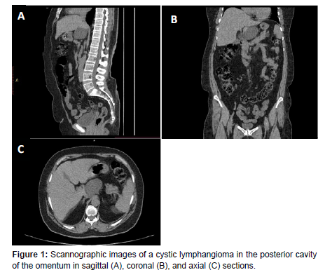
An abdominal computed tomography was performed, revealing a hypodense mass of pure liquid density, unilocular, with a thin and regular wall, without septa or vegetations, measuring 51x51x45 mm (APxTxH), located in the posterior cavity of the omentum, respecting adjacent structures (Figure 1).
Over 80% of lymphangiomas are diagnosed in the first year of life, with rare cases in adults. Gender distribution in adulthood is roughly equal [1].
Cystic lymphangiomas (CL) can develop in various anatomical locations, predominantly in the cervical and axial regions. Intraabdominal cases are rare, comprising less than 5%, and are most commonly located in the mesentery, greater omentum, mesocolon,and retroperitoneum, with even rarer cases in the posterior cavity of the omentum [1,3].
Cystic lymphangiomas are often asymptomatic, with clinical presentations varying based on size and location. Complications may lead to acute scenarios such as cystic hemorrhage, secondary infections, and obstruction of urinary, biliary tracts, and intestines [3].
Diagnostic imaging involves radiological methods, with ultrasound as the primary screening modality, revealing distinct features. Computed Tomography (CT) scans depict a low-density cyst with a smooth shell, and Magnetic Resonance Imaging (MRI) enhances characterization, showcasing low-signal masses in T2-weighted and high-signal masses in T1-weighted sequences [1,2].
Differential diagnoses for cystic lymphangioma include lymphoma, hydatid cysts, ovarian cysts, digestive duplication, mucinous cystadenomas, and mesenteric cysts [1].
The definitive treatment for abdominal cystic lymphangioma is radical excision, even in asymptomatic cases. However, with increasing tumor size, radical resection becomes more challenging, elevating the risk of local recurrence [3].
References
- Houcine Maghrebi, Chaima Yakoubi, Hazem Beji, Feryel Letaief, Sadok Megdich Amin Makni, et al. (2022). Intra-abdominal cystic lymphangioma in adults: A case series of 32 patients and literature review. Ann Med Surg 81: 104460
- Jianchun Xiao, Yuming Shao, Shan Zhu, Xiaodong He (2020) Characteristics of adult abdominal cystic Lymphangioma: a single-center Chinese cohort of 12 cases. Gastroenterol 20: 244
- Mohamed Ben Mabrouk, Malek Barka, Waad Farhat, Fathia Harrabi, Mohamed Azzaza, et al. (2015) Intra-Abdominal Cystic Lymphangioma: Report of 21 Cases. J Cancer Ther 6: 572.
Indexed at, Google Scholar, Crossref
Indexed at, Google Scholar, Crossref
Citation: Zenjali S, Jellal S, Khadija EA, Jamal EF, Rachida S (2024) Adult CysticLymphangioma in Posterior Omentum Cavity. OMICS J Radiol 13: 525.
Copyright: © 2024 Zenjali S, et al. This is an open-access article distributed underthe terms of the Creative Commons Attribution License, which permits unrestricteduse, distribution, and reproduction in any medium, provided the original author andsource are credited.
Select your language of interest to view the total content in your interested language
Share This Article
Open Access Journals
Article Usage
- Total views: 1463
- [From(publication date): 0-2024 - Nov 03, 2025]
- Breakdown by view type
- HTML page views: 1149
- PDF downloads: 314

