Case Report Open Access
Adenocarcinoma Arising from Fistula in Ano
Saalim Nazki, Nisar A. Chowdari, Fazl Q. Parray*, Rauf A Wani and Lubna Samad
SKIMS, Colorectal surgery, 44 rawalpora,govt housing colony,sanat nagar, srinagar,Jammu and Kashmir, 190005, India
- *Corresponding Author:
- Fazl Q. Parray, SKIMS
Colorectal surgery, 44 rawalpora,govt housing colony
sanat nagar, srinagar,Jammu and Kashmir, 190005, India
Tel: 9419008550
E-mail: fazlparray@rediffmail.com
Received date: October 2, 2015 Accepted date: December 1, 2015 Published date: December 7, 2015
Citation: Nazki S, Chowdari NA, Parray FQ, Wani RA, Samad LA (2015) Adenocarcinoma Arising from Fistula in Ano. J Gastrointest Dig Syst 5:370. doi:10.4172/2161-069X.1000370
Copyright: © 2015 Nazki S, et al. This is an open-access article distributed under the terms of the Creative Commons Attribution License, which permits unrestricted use, distribution, and reproduction in any medium, provided the original author and source are credited.
Visit for more related articles at Journal of Gastrointestinal & Digestive System
Abstract
Adenocarcinoma arising from a chronic anorectal fistula is rare of which there are no more than 200 cases in literature. There has been some debate as to whether the fistula is the source of the tumor, or whether the fistula is the presenting feature of the tumor itself. Fistula and associated anal glands are indeed the source of this unusual tumor. A patient of chronic perianal fistula-in-ano was identified to have developed perianal adenocarcinoma from chronic fistula in ano on biopsy postoperatively. Fistula-associated perianal mucinous adenocarcinoma is an uncommon malignant transformation of chronic fistula-in-ano.
Keywords:
Adenocarcinoma
Introduction
The development of carcinoma in a longstanding fistula-in-ano is an interesting though rare complication [1]. Perianal mucinous adenocarcinoma arising from fistula in ano accounts for only 2-3% of all large bowel cancers [2]. It is because of that rarity of the tumor and lack of ample amount of patients for clinical trials there is no definite consensus for its diagnosis and treatment strategies. It is diagnosed late because of absence of tumor inside the bowel and its slow growth deep into the ischioanal fossa and perineum [3]. Early diagnosis is also difficult as it mimics benign inflammatory pathology of anorectal region and biopsies fail to reveal carcinomas at early stage. Such lesions can be misdiagnosed for the more common benign lesions [4]. Diagnosis can confirmed by histopathology and immunohistochemical profile (CK7 positive/CK20 negative) [5].
It has been thought that the carcinomatous changes are due to long continuous sepsis and chronic inflammation but due to its rarity etiological relationshipis not well documented in literature. Although it is a known fact that anal fistula and abscess arise from anal gland and ducts but the origin of carcinoma arising from these ducts and glands is in debate, some literature say that the carcinoma was due to malformation of intestinal tract with rests of cells being source of carcinoma [6]. Though literature also says that the anal fistula is secondary to carcinoma in anorectal region [7]. Kline et al. documented a study where carcinoma from anal fistula was shown to be associated with carcinoma elsewhere in colon or may develop from fistula itself [8]. Study has shown fistula-associated anal adenocarcinoma in Crohn's disease as well [9]. Yamada K et al. in a study concluded anal fistulae are commonly seen in the coloproctology clinic, but special attention to similar conditions associated with malignant disease is needed [10].
Case Report
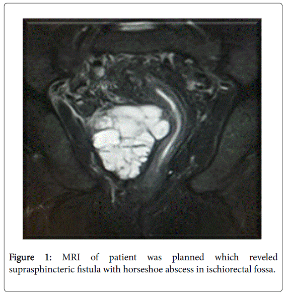
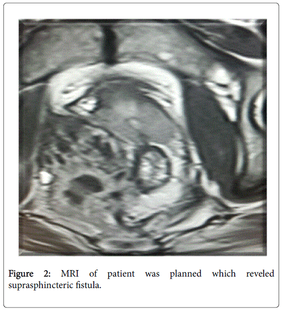
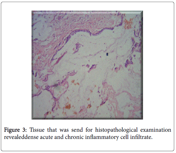
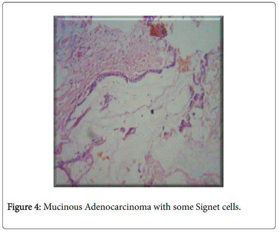
A 45 year male without any comorbidity with a diagnosis of fistulain ano since 16 years; was operated 4 times before reporting to our hospital. 14 years before patient had perianal induration with a mucous discharge from it and was diagnosed as fistula in ano and underwent fistulectomy in a government hospital. The disease recurred back after one year and patient underwent redo-fistulectomy in a private hospital. Patient had infected fistula with a pus discharging from it and fever two years later after the second surgery. On DRE internal opening was felt posteriorly and was confirmed on proctoscopy where an internal opening was seen posteriorly in midline at the level of dentate line and underwent fistulectomy again in the same admission at some other government hospital.Histopathology revealed fistulous tract with inflammatory cells. Patient did well for next 9 years when he came to our hospital with complaints of foul smelling pus discharge from the perianal region. DRE was performed in outpatient department which revealed multiple skin tags, two scars were seen in upper inner quadrant of right gluteal region and one of the scars discharging fecal matter,other external opening was at 5 o’clock position about 5cm from anal verge. Induration was felt inside at around 7 to 10 o’clock position about 4 cm from anal verge. In addition colonoscopy was performed which did not reveal any other positive finding. Crohns and tuberculosis was ruled out in the patient after all the respective investigations and patient was labeled as complex fistula in ano and fistulectomy with curreting of fistulous tract was done. Intra operative findings revealed two tracts coursing along perianal region to form a single tract ending into intersphincteric area, with lot of fibrosis around the tract. Patient came back again to our hospital after 2 years with fresh complaints of pain during defecation, mucousdischarge from perianal region and constipation. DRE was again performed which showed deep fibrosed scar about 8 to 10 cm on side of natal cleft on left side , multiple skin tags present discharge was also seen. Discharge was sent for culture sensitivity which reveled growth of E-coli after 24 hours of incubation. MRI of patient was planned which reveled suprasphincteric fistula with horseshoe abscess in ischiorectal fossa (Figures 1 and 2). Internal drainage of right ischiorectal and supralevator abscess with curettage of unhealthy granulation tissue with external tube drainage of right ischiorectal abscess cavity was done. Tissue that was send for histopathological examination revealeddense acute and chronic inflammatory cell infiltrate with areas of necrosis and focus of Mucinous Adenocarcinoma with some Signet cells (Figures 3 and 4). As the tumor was very large and occupying the adjacent structures as well so it was decided to send patient for proper radiochemotherapy to reduce the size of tumor before a proper surgical decision could be taken.
Discussion
For a malignant change to occur in a fistula in ano it should be long standing, there should be no tumor inside anal canal or rectum unless metastatic and internal opening of fistula in anal canal or rectum should not contain malignantcells [11]. It is documented in literature to rule out that cancerous changes are complication of long standing perianal fistula or fistula is a manifestation of tumor itself. Cancerous changes in fistula are probably due to chronic inflammation, but etiological relationship lacks surety because of the rarity of the disease. Cancerous changes should be suspected in chronicperianal fistula which shows undue induration that does not disappear following a surgical excision. Adequately treated perianal fistula with persistent mucoid discharge and prolonged healing a carcinoma should be suspected. Early diagnosis is difficult to establish due to insidious nature of disease and masking of symptoms by perianal fistula itself. Process of carcinomatous changes contain both indurated and inflammatory zone, it is advised to take multiple biopsies from indurated area and fistula itself instead of inflammatory zone. Previously reported adenocarcinomas areof low grade in about 44% percent cases [12]. One of the study concluded the final management as abdominoperineal resection with wide tumor excision on the gluteal region and reconstruction by bilateral gracilis musculocutaneous flaps [13]. It is importent to look for unusual changes like persistent discharge, gradually increasing perianal mass, bloody discharge, unusual healing or induration in case of perianal fistula. Biopsy should be taken in all cases to be on the safer side.
References
- Kyzer S, Bayer I, Turani H, Chaimoff C et al. (1985) Verrucous squamous carcinoma as a complication of recurrent multiple perianal fistulae. Colo-proctology 7: 104-106.
- Salat A (2006) Anal cancer EurSurg 38:135–138.
- Heidenreich A, Collarini HA, Paladino AM, Fernandez JM, Calvo TO (1966) Cancer in anal fistulas: report of two cases. Dis Colon Rectum 9: 371-376.
- Yang BL, Shao WJ , Sun GD, Chen YQ, Huang JC (2009) Perianal mucinous adenocarcinoma arising from chronic anorectal fistulae: a review from single institution.xInternational Journal of Colorectal Disease 24: 1001-1006.
- Diffaa A, Samlani Z, Elbahlouli A, Rabbani K, Narjis Y, etal. (2011) Primary anal mucinous adenocarcinoma: A case series. Arab Journal of Gastroenterology 1: 48-50.
- Dukes CE, Galvin C (1956) Colloid carcinoma arising within fistulae in the anorectal region. Ann R CollSurgEngl 18: 246-261.
- Skir I (1948) Mucinous carcinoma associated with fistulas of long-standing. Am J Surg 75: 285-289.
- Kline RJ, Spencer RJ, Harrison EGJR (1964) carcinoma associated with fistula-in-ano. Arch Surg 90: 989-994.
- Iesalnieks I, Gaertner WB, Gla H, Strauch U, Hipp M, etal. (2010) Fistula-associated anal adenocarcinoma in Crohn's disease 10: 1643-1648.
- Yamada K, Miyakura Y, Koinuma K, Horie H, Alan T (2014)Leforetal Primary and secondary adenocarcinomas associated with anal fistulae. Surgery Today 44: 888-896.
- Rossner C (1934) Relation of fistula-in-ano to cancer of the anal canal. Trans Am ProcSoc 65-70.
- Kline RJ, Spencer RJ, Harrison EGJR (1964) carcinoma associated with fistula-in-ano. Arch Surg 89: 989-994.
- Erhan Y, Sakarya A, Aydede H, Demir A, Seyhan A, et al. (2003) A Case of Large Mucinous Adenocarcinoma Arising in a Long-Standing Fistula-in-Ano. Digestive Surgery 20:69-71.
Relevant Topics
- Constipation
- Digestive Enzymes
- Endoscopy
- Epigastric Pain
- Gall Bladder
- Gastric Cancer
- Gastrointestinal Bleeding
- Gastrointestinal Hormones
- Gastrointestinal Infections
- Gastrointestinal Inflammation
- Gastrointestinal Pathology
- Gastrointestinal Pharmacology
- Gastrointestinal Radiology
- Gastrointestinal Surgery
- Gastrointestinal Tuberculosis
- GIST Sarcoma
- Intestinal Blockage
- Pancreas
- Salivary Glands
- Stomach Bloating
- Stomach Cramps
- Stomach Disorders
- Stomach Ulcer
Recommended Journals
Article Tools
Article Usage
- Total views: 11422
- [From(publication date):
December-2015 - Nov 21, 2024] - Breakdown by view type
- HTML page views : 10731
- PDF downloads : 691


