Case Report Open Access
Adductor Muscle Atrophy Due to Obturator Nerve Compression by Metastatic Lymph Node Enlargement–A Rare Complication of Recurrent Bladder Cancer
| Dhiren J Shah1*, Allan C Andi1, Keith Ramesar2 and Gillian Watson MT3 | |
| 1Department of Radiology, Guy’s and St. Thomas’ Hospitals NHS Foundation Trust, London, UK | |
| 2Departments of Histopathology, Eastbourne District General Hospital, East Sussex Hospitals NHS Trust, East Sussex, UK | |
| 3Diagnostic Radiology, Eastbourne District General Hospital, East Sussex Hospitals NHS Trust, East Sussex, UK | |
| Corresponding Author : | Janice Dhiren J Shah Department of Radiology Guy’s and St. Thomas’ Hospitals NHS Foundation Trust, London, UK E-mail: dhirenshah@nhs.net |
| Received March 13, 2013; Accepted March 28, 2013; Published April 04, 2013 | |
| Citation: Shah DJ, Andi AC, Ramesar K, Gillian Watson MT (2013) Adductor Muscle Atrophy Due to Obturator Nerve Compression by Metastatic Lymph Node Enlargement–A Rare Complication of Recurrent Bladder Cancer. OMICS J Radiology 2:122. doi: 10.4172/2167-7964.1000122 | |
| Copyright: © 2013 Shah DJ, et al. This is an open-access article distributed under the terms of the Creative Commons Attribution License, which permits unrestricted use, distribution, and reproduction in any medium, provided the original author and source are credited. | |
Visit for more related articles at Journal of Radiology
Abstract
Advanced cancer frequently causes neuropathies or plexopathies if peripheral nerves are invaded. However, isolated obturator mononeuropathy in this setting is exceedingly rare. A 59-year-old male presenting with haematuria went on to have a cystoscopy and trans-urethral resection of a bladder tumour. Histological examination revealed a pT2 grade 3 transitional cell carcinoma, which necessitated a radical cystectomy with ileal conduit. After two cycles of adjuvant chemotherapy he developed severe paraesthesia in his right inner thigh. A pelvic MRI was performed which revealed atrophy of the right adductor muscle group with a proximal obturator node the likely cause of the denervation injury. Neuromuscular symptoms in the context of pelvic cancer should alert the clinician to nerve involvement from either local or metastatic disease. An MRI scan is the ‘gold-standard’ diagnostic test.
| Keywords |
| Metastatic cancer; Malignant neuropathy; Bladder neoplasm; Obturator nerve |
| Introduction |
| The obturator nerve arises from the anterior rami of the second, third, and fourth lumbar nerves; the branch from the third is the largest, while that from the second is often very small. It passes through the fibres of the psoas major muscle emerging from its medial border near the pelvic brim. The obturator nerve is responsible for the sensory innervation of the skin of the medial aspect of the thigh and the motor innervation of the adductor muscles of the lower limb (obturator externus, pectineus, adductor longus, adductor brevis, adductor magnus, gracilis). The hamstring portion of adductor magnus has an ischial tuberosity origin and sciatic nerve innervation in common with the hamstring muscles. Consequently, it would be expected to be unaffected by ‘pure’ obturator nerve pathologies. |
| Obturator nerve palsy is not infrequently seen and this is usually due to therapeutic nerve blocks for spastic conditions of adductor thigh muscles, such as multiple sclerosis and cerebral palsy [1]. Obturator nerve block may also be employed intra-operatively to prevent bladder wall damage by unintentional contractions of the adductor muscles caused by obturator nerve irritation [2]. |
| Pathological obturator nerve injury more commonly occurs as a direct result of pelvic surgery, including radical prostactectomy, gynaecological surgery and orthopaedic surgery [3-6]. Other causes include radiation damage, pelvic trauma, hip pathology, psoas abscesses and retroperitoneal haematoma especially in the context of anticoagulation therapy [7-10]. Malignancy can theoretically affect the obturator nerve in any one of three ways: by direct spread of the primary tumour, by metastatic deposits in the surrounding soft and bony tissues, and by metastatic deposits to the nerve and lumbar plexus itself [9]. |
| Case Report |
| A 59 year-old male presenting with haematuria was investigated with a renal tract ultrasound revealing a 4×3×6 cm tumour arising from the posterior wall of the bladder. The tumour was treated with an uncomplicated transurethral resection (TUR). |
| Histological examination revealed a pT2 grade 3 transitional cell carcinoma with marked squamous metaplasia and areas of complete squamous differentiation. The patient was discussed at a multidisciplinary team meeting, which concluded that further surgery was required. A staging CT scan pre-operatively showed bladder wall thickening consistent with the trans-urethral resection or residual tumour, but there were no significantly enlarged regional or distant metastases. Six weeks after the original procedure, he went on to have a radical cystectomy and ileal conduit formation as a definitive procedure. |
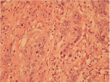
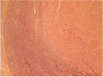
| This resection specimen confirmed the diagnosis of a grade 3 transitional cell carcinoma with squamous differentiation (Figure 1), but also revealed a right peri-vesical lymph node metastasis within the specimen (stage pT4a, N1) (Figure 2). The tumour involved the lower bladder, bladder neck and left side of prostate. The patient had an uncomplicated post-operative course and was discharged home after three days. Subsequently he was commenced on adjuvant chemotherapy with Gemcitabine and Cisplatin, however could only tolerate two cycles. |
| At the next oncological review clinic, three months after his cystectomy, he complained of severe paraesthesia in his right inner thigh as well as pain around his right sacro-iliac joint. Diagnostic confusion resulted from the fact that the patient already had weakness in his right leg from a left middle cerebral artery stroke two years previously. An MRI scan of his lumbo-sacral spine and pelvis was performed three days later. |
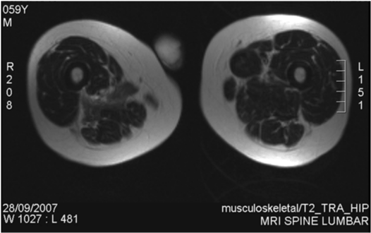
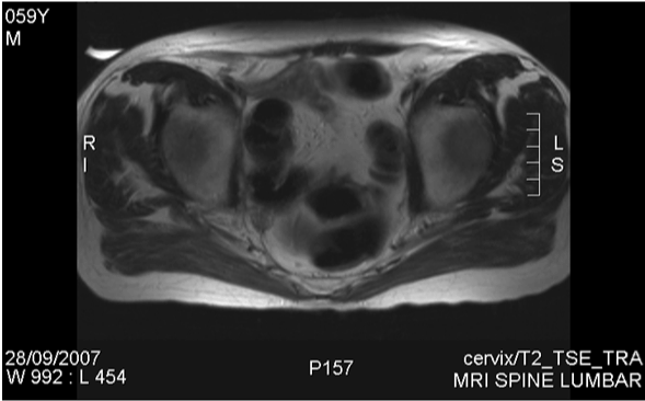
| The MRI scan demonstrated increased signal within the right adductor compartment (with sparing of part of the adductor magnus muscle) and obturator externus muscle on T2-weighted and STIR sequences (Figures 3 and 4). |
| In addition, the muscle bulk of the right adductor compartment was significantly reduced. The cause of these appearances was postulated to be an enlarged right obturator lymph node situated just medial to the obturator neurovascular bundle (Figure 5), which was likely to be metastatic from the bladder cancer. |
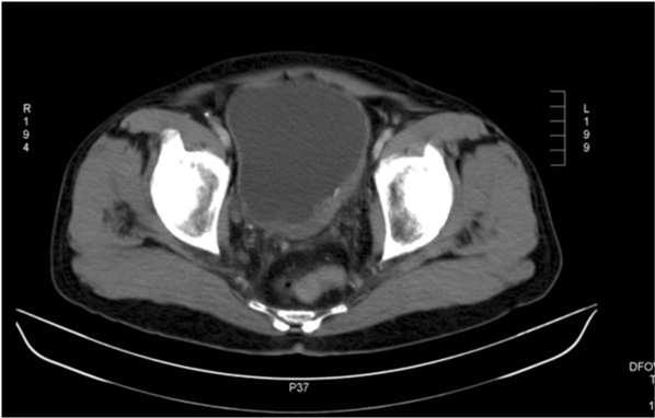
| This lymph node was just discernible on the original staging CT scan (Figure 6), however at the time was only 5 mm in diameter and therefore not considered pathological by size criteria. |
| The lymph node was extremely unlikely to be reactive from the surgery, almost certainly representing malignant involvement. It was deemed inappropriate to obtain a biopsy of this lymph node, and the patient was therefore subsequently managed with palliative care. |
| Discussion |
| The obturator nerve could have been affected by direct invasion, although the denervation may have been explained by compression of the nerve by the lymph node, without necessarily invading it. The latter is a more plausible explanation, considering the infrequent pattern of perineural invasion of transitional cell carcinoma [11]. In support of this, the MRI images from this patient demonstrate a plane of cleavage between the involved lymph node and obturator nerve (Figure 5). Moreover, the presence of the affected node on earlier CT scans would suggest that the neuropathic phenomenon observed relates directly to a progressive pressure effect with enlargement. |
| Bladder cancer commonly metastasises to regional lymph nodes, lung, bone, liver and brain. However, for the lymphadenopathy to cause neuropathy and subsequent muscle denervation is extremely rare. Only three cases of obturator nerve involvement have been reported in the literature in the last 15 years, and these were all from the United States. There have been no reported cases from Europe [12]. |
| The muscles involved were all supplied by the obturator nerve, accounting for the sensory findings of paraesthesia in the medial thigh, and motor findings of adductor muscle atrophy, with sparing of the hamstring portion of adductor magnus (supplied by the sciatic nerve). The findings resulted in the patient being treated with regional palliative radiotherapy for symptom control only, thus obviating the morbidity of more aggressive therapy. |
| This very unusual manifestation of bladder cancer recurrence has never been reported in Europe before. The presence of any neuromuscular symptoms should be treated with suspicion in patients with a history of pelvic cancer, even if apparently resected completely. Prompt radiological investigation with an MRI scan of the affected muscle groups and supplying nerves is advocated for accurate diagnosis [13]. |
References |
|
--
Figures at a glance
 |
 |
 |
| Figure 1 | Figure 2 | Figure 3 |
 |
 |
 |
| Figure 4 | Figure 5 | Figure 6 |
Relevant Topics
- Abdominal Radiology
- AI in Radiology
- Breast Imaging
- Cardiovascular Radiology
- Chest Radiology
- Clinical Radiology
- CT Imaging
- Diagnostic Radiology
- Emergency Radiology
- Fluoroscopy Radiology
- General Radiology
- Genitourinary Radiology
- Interventional Radiology Techniques
- Mammography
- Minimal Invasive surgery
- Musculoskeletal Radiology
- Neuroradiology
- Neuroradiology Advances
- Oral and Maxillofacial Radiology
- Radiography
- Radiology Imaging
- Surgical Radiology
- Tele Radiology
- Therapeutic Radiology
Recommended Journals
Article Tools
Article Usage
- Total views: 16817
- [From(publication date):
June-2013 - Mar 29, 2025] - Breakdown by view type
- HTML page views : 12248
- PDF downloads : 4569
