Case Report Open Access
Acute Myopericarditis due to Hepatitis E Virus Infection: The First Reported Case in the Western Hemisphere
Timothy Dougherty, Showkat Bashir, Mahmuda Khan Jason Adam and Marie Borum*Division of Gastroenterology and Liver Diseases George Washington University, Washington, D.C., USA
- *Corresponding Author:
- Marie L. Borum
MD, EdD, MPH Professor of Medicine Director
Division of Gastroenterology and Liver
Diseases George Washington University
Washington, D.C., USA
Tel: 1-202-741-2160
E-mail: mborum@mfa.gwu.edu
Received date: December 28, 2015 Accepted date: January 28, 2016 Published date: February 5, 2016
Citation: Dougherty T, Bashir S, Adam MKJ, Borum M (2016) Acute Myopericarditis due to Hepatitis E Virus Infection: The First Reported Case in the Western Hemisphere. J Gastrointest Dig Syst 6:386. doi:10.4172/2161-069X.1000386
Copyright: © 2016 Dougherty T, et al. This is an open-access article distributed under the terms of the Creative Commons Attribution License, which permits unrestricted use, distribution, and reproduction in any medium, provided the original author and source are credited.
Visit for more related articles at Journal of Gastrointestinal & Digestive System
Abstract
Hepatitis E is a single stranded RNA virus endemic to parts of Asia and Africa. Presentation ranges from asymptomatic to fulminant . Extrahepatic manifestations include acute pancreatitis, Guillain- Barre syndrome, neuralgic amyotrophy, hemolytic anemia, thrombocytopenia, glomerulonephritis, and mixed cryoglobulinemia.
Keywords
Hepatitis E infection; Acute myopericarditis
Introduction
Hepatitis E is a single stranded RNA virus endemic to parts of Asia and Africa. Presentation ranges from asymptomatic to fulminant hepatic failure. Extrahepatic manifestations include acute pancreatitis, Guillain-Barre syndrome, neuralgic amyotrophy, Hemolytic anemia, Thrombocytopenia, glomerulonephritis, and mixed Cryoglobulinemia [1,2]. Believed to be transmitted via the fecal-oral route, it has a short prodromal phase, and a self-limited symptomatic period lasting days to weeks [3,4] Hepatitis E is a common cause of acute hepatitis in the world, but it is uncommon in the United States where it has typically been encountered in patients returning from developing countries or after consumption of undercooked pork.
Here we report a case of HEV-associated myopericarditis which we believe to be the first involving a patient who lives and travels in the western hemisphere. Our patient’s disease was relatively mild. The other reported cases of HEV-myocarditis have occurred in India and in patients with severe illness [5].
Case Report
A 50-year-old woman with history of Gilbert syndrome presented to our hospital with a two-day history of chest pain, palpitations, dyspnea on exertion, and a single syncopal episode. She had felt chilled but denied fever, abdominal pain, nausea, vomiting, icterus, jaundice, and any change in stool or urine color. She had returned from a two-week trip to Panama ten days earlier and noted a period of rhinorrhea and malaise while she was there.
During the trip, she spent time in the rainforest, pastures, and cities. She denied any contact with live mammals. She had not used any medications, supplements, or herbal preparations. She denied alcohol and drug abuse. Physical exam was notable for mild, fluid-responsive hypotension and a pericardial friction rub. She was free of rashes, arthritis, and fever.
Laboratory studies included an elevated Troponin I at 1.32 ng/mL (reference range 0–0.034) and creatinine phosphokinase MB of 5.4 ng/mL (reference range 0-2.3).
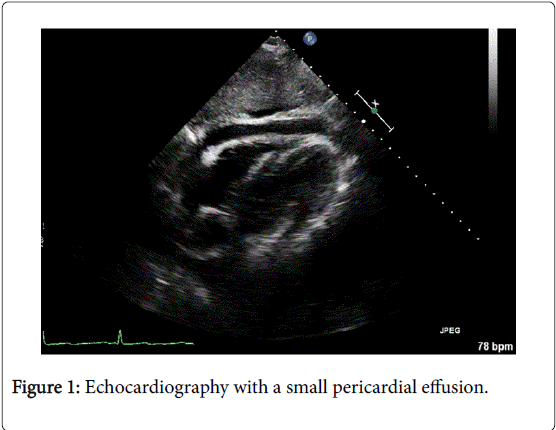
An electrocardiogram revealed generalized PR segment depression. Echocardiography demonstrated a small pericardial effusion. (Figure 1) Computed tomography ruled out pulmonary embolism. These findings support a diagnosis of myopericarditis.
In addition, she was also noted to have elevated transaminases (serum AST 292 units/L, ALT 307 units/L) and bilirubin 1.7 mg/dL (all unconjugated).
Laboratory values are shown in Table 1. A sonogram of the right upper abdomen showed a normal liver and gallbladder. Assays for hepatitis A, B, and C were negative as were those for cytomegalovirus and coxsackie A virus.
| White Blood Cells | 7.05 X 10³ cells/ML |
| Red Blood Cells | 4.32 X 10⁶ cells/ML |
| Hemoglobin | 13.5 g/dL |
| Hematocrit | 41.30% |
| Platelets | 170 X 103 cells/ML |
| AST | 292 units/L |
| ALT | 307 units/L |
| Total Bilirubin | 1.7 mg/dL |
| Direct Bilirubin | 0.0 mg/dL |
| Troponin I | 1.32ng/mL (ref 0 – 0.034) |
| Creatinine Phosphokinase MB | 5.4ng/mL (ref 0-2.3). |
| Prothrombin Time | 14.3 sec |
Table 1: Laboratory values of elevated transaminases.
The patient was treated for myopericarditis with non-steroid antiinflammatory drugs. Her chest pain and dyspnea resolved and EKG changes normalized. Between her discharge and a follow up appointment two weeks later, positive results of an assay for immunoglobulin M directed against HEV were received. Her transaminase activity normalized and she was completely asymptomatic, so testing for HEV RNA was not obtained.
Discussion
This is, to our knowledge, the only case of Hepatitis E myocarditis or pericarditis reported in the Western Hemisphere. Furthermore, it is the only reported case of myocarditis in the setting of a relatively minor HEV-related illness. While cardiac biopsy is the old standard, and cardiac MRI can be useful in the diagnosis, this patient had mild a mild course that did not justify invasive testing. Her cardiac troponin I was elevated, a finding that signifies myocarditis with 89% specificity [6].
The clinical presentation of myopericarditis is variable. While some cases are discovered incidentally, manifestations may range from pleuritic chest pain, decreased exercise tolerance, palpitations, and dyspnea to arrhythmia, dilated cardiomyopathy, and cardiogenic shock. Chest pain might be indistinguishable from ischemic pain; signs of myocarditis can mimic those of acute coronary syndrome. The development of concomitant pericarditis in the setting of myocardial inflammation is common [7].
Viral infection is the most common cause of myocarditis [7,8] and viral hepatitides have been associated with myocarditis [9]. Theories of pathogenesis include viral cytopathic changes, activation of the innate immune system, TNF over-expression, dysregulation of helper and regulatory T lymphocyte populations, and molecular mimicry resulting in autoimmune cardiomyocyte damage [7,10]. Hepatitis C virus in particular has shown evidence of cardiac tropism and has been shown to result in myocarditis and cardiomyopathies in patients with chronic hepatitis C [10]. A few cases of Hepatitis A-related carditis have also been reported [11,12]. Three cases of Hepatitis E-associated myocarditis have been reported in India [5,13]. All of those patients were male and were critically ill. It is not clear why this patient had such a mild course. One possibility is that different virus genotypes may produce milder disease. Although there is significant geographic overlap with respect to the genotype isolates, Genotype 3 is common in North America but not in India and is associated with milder disease [1]. Other potential explanations include variations in host immunity or environmental factors such as selenium deficiency or mercury exposures [7].
This case of a Western hemisphere traveler with Hepatitis Eassociated myocarditis is unique and serves as a reminder that Hepatitis E should be considered in patients who present with elevated liver transaminases and that the virus, known to result in severe disease in South Asia, can have significant consequences in the West as well. It also reminds clinicians that myopericarditis and HEV infection may vary widely in terms of clinical severity; while the severe presentations of these illnesses may be life-threatening, establishing the diagnosis in a mild illness can be valuable and reassuring.
References
- Aggarwal R, Naik S (2009) Epidemiology of hepatitis E: current status. J Gastroenterol Hepatol 24: 1484-1493.
- Bazerbachi F, Haffar S, Garg SK, Lake JR (2015) Extra-hepatic manifestations associated with hepatitis E virus infection: a comprehensive review of the literature. Gastroenterology Reports 1–15.
- Kamar N1, Bendall R, Legrand-Abravanel F, Xia NS, Ijaz S, et al. (2012) Hepatitis E. Lancet 379: 2477-2488.
- Dalton HR, Stableforth W, Thurairajah P, Hazeldine S, Remnarace R, et al. (2009) Autochthonous hepatitis E in Southwest England: natural history, complications and seasonal variation, and hepatitis E virus IgG seroprevalence in blood donors, the elderly and patients with chronic liver disease. Eur J Gastroenterol Hepatol 20:784-790.
- Premkumar M, Rangegowda D, Vashishtha C, Bhatia V, Khumuckham JS, et al. (2015) Acute viral hepatitis e is associated with the development of myocarditis. Case Reports Hepatol 2015: 458056.
- Smith SC, Ladenson JH, Mason JW, Jaffe AS (1997) Elevations of cardiac troponin I associated with myocarditis: experimental and clinical correlates. Circulation 95: 163-168.
- Cooper LT Jr (2009) Myocarditis. N Engl J Med 360: 1526-1538.
- Pollack A, Kontorovich AR, Fuster V, Dec GW (2015) Viral myocarditis--diagnosis, treatment options, and current controversies. Nat Rev Cardiol 12: 670-680.
- Omura T, Yoshiyama M, Hayashi T, Nishiguchi S, Kaito M, et al. (2005) Core protein of hepatitis C virus induces cardiomyopathy. Circ Res 96: 148-150.
- Sanchez MJ, Bergasa NV (2008) Hepatitis C associated cardiomyopathy: potential pathogenic mechanisms and clinical implications. Med Sci Monit 14: RA55-63.
- Yazu T, Miyata Y, Matsuura H, Kimura H, Koga S (1988) [A case of hepatitis A accompanied with acute myocarditis]. Nihon Shokakibyo Gakkai Zasshi 85: 1304-1307.
- Jagtap R, Sethi R, Jeloka T (2008) Hepatitis A leading to myocarditis. J Assoc Physicians India 56: 391-392.
- Goyal BK, Mishra DK, Kawar R, Kalmath BC, Sharma A, et al. (2009) Gautam S. Hepatitis E associated myocarditis: an unusual entity. Bombay Hospital Journal 51:361–362.
Relevant Topics
- Constipation
- Digestive Enzymes
- Endoscopy
- Epigastric Pain
- Gall Bladder
- Gastric Cancer
- Gastrointestinal Bleeding
- Gastrointestinal Hormones
- Gastrointestinal Infections
- Gastrointestinal Inflammation
- Gastrointestinal Pathology
- Gastrointestinal Pharmacology
- Gastrointestinal Radiology
- Gastrointestinal Surgery
- Gastrointestinal Tuberculosis
- GIST Sarcoma
- Intestinal Blockage
- Pancreas
- Salivary Glands
- Stomach Bloating
- Stomach Cramps
- Stomach Disorders
- Stomach Ulcer
Recommended Journals
Article Tools
Article Usage
- Total views: 10136
- [From(publication date):
February-2016 - Nov 21, 2024] - Breakdown by view type
- HTML page views : 9456
- PDF downloads : 680

