Acute Appendagitis: An unusual cause of Acute abdomen
Received: 29-Mar-2023 / Manuscript No. roa-23-93106 / Editor assigned: 01-Apr-2023 / PreQC No. roa-23-93106 (PQ) / Reviewed: 15-Apr-2023 / QC No. roa-23-93106 / Revised: 18-Apr-2023 / Manuscript No. roa-23-93106 (R) / Published Date: 25-Apr-2023 DOI: 10.4172/2167-7964.1000436
Abstract
Acute epiploic appendagitis is a rare but not exceptional pathology that has been brought out of anonymity by cross-sectional imaging. Its identification is easy in front of pathognomonic signs allowing a diagnosis of certainty and a management by a conservative treatment without hospitalization nor surgery.
Keywords
Appendagitis, acute abdomen, CT scan
Clinical Image
Acute epiploic appendagitis is an uncommon cause of abdominal pain [1]. It is a rarely reported condition (1.3% of abdominal pain explored by CT) caused by spontaneous torsion of one or more epiploic appendages.
Epiploic appendages (EA) are small, pedunculated, movable protrusions of fat located on the colonic wall and range in size from 0.5 to 5 cm. Although they can occasionally reach 15 cm in diameter [2]. They are found in the rectosigmoid junction (57%), ileocecal region (26%), ascending colon (9%), transverse colon (6%) and descending colon (2%) [1]. Their vascularization is precarious from that of the colon.
Epiploic appendagitis can be primary or secondary. Primary epiploic appendicitis is caused by spontaneous torsion or venous thrombosis of the involved epiploic appendix. Secondary epiploic appendage is associated with inflammation of adjacent organs, such as diverticulitis, appendicitis or cholecystitis [2].
The consequence of infarction (of venous origin) is fat necrosis at the origin of a lipophagic resorption reaction. This may become chronic by a nodular fibrous transformation and calcification realizing the aspect of “peritoneal mouse” mobile in the peritoneal cavitý [3].
Patients with epiploic appendagitis most often present with constant abdominal pain, often persistent variable in location and rarely associated with accompanying signs. The biology is aspecific [1].
Improved imaging techniques including ultrasound and computed tomography (CT) have increased the possibility of making à correct diagnosis of EA before surgical indication [2].
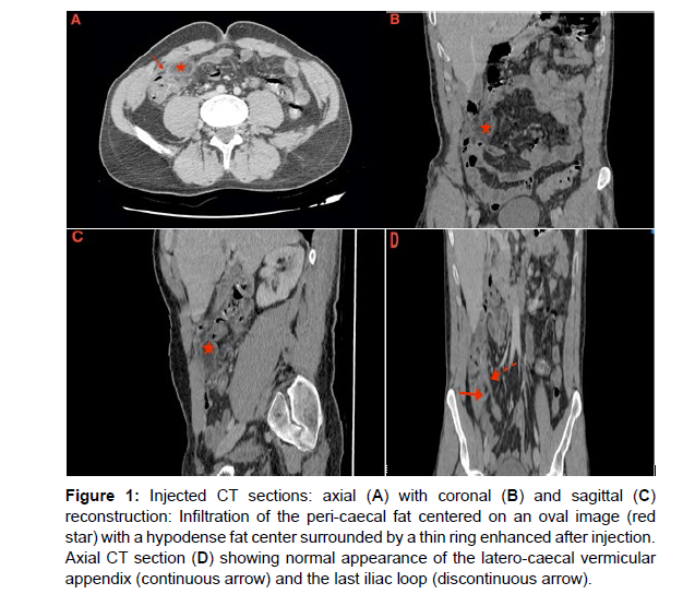
On CT scan, the lesion appears as a fatty mass connected to the serosal surface of the colon, in most cases less than 5 cm in diameter, of higher density than normal fat in its center with an oval “shuttle” shape, bounded by a thin ring taking contrast after injection [3].
Appendagitis is a self-limiting condition and conservative treatment with analgesics is usually sufficient [1] (Figure 1).
Figure 1: Injected CT sections: axial (A) with coronal (B) and sagittal (C) reconstruction: Infiltration of the peri-caecal fat centered on an oval image (red star) with a hypodense fat center surrounded by a thin ring enhanced after injection. Axial CT section (D) showing normal appearance of the latero-caecal vermicular appendix (continuous arrow) and the last iliac loop (discontinuous arrow).
Conflict of Interest
The authors are contributed equally and declare no competing interest
Guarantor of Submission
The corresponding author is the guarantor of submission
References
- Subramaniam R (2006) Acute appendagitis: emergency presentation and computed tomographic appearances. Emerg Med J 23: e53.
- Martineza CAR, Palmab RT, Silveira Júniorc PP, Sato DT, Rodrigues MR, et al. (2013) Primary epiploic appendagitis. J Coloproctol 33: 161-166.
- De Brito P, Gomez MA, Besson M, Scotto B, Huten N, et al. (2008) Frequency and epidemiology of primary epiploic appendagitis on CT in adults with abdominal pain. J Radiol 89: 235- 243.
Indexed at, Google Scholar, Crossref
Citation: ABIDE Z, KHOUCHOUA S, ELAOUFIR O, JROUNDI L, LAAMRANI FZ(2023) Acute Appendagitis: An unusual cause of Acute abdomen. OMICS J Radiol12: 436. DOI: 10.4172/2167-7964.1000436
Copyright: © 2023 ABIDE Z, et al. This is an open-access article distributed underthe terms of the Creative Commons Attribution License, which permits unrestricteduse, distribution, and reproduction in any medium, provided the original author andsource are credited.
Share This Article
Open Access Journals
Article Tools
Article Usage
- Total views: 1322
- [From(publication date): 0-2023 - Mar 29, 2025]
- Breakdown by view type
- HTML page views: 1090
- PDF downloads: 232

