Make the best use of Scientific Research and information from our 700+ peer reviewed, Open Access Journals that operates with the help of 50,000+ Editorial Board Members and esteemed reviewers and 1000+ Scientific associations in Medical, Clinical, Pharmaceutical, Engineering, Technology and Management Fields.
Meet Inspiring Speakers and Experts at our 3000+ Global Conferenceseries Events with over 600+ Conferences, 1200+ Symposiums and 1200+ Workshops on Medical, Pharma, Engineering, Science, Technology and Business
Case Report Open Access
Acute Anterior Wall Myocardial Infarction during Pregnancy in a 36-Year Old Patient
| Jeroen HM van den Broek, Leo Veenstra, Jan CA Hoorntje* and Timo Lenderink | |
| Department of cardiology, Atrium MC, Heerlen, Netherlands | |
| Corresponding Author : | Jan CA Hoorntje Department of cardiology, Atrium MC, Heerlen, Netherlands Tel: +31651955173 E-mail: j.hoorntje@atriummc.nl |
| Received June 30, 2014; Accepted July 21, 2015; Published July 27, 2015 | |
| Citation: van den Broek JHM, Veenstra L, Hoorntje JCA, Lenderink T (2015) Acute Anterior Wall Myocardial Infarction during Pregnancy in a 36-Year Old Patient. J Preg Child Health 2:183. doi:10.4172/2376-127X. 1000183 | |
| Copyright: © 2015 van den Broek JHM, et al. This is an open-access article distributed under the terms of the Creative Commons Attribution License, which permits unrestricted use, distribution, and reproduction in any medium, provided the original author and source are credited. | |
| Related article at Pubmed, Scholar Google | |
Visit for more related articles at Journal of Pregnancy and Child Health
Abstract
Acute myocardial infarction during pregnancy is a rare clinical condition. We present a case of a 36-year old woman in her 36th week of pregnancy with an acute anterior ST-elevated myocardial infarction (STEMI) caused by an occlusive proximal LAD. First treatment was thrombosuction, followed by placement of a bioresolvable vascular scaffold. Because of post procedural fetal bradycardia an emergency cesarean section was performed on the same day, despite all antithrombotic medications.
| Keywords |
| Myocardial infarction; STEMI; Pregnancy |
| Introduction |
| Acute myocardial infarction during pregnancy is a rare condition, estimated to occur in 28 to 62 per 100.000 deliveries with the majority (72%) of patients aged over 30 years [1,2]. Although different pathophysiological processes can lead to myocardial infarction, coronary artery atherosclerotic disease is most common, also during pregnancy [1]. |
| Myocardial infarction, especially extended anterior infarction can lead to hemodynamic instability and in this case is a threat to both maternal and fetal life, which reemphasizes the need for urgent intervention. |
| The potential of bioresorption and restoration of endothelial and vascular function makes the recently introduced bioresolvable vascular Scaffold (BVS) a promising treatment option, especially in young people. Research shows a similar rate of adverse cardiac events [3]. Though BVS seems to be suitable in the case of acute ST-segment elevation myocardial infarction (STEMI) [4], the use of a BVS in STEMI during pregnancy has not yet been reported. |
| Case Report |
| A 36-year old woman – with no previous cardiovascular disease – was admitted to the labor ward in her 36th week of gestation. Presentation symptoms were chest discomfort radiating to the left arm, dyspnea, and nausea and vomiting, existing for several hours. Cardiovascular risk factors included smoking and a family history of cardiovascular disease. Physical examination revealed no obvious cardiac abnormalities; blood pressure was 120/85 with a heart rate of 75. |
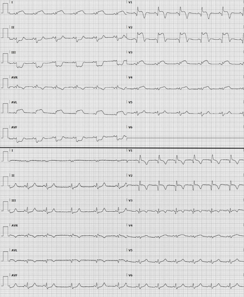
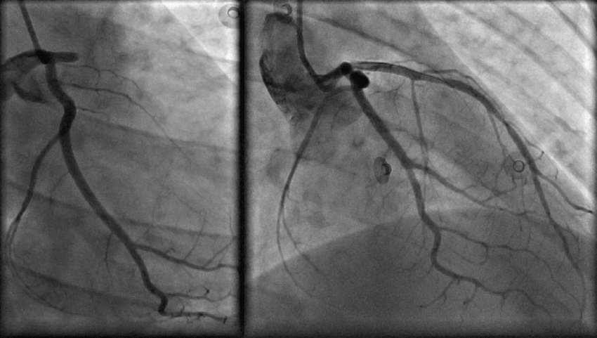
| A 12-lead ECG showed extensive anterior ST-elevation myocardial infarction (Figure 1; upper panel). Bedside cardiac ultrasound showed wall motion abnormalities of a large part of the anterior, apical and septal wall. Aspirin 300 mg and ticagrelor 180 mg were administered orally and i.v. heparin (7000 IU). Emergency coronary angiography was performed and a proximally occluded LAD coronary artery was found (Figure 2; left panel), the other coronary arteries showed no abnormalities. Differential diagnosis includes thrombosis or spontaneous coronary artery dissection (SCAD). Percutaneous coronary intervention (PCI) was performed with thrombosuction and placement of a BVS (Figure 2; right panel). Because of distal thrombosis of the vessel, tirofiban was administered i.c. Serum markers were highly elevated, left ventricular ejection fraction, estimated with ultrasound was 35%. Post procedural electrocardiography showed ST-resolution with intraventricular conduction delay and QR pattern on the anterior wall leads (Figure 1; lower panel). During the procedure the patient developed hypotension, after which the fetus showed a repetitive bradycardia on CTG-monitoring. |
| After the intervention, blood pressure stabilized at 100/60 mmHg, a total of 2000 ml of plasma expander and sodium chloride were given. Four hours post-PCI, the fetus showed another bradycardia lasting for five minutes, after which was decided to perform a cesarean section, despite all the antithrombotic drugs administered. Section was successful but complicated by severe postoperative hematoma of the abdominal wound and anemia. Later the patient showed signs of acute heart failure (pulmonary edema and low blood pressure), therefore intravenous inotropy was started and furosemide was administered intravenous once to twice daily during four days. |
| The patient remained on the coronary care unit for 5 consecutive days. Cardiac ultrasound before discharge showed an improved ejection fraction of 45% with wall motion abnormalities anterior, septal and apical and no ventricular thrombi. |
| Discussion |
| Acute myocardial infarction in young women, especially during pregnancy is rare. Differential diagnosis should include spontaneous coronary artery dissection, especially during pregnancy [1]. Guidelines are clear about the preferred treatment, emergency revascularization with primary PCI including all related antithrombotic medications. |
| Because it is such a rare condition, there are no data about the safety of administering high doses of antithrombotic drugs during pregnancy. Especially when delivery is expected on short term or when a cesarean section might be considered, antithrombotic treatment might lead to increased blood loss. Opposed to the cases reported by Wassing et al. [5] treated with thrombolysis, percutaneous intervention offers a better option with less bleeding risk and adjustable platelet aggregation inhibition with tirofiban. |
| Depending on the type of stent that is placed, prolonged antithrombotic therapy with ticagrelor might be indicated. Placement of a bare metal stent (BMS) does not require additional antithrombotic treatment after the acute phase, but might not be the desirable solution in young, otherwise healthy women because of the increased risk of instent restenosis [6-8]. In this case, considering the age of the patient, a BVS was placed, which requires prolonged antithrombotic therapy with ticagrelor. |
| There was need for emergency cesarean section only 4 hours after PCI procedure, which was performed successfully. The full antithrombotic treatment unfortunately led to a severe, but not life threatening hematoma around the abdominal wound. |
| Post procedural hemodynamic instability, which is common in anterior wall infarction, caused fetal distress and in this case the decision to perform an emergency cesarean section. Fluid suppletion in response to post procedural hypotension has led to pulmonary edema and the need for treatment with diuretics. Possibly earlier administration of intravenous inotropes could have prevented this. |
| In conclusion, acute myocardial infarction during pregnancy is a rare but severe clinical entity which poses both maternal and fetal life at risk. PCI with placement of the recently introduced BVS was successful but consecutive cesarean section was complicated by severe but not lifethreatening wound hematoma. Although complicated, both mother and daughter did well during follow up. |
| Acknowledgments |
| There are no acknowledgments. |
| Conflict of Interest |
| There are no conflicts of interest. |
References
- Roth A, Elkayam U (2008)Acute myocardial infarction associated with pregnancy. J Am CollCardiol:52.
- James AH, Jamison MG, Biswas MS, Brancazio LR, Swamy GK, et al. (2006) Acute myocardial infaction in pregnancy: A united states population-based study. Circulation 113:1564-1571.
- Simsek C, Karanasos A, Magro M, Garcia-Garcia HM, Onuma Y, et al.(2014)Long-term invasive follow-up of the everolimus-eluting bioresorbable vascular scaffold: Five-year results of multiple invasive imaging modalities. EuroIntervention:10.
- Diletti R,Karanasos A, Muramatsu T (2014)Everolimus-eluting bioresorbable vascular scaffolds for treatment of patients presenting with ST-segment elevation myocardial infarction: BVS STEMI first study. Eur Heart J 35:777-786.
- Wassing JGP, Polak PE, de Groot CJM, Roos-Hesselink JW (2005)Acute coronary syndrome during pregnancy. Neth Heart J 13:360-365.
- Sabaté M, Brugaletta S, Cequier A, Iñiguez A, Serra A, et al. (2014) The EXAMINATION trial (Everolimus-Eluting Stents Versus Bare-Metal Stents in ST-Segment Elevation Myocardial Infarction): 2-year results from a multicenter randomized controlled trial. JACC CardiovascInterv7:64-71.
- Wallace EL, Abdel-Latif A, Charnigo R (2012) Meta-analysis of long-term outcomes for drug-eluting stents versus bare-metal stents in primary percutaneous coronary interventions for ST-segment elevation myocardial infarction. Am J Cardiol109:932-940.
- Merken JJ, Majidi M, Altintas S, Hoorntje JCA (2014)Severe spontaneous coronary artery dissection in a 42-year-old male: A treatment strategy challenge. Int J Cardiol177:1049-51.
Figures at a glance
 |
 |
| Figure 1 | Figure 2 |
Post your comment
Relevant Topics
Recommended Journals
Article Tools
Article Usage
- Total views: 14070
- [From(publication date):
August-2015 - Jul 02, 2025] - Breakdown by view type
- HTML page views : 9514
- PDF downloads : 4556
