Acute Airway Obstruction in Rheumatoid Arthritis
Published Date: 27-Dec-2016 DOI: 10.4172/2161-119X.1000282
Abstract
Rheumatoid arthritis is cause of laryngeal stridor. This symptom may occur as a result of the upper airway edema or adduction defects of the vocal folds (with/without cricoarytenoid joint involvement). Others manifestations of this inflammatory disease in the laryngeal level are hoarseness, cough, respiratory distress, foreign body sensation, dysphagia or odynophagia
Introduction
Rheumatoid Arthritis (RA) is a systemic disease. When RA affects the cricoarytenoid joint, this may lead to acute stridor or dyspnoea, and potentially life-threatening complications. We report a case of RA patient with acute laryngeal stridor [1].
Case Presentation
A 46 year old woman who was previously diagnosed with RA (and treated with Methotrexate), came to the Emergency Department of our hospital for hoarseness, dysphagia, foreign body sensation dyspnoea and stridor of 2 days. No fever.
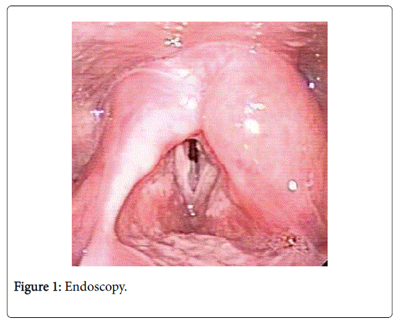
On the clinical examination, there were no lesions or edemas in the oropharynx or oral cavity. Endoscopy showed fixed adduction of the left vocal cord and paresia of the right vocal cord, besides edema and redness of the arytenoids, posterior commissure and subglottic area (Figure 1).
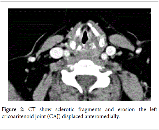
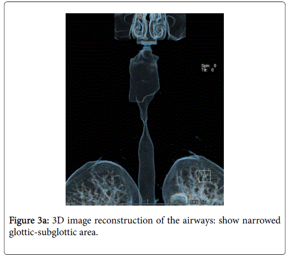
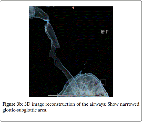
A Computed Tomography (CT) scan was performed. The imaging showed sclerotic fragments and erosion the left cricoaritenoid joint (CAJ) displaced anteromedially (Figure 2), swollen of falses and trues vocal cords and narrowed subglottic area (Figures 3A and 3B).
After IV steroids, the endoscopic examinations two days later showed an improved movility of the vocal cords and absence of edema or laryngeal swelling.
Discussion
RA is a chronic multisystemic disease charactized by inflammation and damage in the sinovial and joint tissues. It can affect all joints in the body. RA occurs in 0.5% to 3% of population [1] and women tend to be affected 3 times more often than men [2]. Peak onset of RA is in the fourth and fith decades [3].
There are extra-articular features (20% of the cases including eyes, lungs, cardiovascular and nervous systems…). Rheumatoid arthritis can affect the ear, nose, and throat, and involve the larynx (from 13 to 75% in different series and between 45 and 88% in postmortem studies [3,4] and 15% of patiens are having laryngeal involment as unique manifestation of the RA. Laryngoscopic examination shows edema, myositis of intrinsic laryngeal muscle, hiperemia, inflammation and swelling of the arytenoids, interarytenoid mucosa, aryepiglottic folds and epiglottis, and impaired mobility or fixation (in the median, paramedian or lateral position of one or both vocal cords) and ankilosis of cricoarytenoid joint (the cricoarytenoid joint is a diarthrosis with a sinovial membrane. When the joint is involved, the inflammation starts with a sinovial lining and spreads through the articular surface producing fibrosis and anquilosis), rheumatoid nodules and Bamboo nodes (in submucosal space of the middle portion of the vocal cords) [1,5].
Common symtoms are hoarseness, sensation of a foreign body in the throat, dyspnoea, throat pain radiating to the ears, stridor or odino-dysphagia [6,7]. There are others causes of dyspnoea in a patient with rheumatoid arthritis: for systemic manifestations ( pericardial or pleural effusion, pulmonary hypertension, pulmonary nodules, intersticial disease, bronchiolitis obliterans-organizing pneumonia BOOP, Caplan´s syndrome, atlantoaxial subluxation) or for the treatment (Metrotrexate fibrosis, asthma or pneumonitis induced by non- steroidal anti-inflammatory drugs or penicillamine bronchiolitis). It could also be entirely incidental [8-10].
Others causes of stridor in RA are: Ischemic neuropathy (for vasculitis) cervico-medullar compresión, laryngeal amiloidosis or atrophy of the laryngeal muscle associated to demyelinization and degeneration of recurrent and vagus nerves [4,6].
Radiologic signs can preceded clinical symptoms [3]. The first radiological sign in CT is the thickening and increased space of the articular cartilage (for the increase of synovial liquid and/or the apposition of sinovial membrane). Brazeau-Lamontagne [11] proposed a CT stating:
Level I: There is thickening of the CAJ
Level II: There is erosion of the CAJ
Level III: There is luxation of the CAJ
Level IV: There is luxation of the larynx
Others radiologic findings are narrowing in the piriform sinuses, decrease in the CAJ space, narrowing and/or increase in the CTJ density (of the cricothyroid joint) and/or CAJ [3]. There has been described also inflammatory subglottic mass [12].
The treatment in the acute phase consists of administration of steroids systemically or locally into the joint (alone or with parenteral treatment) [2]. When there is a compromise of the upper airway and in cases with bilateral CJ involvement, the traqueotomy should be performed [1].
Acknowledgement
Laryngeal manifestation of RA are several: edema or fixation of cricoarytenoid joint, mucosal eritema and edema, myositis, rheumatoid nodules and Bamboo nodes. When the RA involvement the CAJ may be cause of vocal folds paralysis.
References
- Amdan AL, Sarieddine D (2013) Laryngeal manifestations of rheumatoid arthritis. Autoimmune Dis 2013: 1-6.
- Leicht MJ, Harrington TM, Davis DE (1987) Cricoarytenoid arthritis: A cause of laryngeal obstruction. Ann Emerg Med 16: 885-888.
- Greco A, Fusconi M, Macri A, Marinelli C, Polettini E, et al. (2012) Cricoiarytenoid joint involvement in rheumatoid arthritis: Radiologic evaluation. Am J Otolaryngol 33:753-755.
- Abe K, Mitsuka T, Yamaoka A, Yamashita K, Yamashita M, et al. (2013) Sudden glottic stenosis caused by cricoarytenoid joint involvement due to rheumatoid arthritis. Intern Med 52: 2469-2472.
- Voulgari PV, Papazisi D, Bai M, Zagorianakou P, Assimakopoulos D, et al. (2005) Laryngeal involvement in rheumatoid arthritis. Rheumatol Int 25: 321-325.
- Peters JE, Burke CJ, Morris VH (2011) Three cases of rheumatoid arthritis with laryngeal stridor. Clin Rheumatol 30: 723-727
- Bamshad M, Rosa U, Padda G, Luce M (1989) Acute upper airway obstruction in rheumatoid arthritis of the cricoarytenoid joints. South Med J 82: 507-510.
- Bossingham DH, Simpson FG (1996) Acute laryngeal obstruction in rheumatoid arthritis. BMJ 312: 295-296.
- Lipsky PE (2009) Rheumatoid arthritis. In: Fauci AS, Braunwald E, Kasper DL, et al. (eds.) Harrison’s Principles of Internal Medicine.17th edn. New York: McGraw Hill pp: 2083-2092.
- Lin HY, Chen CC, Lee YK, Su YC (2012) Dyspnea caused by atlantoaxial subluxation in a patient with rheumatoid arthritis. Case Rep Emerg Med 2012: 1-2.
- Brazeau-Lamontagne L, Charlin B, Levesque RY, Lussier A (1986) Cricoarytenoiditis: CT assessment in rheumatoid arthritis. Radiology 158: 463-466.
- Haben CM, Chagnon FP, Zakhary K (2005) Laryngeal manifestation of autoimmune disease: rheumatoid arthritis mimicking a cartilaginous neoplasm. J Otolaryngol 34: 203-206.
Share This Article
Recommended Journals
Open Access Journals
Article Tools
Article Usage
- Total views: 6322
- [From(publication date): 0-2016 - Apr 07, 2025]
- Breakdown by view type
- HTML page views: 5477
- PDF downloads: 845


