Research Article Open Access
Achilles Tendon Rupture and Abnormal Lipid Profile: A Descriptive Clinical Laboratory and Histology Study
Loisel François1*, Hugo Kielwasser1, Grégoire Faivre1, Kantelip Bernadette2 and Obert Laurent11Orthopaedic and Traumatology Surgery Service, University Hospital of Besançon, Besançon France Intervention, Innovation, Imagery, Engineering in Health, Medical and Pharmacology Section, University of Franche-Comté, Besançon, France
2Department of Anatomy and Cell Pathology, CHU Jean Minjoz, Boulevard Alexandre Fleming Besançon, France
- Corresponding Author:
- François L
Orthopaedic and Traumatology Surgery Service
University Hospital of Besançon 25000
Besançon France Intervention, Innovation, Imagery
Engineering in Health (EA 4268), Medical and Pharmacology Section
IFR 133, University of Franche-Comté 25000, Besançon, France
Tel: (33) 06 31 98 75 32
E-mail: francois_loisel@yahoo.fr
Received Date: October 20, 2016; Accepted Date: November 05, 2016; Published Date: November 11, 2016
Citation: François L, Kielwasser H, Faivre G, Bernadette K, Laurent O (2016) Achilles Tendon Rupture and Abnormal Lipid Profile: A Descriptive Clinical Laboratory and Histology Study. Clin Res Foot Ankle 4:214. doi:10.4172/2329-910X.1000214
Copyright: © 2016 François L, et al. This is an open-access article distributed under the terms of the Creative Commons Attribution License, which permits unrestricted use, distribution, and reproduction in any medium, provided the original author and source are credited.
Visit for more related articles at Clinical Research on Foot & Ankle
Abstract
Introduction: Several human and animal studies have revealed a link between abnormal serum lipid levels and Achilles tendon rupture. However, clinicians have not detected macroscopic amounts of fatty tissue during the repair of ruptured tendons.
Material and Methods: A lipid profile and evaluation of tendon lipomatosis were performed in a group of 65 patients with Achilles tendon rupture recruited over a two-year period at two French hospitals. Cardiovascular and tendon rupture risk factors were inventoried.
Results: Ten patients had a history of hypercholesterolemia, seven of whom were undergoing statin treatment (15%). Two patients had known risk factors for tendon rupture (3%): one was taking inhaled corticosteroids and the other suffered from hyperuricemia. Total cholesterol was normal in 63% of cases; triglycerides were normal in 67% of cases and HDL cholesterol was normal in 54% of cases. If cardiovascular risk factors were taken into account, the portion of high-LDL cholesterol went from 17% to 23%, but this was not significant. Pathology analysis found four cases of tendon lipomatosis among the 36 collected samples (11%).
Discussion: Since this study did not include a control group, we cannot make any conclusions about the lack of relationship between abnormal lipid profile and tendon rupture. The prevalence of hypercholesterolemia in our study population was similar to that of the general population. Although high cholesterol levels have been implicated by some authors, cholesterol accumulation within the tendon is probably not directly responsible for weakening it; however, an increase in atheromatous plaque in tendon blood vessels may lead to hypoxia.
Keywords
Achilles tendon rupture; Cardiovascular risk factor; Connective tissue disease; Foam cell; Hypercholesterolemia; Lipomatosis
Introduction
Achilles tendon rupture is a rare condition, but its incidence has greatly increased over the past ten years. Although the risk factors are well known, they cannot explain every rupture.
Research into tendinous xanthomas and Type IIa familial dyslipidemia [1] and the histological study of ruptured tendons [2] (tendon lipomatosis) can help us better understand which populations are at risk. These recent studies seem to suggest that hypercholesterolemia is a predisposing factor for tendon rupture.
Drug vigilance organizations have increasingly pointed to statins as being an additional predisposing factor, in combination with hypercholesterolemia [3].
The purpose of this study was to evaluate cholesterol levels and tendon lipomatosis in patients with Achilles tendon rupture through a prospective, continuous, descriptive study carried out in two French hospitals.
Material and Methods
Patients were enrolled for two consecutive years at the University Hospital (CHU) of the author and a Peripheral Hospital (CHR) through the emergency department or orthopedic consultations. Sixtyfive patients were enrolled in all (5 at the CHR and 60 at the CHU).
Inclusion criteria consisted of subcutaneous Achilles tendon rupture, with or without associated trauma. Patients with open tendon lacerations were excluded. Every enrolled patient was given an information sheet and was asked to sign an informed consent form. A fasting serum lipid profile was performed either at the hospital or an outside facility. This lipid profile measured total cholesterol, triglycerides, and HDL-cholesterol and calculated LDL-cholesterol.
The unit of measurement was grams per liter. Each patient was questioned about personal or familial medical history (especially hypercholesterolemia), drugs taken at the time of the rupture or in the months prior, patient's height and weight, and smoking habits. These parameters were used to calculate the cardiovascular risk factors and reveal known causes of tendon rupture.
Other parameters were recorded, such as the history of chronic tendinopathy and mechanism of rupture. The latter consisted of either direct impact to the posterior aspect of the leg (e.g. sliding tackle), loading upon foot strike (running or standing start) or no specific trauma event (walking). Any patients with a history of spinal disc disease, inguinal or hiatal hernia and meniscus injury were grouped into the category "history of collagen abnormality".
Recurrence of tendon rupture and bilateral ruptures were included in this patient subgroup. If the patient was operated on, the tendon margins (debridement, surgical remnants) were analyzed histologically to look for lipomatosis. The variable-length tendon sample (a few millimeters to several centimeters) was not fixed but placed in a sterile, saline-filled flask. The vascularity of the tendon sample was not evaluated.
Results
The various epidemiological characteristics are given in (Table 1). Of the 65 enrolled patients, 7 were treated conservatively based on the decision of the on-call surgeon; there was one woman and six men with a mean age of 52 years.
| Mean age | Gender ratio M/F | BMI |
|---|---|---|
| 44 [19 - 74] | 3.64 | 26.2 [19.1 - 48.5] |
| Type of trauma | ||
| Direct impact = 2 | Loading upon foot strike = 57 | No injury event = 6 |
| Smoking | ||
| 19 smokers | 39 nonsmokers | 7 no information |
Table 1: Epidemiological features of the study population.
Ten patients had a history of hypercholesterolemia, with seven of them undergoing statin treatment (15%).
Among these seven patients taking statins, one patient had an additional known tendon rupture risk factor (hyperuricemia). One other patient had a known risk factor for tendon rupture (inhaled corticosteroids use).
Three patients had a contralateral recurrence of tendon rupture, one patient had ipsilateral recurrence after failed conservative treatment, and one patient had signs of tendinopathy before the rupture occurred.
Nineteen patients had a history of abnormal collagen findings (29%). One patient had a simultaneous bilateral Achilles tendon rupture.
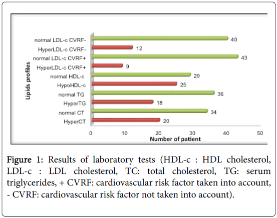
Figure 1 shows the results of the laboratory tests. Eleven of the 65 patients declined the lipid profile. Two patients had elevated triglycerides (greater than 4 g/L) that made LDL cholesterol calculation impossible.
Total cholesterol (TC) was normal in 63% of cases; triglycerides (TG) were normal in 67% of cases and HDL cholesterol was normal or high in 54% of cases. If cardiovascular risk factors were taken into account (+/- CVRF), the portion of high LDL cholesterol went from 17% (CVRF+) to 23% (CVRF-), but this was not significant.
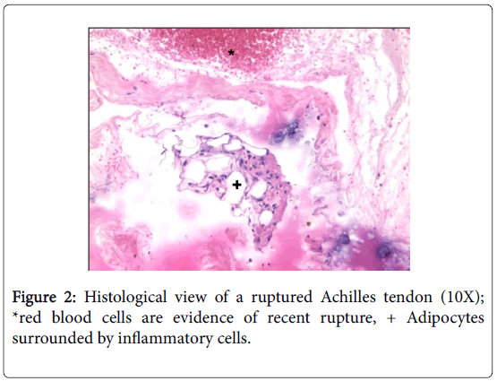
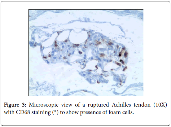
Pathology analysis found four cases of tendon lipomatosis among the 36 collected samples (11%) (Figures 2 and 3).
Discussion
The limitations of this study are its descriptive, uncontrolled, and nonrandomized aspect. On the other hand, it is the first study coupling both blood cholesterol with an histological analysis of ruptured tendons in a target population.
Since this was a descriptive study, we could not determine if a statistical relationship existed between tendon rupture and cholesterol levels. A control group would have been required for statistical comparison. But what would be the make-up of a tendon rupture control group? An age and gender-matched group of patients free of tendon injury or historical collagen abnormalities would be required. But tendon disease, especially tendon rupture, is often insidious. As a consequence, the risk that "diseased" patients will be included in the "healthy" group is high.
One solution would be to perform MRI imaging to detect tendon injury or signs of previous tendon remodeling before including any patient in the injury-free control group. Another solution would be to compare the presence of pathological cholesterol levels in a subgroup of patients with no risk factors or history of collagen abnormalities with those in a group of patients who have these characteristics. Unfortunately, we found no significant differences when comparing these subgroups. Could the patients in our series be compared with patients in existing series in terms of the prevalence of dyslipidemia? The French multicenter study performed by Ferrières [4] analyzed the change in cholesterol levels in a representative sample of the French population from 1996 to 1997 and from 2006 to 2007.
Table 2 compares the main results of the Ferrières study with those of our own. We applied the criteria of hypercholesterolemia and absence of dyslipidemia from the Ferrières study to the results of the current study. Our series had a comparable prevalence of hypercholesterolemia (36.9% for Ferrières and 33.3% for our study) and comparable use of lipid-lowering drugs (12.5% for Ferrières and 10.7% for our study). However, there are limitations to this comparison with the multicenter study: the laboratory tests used different material, there was a wider age-range in our series than in that of Ferrières (19−74 years), and there were biases specific to the Ferrières study (French population sample from three regions only, selection bias, socio-economic disparities).
| Type of Study | Ferrières study | Current tendon rupture study | |
|---|---|---|---|
| 1996-1997 (n= 3508) | 2006-2007 (n= 3597) | (n= 65) | |
| BMI | 26.2 | 26.1 | 26.2 |
| Smokers | 22.1% | 18.9% | 32% (19/58) |
| Hypercholesterolemia | 41.7% | 36.9% | 33.3% (18/54) |
| Cholesterol-lowering drugs | 10.4% | 12.5% | 10.7% (7/65) |
| Normal lipid profile | 45% | 51.5% | 38.8% (21/54) |
Table 2: Lipid profile in the current series relative to general population.
The relationship between cholesterol and tendons has been studied for the past 40 years. The first studies mainly focused on specific cases of family dyslipidemia or tendon xanthomas [1,5,6] and seemed to reveal a link between tendon disorders and hypercholesterolemia. Later on, animal studies confirmed the deleterious role of a cholesterol-rich environment on embryonic rat fibroblasts [7], rat tail tendon and skin [8] and tendon biomechanics [9]. Two main human studies have found a relationship between tendon rupture and hypercholesterolemia. Mathiak [5] found elevated total cholesterol in 26 of 31 patients (83%) with ruptured Achilles tendons. Six of them had a history of hypercholesterolemia (14%). Ozgurtas [6] measured serum cholesterol levels within six hours of the tendon rupture in 47 non-fasted patients. Their control group was older, and they did not evaluate cardiovascular risk factors or perform a histological analysis. In addition, the gender ratio was different in the control group than in the rupture group, which could explain the differences found. Thus the link between hypercholesterolemia and the Achilles tendon has not been demonstrated in published studies (Table 3).
| Authors | Number of subjects / control group | Age (mean value) | Gender ratio M/F | High TC | High TG | Low HDL | High LDL | Histology |
|---|---|---|---|---|---|---|---|---|
| Mathiak | 41 / 0 | 40.2 | 1.9 | 26 (83%) | Not evaluated | Not evaluated | Not evaluated | No tendinousxanthomas (macroscopic) |
| Ozgurtaz | 47 / 26 | 25.7 / 32.6 | 7.8/3.3 | 35 (74.5%) / 2 (7.7%) | 18 (38.3%) / 6(23.1%) | 20 (42.6%) / 6 (23.1%) | 33 (70.2%) / 2 (7.7%) | Not evaluated |
| Current series | 65 / 0 | 44 | 3.6 | 20 (37%) | 18 (33%) | 25 (46%) | 12 (23%) | 4 cases of lipid inclusion in the 36 samples |
Table 3: Comparison with published results.
Since the 1970s, research teams have looked into the presence of lipids or cholesterol in tendons with the goal of explaining tendon ruptures [7-9]. The most extensive tendon histology study was published in 1991 by Kannus [2]. In this study, ruptured tendons from 891 patients were evaluated histologically, finding a high prevalence of hypoxic degeneration (43%) in ruptured tendons, which was consistent with the blood vessel changes found in 62% of cases. Mucoid degeneration was the second most common histological change observed (20%). Lipomatosis was observed in 8% of Achilles tendon rupture cases. These three main types of lesions were also present in the non-ruptured tendon control group, but in significantly lower amounts.
Our results also showed a preponderance of patients taking statins compared to the other risk factors for rupture; lipid-lowering drugs are likely an underestimated risk factor for tendon rupture.
It seemed pertinent to determine if tendon ruptures fell within a larger context of collagen abnormalities. We found that 29% of patients (19/65) had a history of collagen abnormalities. The histological, biochemical composition and developmental mechanisms can help us draw parallels between inguinal hernias, intervertebral disc disease, meniscus injuries and tendon ruptures [10–16]. Few studies have looked into the pathological similarities of collagen-rich organs. These elements (intervertebral discs and tendons, but not meniscus or hernia) have been examined in the context of a study on the aging of soft tissues and changes in musculoskeletal function [17]. Only Maffulli's group was able to demonstrate a statistical relationship between ipsilateral Achilles tendon rupture and sciatica in a controlled, retrospective study [18]. The authors suggested that disorganization of nerve afferents in the lower limb during sciatica can cause poor coordination of ankle movements. They also suggested that intervertebral discs and tendons are subjected to the same injury mechanisms, namely collagen degradation and hypovascularity.
There was one case of bilateral Achilles tendon rupture in our study in a patient who only had statin treatment as a risk factor. Upon performing a review of literature for causes and circumstances of the occurrence of bilateral tendon ruptures, we found that the causes were the same as for unilateral ruptures and that the causes were not related to each other [19–30].
In our population of patients with tendon ruptures, the rate of hypercholesterolemia was not higher, although 71% of patients had at least one abnormal result in their lipid profile. This led us to explore how lipids end up in the tendon. One hypothesis for tendon lipomatosis is that immature stem cells are transformed into adipocytes in response to hypoxia [31]. It has been shown in rabbit tendons that immature tenocytes are able to differentiate into different cell types (mature tenocytes, adipocytes, chondrocytes or osteocytes) depending on the type of loading applied. Physiological loading favors the growth of the tenocyte population while excessive loading favors non-tendon cell lines [32]. To summarize, tendon lipomatosis could be explained by regional tendon hypoxia and/or excessive tendon loading; in both cases, immature cells differentiate into adipocytes through a still undetermined mechanism.
The potential role of hypercholesterolemia in tendon rupture is raised increasingly often, but controlled studies are few and biased. The current study’s methodology did not allow us to draw any conclusions about the possible relationship between hypercholesterolemia and Achilles tendon rupture. The prevalence of hypercholesterolemia in our population (33%) and in the general population (37%) is similar. This suggests that cholesterol is neither a risk factor nor a protective factor for Achilles tendon rupture.
Faced with a patient with both a ruptured Achilles tendon and high cholesterol, the study helps to explain in more detail the physiopathology of rupture: the likely etiology estimated as statins and the physiological hypovascularisation aggravated by atherosclerosis. Treatment of tendon rupture remains unchanged as well as the need to maintain, if necessary, a cholesterol-lowering therapy in the context of cardiovascular prevention. The risk/benefit ratio is apply in the same way to statins than for other tendon ruptures risk factors (fluoroquinolones, corticosteroids).
Future research to prove or disprove this hypothesis must consist of a prospective, controlled study. Abnormal lipid levels can be evaluated in a clinical medicine laboratory in a group of patients with tendon rupture and another group of healthy patients, with no history of collagen diseases. In fact, we have seen that tendon ruptures, intervertebral disc disease, hernias and meniscus injuries are components of the same pathophysiological conditions, with collagen changes being the first sign of injury. Unfortunately, such a study will be difficult to implement because of the low prevalence of tendon rupture, asymptomatic nature of tendon injuries and frequency of associated lesions.
Tendon lipomatosis was present in only a minimal portion of our population, which is consistent with published studies. In cases where cholesterol was thought to be a predisposing factor for ruptures, the tendon was not weakened by direct accumulation inside of it. The most likely pathophysiological hypothesis is that hypercholesterolemia contributes to the formation of atheromatous plaque in blood vessels, leading to tendon hypoxia. Statins are an underestimated drug class, as they are responsible for about twice as many tendon ruptures as classic risk factors.
Acknowledgements
The authors wish to thank Joanne Archambault, PhD, for editorial assistance in the preparation of this article.
Conflict of Interest
None reported.
Conflict of Interest
None reported.
References
- Klemp P, Halland AM, Majoos FL, Steyn K (1993) Musculoskeletal manifestations in hyperlipidaemia: a controlled study. Ann Rheum Dis 52:44-48.
- Kannus P, Józsa L (1991) Histopathological changes preceding spontaneous rupture of a tendon. A controlled study of 891 patients. J Bone Joint Surg Am 73:1507-1525.
- Marie I, Delafenêtre H, Massy N, Thuillez C, Noblet C(2008) Tendinous disorders attributed to statins: a study on ninety-six spontaneous reports in the period 1990-2005 and review of the literature. Arthritis Rheum 59:367-72.
- Ferrières J, Bongard V, Dallongeville J, Arveiler D, Cottel D, et al. (2009) Trends in plasma lipids, lipoproteins and dyslipidaemias in French adults, 1996-2007. Arch Cardiovasc Dis 102:293-301.
- Mathiak G, Wening JV, Mathiak M, Neville LF, Jungbluth K (1999) Serum cholesterol is elevated in patients with Achilles tendon ruptures. Arch Orthop Trauma Surg 119: 280-284.
- Ozgurtas T, Yildiz C, Serdar M, Atesalp S, Kutluay T(2003) Is high concentration of serum lipids a risk factor for Achilles tendon rupture? ClinChimActa 331:25-28.
- Crouse JR, Grundy SM, Ahrens EH (1972) Cholesterol distribution in the bulk tissues of man: variation with age. J Clin Invest 51:1292-1296.
- Tall AR, Small DM, Lees RS (1978) Interaction of collagen with the lipids of tendon xanthomata. J Clin Invest 62:836-846.
- Józsa L, Réffy A, Bálint JB (1984) The pathogenesis of tendolipomatosis; an electron microscopical study. IntOrthop 7:251-255.
- Franz MG (2008) The biology of hernia formation. SurgClin North Am 88:1-15.
- Rannou F, Mayoux-Benhamou MA, Poiraudeau S, Revel M (2004) Intervertebral disc and adjacent structures of the lumbar spine: anatomy, biology, physiology and biomechanics. EMC RhumatologieOrthopédie 1:487-507.
- Sarver JJ, Elliott DM (2004) Altered disc mechanics in mice genetically engineered for reduced type I collagen. Spine 29:1094-1098.
- Verdonk R, Almqvist F (2005) Injuries to the knee menisci. EMCRhumatologieOrthopédie 2:592-613.
- Wilson CG, Vanderploeg EJ, Zuo F, Sandy JD, Levenston ME (2009) Aggrecanolysis and in vitro matrix degradation in the immature bovine meniscus: mechanisms and functional implications. Arthritis Res Ther 11:R173.
- Ishihara G, Kojima T, Saito Y, Ishiguro N (2009) Roles of metalloproteinase-3 and aggrecanase 1 and 2 in aggrecan cleavage during human meniscus degeneration.Orthop Rev 1: e14.
- Fuller ES, Smith MM, Little CB, Melrose J (2012) Zonal differences in meniscus matrix turnover and cytokine response. OsteoarthrCartil20:49-59.
- Buckwalter JA, Woo SL, Goldberg VM, Hadley EC, Booth F, et al. (1993) Soft-tissue aging and musculoskeletal function. J Bone Joint Surg Am 75:1533-1548.
- Maffulli N, Irwin AS, Kenward MG, Smith F, Porter RW (1998) Achilles tendon rupture and sciatica: a possible correlation. Br J Sports Med 32:174-177.
- Dickey W, Patterson V (1987) Bilateral achilles tendon rupture simulating peripheral neuropathy: unusual complication of steroid therapy. J R Soc Med 80:386-387.
- Green JB, Skaife TL, Leslie BM (2012) Bilateral distal biceps tendon ruptures. J Hand Surg Am 37:120-123.
- Wani NA, Malla HA, Kosar T, Dar IM (2011) Bilateral quadriceps tendon rupture as the presenting manifestation of chronic kidney disease. Indian J Nephrol 21:48-51.
- Haines JF (1983) Bilateral rupture of the Achilles tendon in patients on steroid therapy. Ann Rheum Dis 42:652-654.
- Audenaert E, Van Nuffel J, Deroo K, Vuylsteke M, Verdonk R (2004) Bilateral simultaneous traumatic Achilles tendon rupture. Foot Ankle Surg 10:49-50.
- Shukla DD (2002) Bilateral spontaneous rupture of achilles tendon secondary to limb ischemia: a case report. J Foot Ankle Surg 41:328-329.
- Baruah DR (1984) Bilateral spontaneous rupture of the Achilles tendons in a patient on long-term systemic steroid therapy. Br J Sports Med 18:128-129.
- Rubin G, Haddad E, Ben-Haim T, Elmalach I, Rozen N (2011) Bilateral, simultaneous rupture of the quadriceps tendon associated with simvastatin. Isr Med Assoc J 13:185-186.
- Neubauer T, Wagner M, Potschka T, Riedl M (2007) Bilateral, simultaneous rupture of the quadriceps tendon: a diagnostic pitfall? Report of three cases and meta-analysis of the literature. Knee Surg Sports TraumatolArthrosc 15:43-53.
- Potasman I, Bassan HM (1984) Multiple tendon rupture in systemic lupus erythematosus: case report and review of the literature. Ann Rheum Dis 43:347-349.
- Carmont MR, Highland AM, Blundell CM, Davies MB (2009) Simultaneous bilateral Achilles tendon ruptures associated with statin medication despite regular rock climbing exercise. PhysTher Sport 10: 150-152.
- Nesselroade RD, Nickels LC (2010) Ultrasound diagnosis of bilateral quadriceps tendon rupture after statin use. West J Emerg Med 11:306-309.
- Fink T, Abildtrup L, Fogd K, Abdallah BM, Kassem M, et al. (2004) Induction of adipocyte-like phenotype in human mesenchymal stem cells by hypoxia. Stem Cells 22:1346-1355.
- Zhang J, Wang JH C (2010) Mechanobiological response of tendon stem cells: implications of tendon homeostasis and pathogenesis of tendinopathy. J Orthop Res 28:639-643.
Relevant Topics
Recommended Journals
Article Tools
Article Usage
- Total views: 12233
- [From(publication date):
November-2016 - Apr 07, 2025] - Breakdown by view type
- HTML page views : 11361
- PDF downloads : 872


