Research Article Open Access
Aberration in the Path of Eruption of the Mandibular Permanent Second Molar
Nir Shpack1, Tamar Finkelstein1, Yon H Lai2, Mladen M Kuftinec2, Alexander Vardimon1 and Yehoshua Shapira1*
1Departmentof Orthodontics, The Maurice and Gabriela Goldschleger School of Dental Medicine, Tel Aviv University, Israel.
2Department of Orthodontics, New York University College of Dentistry, New York, USA.
- *Corresponding Author:
- Yehoshua Shapira
Departmentof Orthodontics
The Maurice and Gabriela Goldschleger School of Dental Medicine
Tel Aviv University, Tel Aviv 69978 Israel
Tel: +972-2-5335531
Fax: +972-2-5335532
E-mail: yehoshua.shapira@gmail.com
Received Date: November 18, 2013; Accepted Date: December 07, 2013; Published Date: December 09, 2013
Citation: Shpack N, Finkelstein T, Lai YH, Kuftinec MM, Vardimon A, et al. (2013) Aberration in the Path of Eruption of the Mandibular Permanent Second Molar. J Interdiscipl Med Dent Sci 1:103. doi: 10.4172/2376-032X.1000103
Copyright: © 2013 Shpack N, et al. This is an open-access article distributed under the terms of the Creative Commons Attribution License, which permits unrestricted use, distribution, and reproduction in any medium, provided the original author and source are credited
Visit for more related articles at JBR Journal of Interdisciplinary Medicine and Dental Science
Abstract
Objectives: The purpose of this report isto describe aberration in the eruption path of the mandibular permanent second molars. Usually they present with an un explainable angulation during their pre-eruptive stages, resulting in impaction. In addition, the prevalence and distribution of mandibular second molar impaction in orthodontically treated children is presented and discussed.
Materials and methods: Panoramic radiographs of 3500 consecutively treated patients, age 11-15 years, were used to evaluate the eruption stage,as well as the angulation and space available for the mandibular permanent second molars.
Results: A total of 62 impacted mandibular second molars were detected in 49 patients, presenting a prevalence rate of 1.4%. 36 (58%) of these were unilateral and 26 (42%) were bilateral, most of them (88%) mesially angulated. The unexpected and unpredictable change in the eruption path of four mandibular second molars, resulting in their impaction, is presented.
Conclusions: Change in the path of eruption of the mandibular second molar was detected from sequential radiographic examination. The great majority of them were mesially angulated. The factors causing their angulation change that may lead to impaction are still obscure. A prevalence of 1.4% for mandibular second molar impaction was found in the large sample analyzed in our study
Keywords
Mandibular; Second molar; Impaction; Angulation; Aberration
Introduction
Tooth impaction is not a rare developmental dental anomaly. It may involve any tooth in the human dentition. The frequency of impacted teeth in decreasing order is the mandibular and maxillary third molars, maxillary canines, mandibular and maxillary second premolars and maxillary central incisors [1-3].
While third molar impaction is a relatively common occurrence, mandibular second molars are not frequently impacted. Thus, little information is available in the dental literature and, apart from case reports, only a few clinical studies have been published [4-8].
In studies reporting on the incidence of impacted teeth in relatively large samples, no impacted mandibular second molars were reported [1-3].
However, in studies dealing specifically with impacted MM2’s an estimated prevalence of 0.03% in the general population [6,9], and 0.65% in the Taiwanese general population were reported [10]. A prevalence of 0.58%-1% was reported in ethnic Hong Kong Chinese orthodontic school children [11,12], while a prevalence of 2.36% was found for MM2 impaction in orthodontically treated ethnic Chinese- American patients [13].
A smaller prevalence (1.4%) was found in Israeli orthodontically treated white children [14], and quite similar (1.36%) in young Caucasian Italian orthodontic patients [15].
Second molar impaction occurs more often in the mandible than in the maxilla [7]. Unilateral impaction is more common than bilateral and it has been reported more frequently in males. Unlike numerous other developmental dental anomalies, the right side appears to be involved more often than the left [6,7].
The mandibular second molar impactions were reported in three forms or angulations: mesially or distally inclined and vertically positioned. Most commonly they are found in mesial angulation, and considerably less in distal angulation or in vertical position [6].
Insufficient space in the posterior buccal region and crowding both in the molars area and in the anterior segment were suggested as the main reasons for mandibular second molar impactions [16]. The posterior space shortage may be a result of reduced amount of mandibular growth, resulting by short jaw length [17]. According to Ricketts, space is created by forward direction of tooth eruption rather than resorption at the anterior border of the ramus [18]. Begg claimed that the lack of space was a result of insufficient mesial migration of the dentition in modern humans, due to lack of attrition of the teeth [19].
However, sometimes more than adequate space is present in the posterior region to allow the normally developing mandibular second molar to erupt uneventfully. It is, therefore, very unlikely and unpredictable that in these cases the tooth might suddenly, for unknown reasons, change its angulation and become impacted. This phenomenon may occur on one side of the dental arch while on the contra-lateral side, the tooth will erupt normally as expected.
This study was undertaken to evaluate 1) the path of eruption in cases of mandibular permanent second molar impaction in orthodontically treated individuals, 2) the association between the angle of the impacted second molar to the adjacent first molar, 3) the available space between the mandibular first molar (MM1) and the anterior border of the mandibular ramus, as observed on the traditional, 2-dimentional radiograph. The third molars typically were not sufficiently developed at the time of our observations and measurements in this study.
Materials and Methods
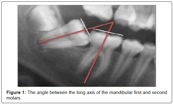
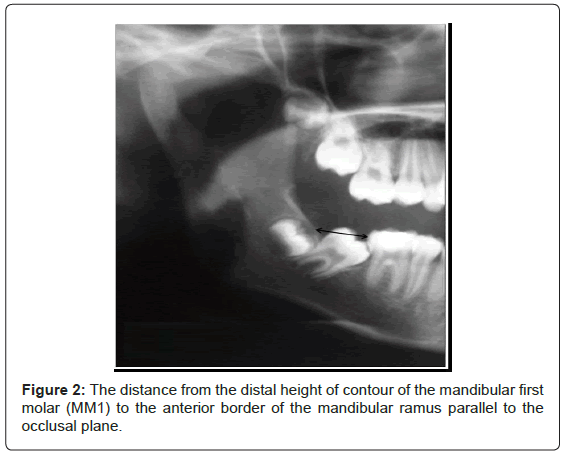
The material for this study consisted of 3,000 consecutive panoramic radiographs of patients referred for orthodontic treatment at Tel Aviv University School of Dental Medicine and 500 from the private office of one of the authors (YS). The patient’s age was 11-15 years (mean age 12.48 years). The panoramic radiographs were analyzed for the presence, position and eruption path of the mandibular permanent second molars. The angle between the long axis of the mandibular first and second molars (Figure 1) presenting the inclination of the impacted second molars was measured using a cephalometric protractor, and the distance from the distal height of contour of the mandibular first molar (MM1) to the anterior border of the mandibular ramus parallel to the occlusal plane was measured with digital ruler (Figure 2). All measurements were taken twice by the same examiner (TF), and the average of these measurements was calculated.
For the purpose of this study, impaction of the mandibular second molar was defined as its failure to erupt to the occlusal level beyond the normal time of its eruption. The inclusion criteria were: 1) Full eruption of MM2 on one side while the contralateral MM2 had not emerged even though three quarters of one root was developed. 2) Abnormal mesial inclination of the impacted MM2 with its crown stuck under the distal surface contour of the first molar. These patients were included in an earlier study on mandibular second molar impaction [20].
Results
Forty-nine patients, 23 (47%) males and 26 (53%) females were found with a total of 62 impacted mandibular second molars, presenting a prevalence rate of 1.4%. 36 (58%) were unilateral and 26 (42%) were bilateral. Among the 36 patients with unilateral impaction, 20 (55.5%) were on the right side and 16 (44.5%) on the left side. In 43 (88%) of the patients, the impacted second molars were mesially angulated, in only 2 they were distally inclined and vertical impactions were present in 4 patients. The angle of mesially impacted mandibular second molars ranged between 30 to 70 degrees. The distance from the MM1distal height of contour to the anterior rim of the ramus was 13.04 + -2.7 mm and 18.28 + -3.86 mm (p<0.001) in the impacted and non- impacted sides, respectively.
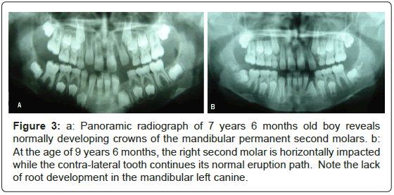
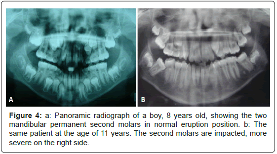
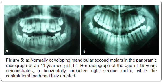
Initial panoramic radiographs of four patients presented normally developing mandibular second molars, while follow up panoramic radiographs taken 3-4 years later showed a change in their eruption path, resulting in mesial angulation and impaction (Figures 3-5).
Figure 3: a: Panoramic radiograph of 7 years 6 months old boy reveals normally developing crowns of the mandibular permanent second molars. b: At the age of 9 years 6 months, the right second molar is horizontally impacted while the contra-lateral tooth continues its normal eruption path. Note the lack of root development in the mandibular left canine.
Discussion
Crowding and arch length-deficiency in the posterior region of the mandibular arch were suggested as the main local factors causing mandibular second molar impaction [4,6]. It is most likely that the major causative factors for second molar impaction are the smaller available space for the second molar on the impacted side, namely the distance between the mandibular first molar and the anterior border of the ascending ramus. In addition, the developing mesial root, previously found to be shorter than the distal root, is probably causing a long axis rotation and mesial angulation of the second molar [20].
Mandibular second molar impaction is usually detected in a routine panoramic radiograph taken for pedodontic and orthodontic diagnosis and treatment planning and is rarely the main reason for referral to the orthodontist.
It has been speculated that the second molars have a constant path of eruption until they contact the adjacent teeth (typically the first molar) and then they erupt. Evidently as shown in the present report, a normally developing lower second molar may change its path of eruption and consequently its angulation, for unknown reasons, and become impacted on one side of the dental arch, while on the contralateral side, the tooth erupts normally.
This phenomenon has been illustrated in the following panoramic radiographs, taken during the early mixed dentition for diagnostic purposes and were followed up to here.
The panoramic radiograph of 7 years 6 months old boy reveals seemingly normally developing crowns of the mandibular permanent second molars that are expected to follow a normal path of eruption into the dental arch (Figure 3a). A follow up radiograph of the same patient at the age of 9- years 6- months showed the mandibular right second molar horizontally impacted while the left one continued its normal development. Interestingly, the roots of the impacted right second molar appear to be more developed compared with its contralateral tooth and its mesial root length is somewhat shorter than the distal one (Figure 3b). It has been suggested that this differential growth of the roots may contribute to a mesial inclination and impaction [14].
The panoramic radiograph of another boy, age 8 years, clearly demonstrated the two mandibular permanent second molars in normal developmental positions (Figure 4a). They did not cause a suspicion of future pathology and were predicted to erupt uneventfully into the dental arch. However, a follow up panoramic radiograph of the same patient at the age of 11 years presented an eruption aberration of both second molars, more severe on the right side (Figure 4b).
Normally developing mandibular second molars were detected in the panoramic radiograph of an 11-year-old girl referred for orthodontic consultation (Figure 5a). However, her panoramic radiograph taken five years later, at the age of 16 years, clearly demonstrated a change in the eruption path and consequently in inclination of the right second molar, which became horizontally impacted. The contra-lateral tooth had uneventfully and fully erupted into the dental arch (Figure 5b).
A normally developing mandibular second molar has previously been reported in an 8-year old boy with sufficient space for its eruption into the dental arch. This tooth has changed its eruption path and consequently angulation within 3.5 years interval and become horizontally impacted for unknown reasons [20].
All the above cases represent initial normally developing mandibular second molars with sufficient eruptive space, expected to erupt into the dental arch uneventfully. Unexpectedly and for unknown reason, some of these may change their path of eruption, tip mesially and become impacted.
A possible explanation for this phenomenon is the “guidance” theory, first suggested for the maxillary canine, which requires the guidance of the lateral incisor root for its normal eruption path [21]. Similarly, it could well be that the lower second molar requires the guidance of the first molar root during the course of its eruption. However, we could not conclude that lack of contact between the mandibular second molar and first molar is the reason for impaction of the former. Moreover, unlike reports suggesting a close association between arch length deficiency and second molar impaction [6], excess space between the developing second molar and the first molar root may allow a more mesial inclination of the second molars, resulting in their impaction under the distal bulge of the first molar crown [20]. Availability of space between the mandibular first and second molars in the early stages of development is not an insurance that the second molar will normally erupt into the dental arch.
Ethnic Chinese population present larger tooth size compared with whites may partially explain the higher prevalence of MM2 impaction [12,22]. In a study on genetic traits in MM2 impaction, Chinese-American children had a higher prevalence (2.3%) of MM2 impaction compared with white Israeli population (1.4%) [14]. Similar results (1.36%) to the white Israeli population were reported in young Caucasian Italian orthodontic patients [15].
No gender differences were found in our study with almost equal number of males [5] and females presenting MM2 impaction. This is contrary to a previous report where boys had more eruption disturbances of MM2's than girls [6].
The developmental mesial angulation of the second molars might be an indication for increased risk of possible future impaction. Occasionally, considerable variation in the degree of their angulation from one patient to another and even between the right and left side of the same patient are detected. Most of the impacted MM2's in our sample of orthodontic population (88%) were mesially angulated ranging from 30 to 70 degrees (mean 47 degrees), quite similar to the angles between 31 and 60 degrees reported for the general population of Chinese in Taiwan [10], but higher than the 15 to 65 degrees angulation reported for white English people [23].
MM2 angulation of 24 degrees or larger were reported to have a high risk of impaction [23]. Observations made from this study do not support an existence of a “threshold” mesial angulation value that predictably leads toward impaction of molars. Such critical or threshold values have been suggested in predicting impactions of the maxillary canines and of the second premolars.
The developing second molars continually change their angular positions and undergo pre-eruptive rotational movements taking place when the incompletely developed second molar comes into contact with the first molar. This rotational movement is important, because if it fails to occur, it may end up in mesio-angular impaction.
The second molar may occasionally, but not always, demonstrate a spontaneous correction of the aberrated path of eruption. That is, upright itself provided that the third molar bud is not developing on top of, or pressing against the erupting second molar. On the other hand, the second molar may further incline mesially, resulting in oblique or even horizontal impaction.
It is not yet clear why normally erupting second molars suddenly change their angulation and become impacted. In an uncrowded mandibular dental arch with well aligned teeth, it is expected that the second molars will erupt uneventfully. However, based on our observations, the eruption path of some of the mandibular second molars is aberrated, particularly those with a clinically significant increased pre-eruptive mesial angulation [5].
Mandibular third molar is the most frequently impacted tooth in the human dentition and is a major problem in the dental practice. Several studies have been conducted to possibly predict whether or not a developing third molar will erupt or become impacted [24]. Suggested risks for their impaction were increased mesial angulation during early and late adolescence, insufficient space for the developing third molar between the second molar and the mandibular ramus, and their complex root development. It was concluded that accurate prediction from radiographic measurements is not possible [24]. However, an equation was suggested to predict the odds of mandibular third molar impaction [25], and a model for rapid prediction of the potential of a third molar eruption or impaction was also proposed [26,27].
The causes of second molar impaction and aberration of their eruption path have not been studied extensively. The rationale behind atypical angular changes of the developing and erupting second molar should be explained by additional studies.
A new study has been recently undertaken by our group to develop an equation for predicting odds of mandibular second molar impaction considering risk factors such as the degree of its mesial angulation, differential root development and space available between the first molar and the anterior border of the ramus.
Diagnosis and treatment planning protocols must determine not only the presence, but also the position of the mandibular second molar. The pediatric dentists who first examine and treat the young children in their practice should be aware of the aberration phenomenon of the mandibular second molar and follow up its path of eruption. Early diagnosis and identification of this problem is based only on radiographs.
No orthodontic treatment is considered successfully completed until these teeth are fully erupted and in functional occlusion. Therefore, early detection of an arrested or failed eruption of a mandibular second molar is imperative, because early corrective measures may eliminate its potential impaction and reduce the need for complicated therapy.
Conclusion
A prevalence of 1.4% was found for mandibular second molar impaction in orthodontically treated patients with majority of them mesially angulated. The aberration in the path of eruption of the impacted mandibular second molars from sequential radiographic examination from several cases has been presented. However, the factors causing a change in their angulation that could lead to impaction are still obscure and require additional studies.
Acknowledgements
We thank Mr. Amir Shapira for his valuable assistance in the preparation of this manuscript.
References
- Dachi SF, Howell FV (1961) A survey of 3, 874 routine full-month radiographs. II. A study of impacted teeth. Oral Surg Oral Med Oral Pathol 14: 1165-1169.
- Kramer RM, Williams AC (1970) The incidence of impacted teeth. A survey at Harlem hospital. Oral Surg Oral Med Oral Pathol 29: 237-241.
- Aitasalo K, Lehtinen R, Oksala E (1972) An orthopantomographic study of prevalence of impacted teeth. Int J Oral Surg 1: 117-120.
- Johnsen DC (1977) Prevalence of delayed emergence of permanent teeth as a result of local factors. J Am Dent Assoc 94: 100-106.
- Evans R (1988) Incidence of lower second permanent molar impaction. Br J Orthod 15: 199-203.
- Varpio M, Wellfelt B (1988) Disturbed eruption of the lower second molar: clinical appearance, prevalence, and etiology. ASDC J Dent Child 55: 114-118.
- Wellfelt B, Varpio M (1988) Disturbed eruption of the permanent lower second molar: treatment and results. ASDC J Dent Child 55: 183-189.
- Vedtofte H, Andreasen JO, Kjaer I (1999) Arrested eruption of the permanent lower second molar. Eur J Orthod 21: 31-40.
- Grover PS, Lorton L (1985) The incidence of unerupted permanent teeth and related clinical cases. Oral Surg Oral Med Oral Pathol 59: 420-425.
- Fu PS, Wang JC, Wu YM, Huang TK, Chen WC, et al. (2012) Impacted mandibular second molars. Angle Orthod 82: 670-675.
- Davis PJ (1988) Findings from 1163 panelipse radiographs taken of 12-year-old children living in Hong Kong. Community Dent Health 5: 243-249.
- Cho SY, Ki Y, Chu V, Chan J (2008) Impaction of permanent mandibular second molars in ethnic Chinese school children. J Can Dent Assoc 74: 521.
- Shapira Y, Finkelstein T, Lai YH, Kuftinec MM, Vardimon A, et al. (2012) Prevalence and characteristic features of mandibular second molar impaction in Chinese-American school children. Acta Stomatol Croatica 46: 215-221.
- Shapira Y, Finkelstein T, Shpack N, Lai YH, Kuftinec MM, et al. (2011) Mandibular second molar impaction. Part I: Genetic traits and characteristics. Am J Orthod Dentofacial Orthop 140: 32-37.
- Cassetta F, Altieri F, Di Mambro A, Galluccio G, Barbato E (2013) Impaction of permanent mandibular second molar: a retrospective study. Med Oral Patol Oral Cir Bucal 18: e564-568.
- Buchner HJ (1973) Correction of impacted mandibular second molars. Angle Orthod 43: 30-33.
- Bjork A, Jensen E, Palling M (1956) Mandibular growth and third molar impaction. Acta Odont Scand14: 231-271.
- Ricketts RM (1972) A principle of arcial growth of the mandible. Angle Orthod 42: 368-386.
- Begg PR (1977) Begg Orthodontic Theory and Technique. (2nd edn) W.B.Saunders Co. Philadelphia, London, Toronto.
- Shapira Y, Borell G, Nahlieli O, Kuftinec MM (1998) Uprighting mesially impacted mandibular permanent second molars. Angle Orthod 68: 173-178.
- Becker A, Zilberman Y, Tsur B (1984) Root length of lateral incisors adjacent to palatally-displaced maxillary cuspids. Angle Orthod 54: 218-225.
- Ling JY, Wong RW (2006) Tanaka-Johnston mixed dentition analysis for southern Chinese in Hong Kong. Angle Orthod 76: 632-636.
- Sonis A, Ackerman M (2011) E-space preservation. Angle Orthod 81: 1045-1049.
- Richardson ME (1977) The etiology and prediction of mandibular third molar impaction. Angle Orthod 47: 165-172.
- Behbehani F, Artun J, Thalib L (2006) Prediction of mandibular third-molar impaction in adolescent orthodontic patients. Am J Orthod Dentofacial Orthop 130: 47-55.
- Ventä I (1993) Predictive model for impaction of lower third molars. Oral Surg Oral Med Oral Pathol 76: 699-703.
- Ventä I, Schou S (2001) Accuracy of the Third Molar Eruption Predictor in predicting eruption. Oral Surg Oral Med Oral Pathol Oral Radiol Endod 91: 638-642.
Relevant Topics
- Cementogenesis
- Coronal Fractures
- Dental Debonding
- Dental Fear
- Dental Implant
- Dental Malocclusion
- Dental Pulp Capping
- Dental Radiography
- Dental Science
- Dental Surgery
- Dental Trauma
- Dentistry
- Emergency Dental Care
- Forensic Dentistry
- Laser Dentistry
- Leukoplakia
- Occlusion
- Oral Cancer
- Oral Precancer
- Osseointegration
- Pulpotomy
- Tooth Replantation
Recommended Journals
Article Tools
Article Usage
- Total views: 16707
- [From(publication date):
December-2013 - Jul 13, 2025] - Breakdown by view type
- HTML page views : 12009
- PDF downloads : 4698

