A Unique Case of Multiple Ossifying Fibromas in Sublingual Space
Received: 03-Feb-2020 / Accepted Date: 18-Feb-2020 / Published Date: 25-Feb-2020 DOI: 10.4172/2167-7964.1000312
Abstract
Purpose: Ossifying fibroma is a rare benign tumor of the mandible and maxilla most commonly seen in females in 4th decade of life. Extra-osseous ossifying fibroma is a rare entity described in the literature. We present this rare case of extra-osseous ossifying fibroma in the sublingual space to highlight the importance of collaborative approach to the correct diagnosis.
Case report: We describe a case of 46 year old female that presented with the complaints of painless swelling over the floor of the mouth since 1 year. Computed Tomography showed the multiple high density lesions in the sublingual space attached to the left medial aspect of the mandible at the myelohyoid line with no obvious extension or soft tissue component. Following this, the patient was posted and planned for total surgical excision under general anesthesia which showed that the mass lesion was in the left sublingual space without mandibular involvement. Diagnosis was confirmed based on the histopathological report which showed it to be an ossifying fibroma.
Conclusions: Ossifying fibroma is a rare bony tumor of the mandible with different morphological features that are often mistaken for other benign Osseo fibrous lesions. Hence, a multidisciplinary approach including clinical, radiological and an accurate histopathological report post-operatively are mandatory for correct diagnosis and appropriate management.
Keywords
Fibro-osseous; Tumor; Ossifying fibroma; Extra-osseous; Sublingual
Introduction
Ossifying fibroma is a rare benign tumor of mesodermal origin arising from the mandible and maxilla. One of the study showed that ossifying fibroma was seen arising from the parapharyngeal and masticator space [1]. Another study showed similar mass lesion in the nasopharynx [2]. However, we didn’t find literature in our research that shows an ossifying fibroma arising in the sublingual space. It most commonly seen in females between 2nd and 4th decade of their life. Different studies report high variability in incidence rate with one such literature representing 0 to 5.5% of all tumors [3]. Recently, mutation in a tumor suppressor gene HRPT2 a protein product known as para fibronin is thought to tumor formation [4].
It behaves like a benign bony neoplasm, though it has been categorized under fibro-osseous lesions including the orofacial region. On histopathological examination, ossifying fibroma is characterized by variable amounts of inactive looking epithelium embedded in mature fibrous connective tissue stroma [5].
Therefore, the present study is aimed to describe a rare and unique case of multiple ossifying fibroma arising in the sublingual space and discuss the clinical, radiological and histopathological features of a rare variant.
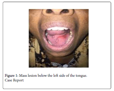
A 46 year female, with no past significant medical or familial records, presented to the department of otorhinolaryngology and was referred to Department of Radiology in Dr. D Y Patil Medical college, Pune for further evaluation. The patient came with chief complaint of swelling over the floor of the mouth since 1 year (Figure 1). It gradually increased in size and was associated with difficulty in swallowing and articulation. No history of associated pain, trauma, weight loss, oral surgery or tooth extraction. On clinical examination, the lesion appeared as a well-defined rounded swelling arising from the floor of the mouth, pushing tongue upwards and towards the right. On palpation, it was bony hard in consistency with a smooth surface. It was non compressible, non-tender and immobile. No associated palpable lymph nodes noted in the neck.
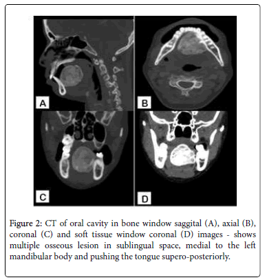
USG neck was inconclusive as the lesion showed dense posterior acoustic shadowing, thus could not be well assessed. On Multi-detector Computed tomography of the neck showed multiple well defined lobulated bone density lesions in the oral cavity at the floor of the mouth in left sublingual space, measuring approximately 4 (AP) x 3.6 (CC) x 3.3 (TR) cm. Lesion appeared to be attached to the left medial aspect of mandible at the mylohyoid line (Figure 2). Based on the imaging findings, the differentials given were a giant peripheral ossifying fibroma vs. an ossifying soft tissue lesion.
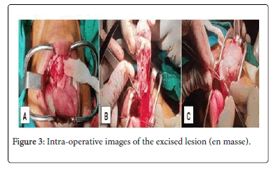
The patient was planned for total surgical excision and was performed under general anaesthesia. On surgery, no mandibular involvement was seen and the lesion was removed en mass (Figure 3).
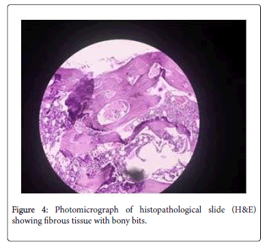
On histopathology, Fibrous tissue with bony bits. Foci of oval to spindle cells with intervening fibrous tissue and calcification with areas of ossification noted (Figure 4). The diagnosis of ossifying fibroma was hence established. The patient underwent routine follow up for 1 year with no signs of recurrence.
Discussion
Montgomery coined the term “ossifying fibroma” in 1927 [6]. Central ossifying fibroma is more common in females than in males with different studies showing different results with female to male ratio ranging from 2.8:1 to 5:1 [7]. Mostly seen in mandible 70-90% (pre molar and molar region) followed by maxilla, ethmoid and orbital regions. Its etiology is unknown, but it is hypothesized that, developmental or traumatic cause originating from periodontal ligament because of their capacity to produce cementum and osteoid material [8]. Few studies have shown chromosomal abnormalities such as translocation and deletion of codings in chromosome 2 [9,10]. Ossifying fibroma is often a well-defined lesion with moderate to high density, bordered by peripheral calcified shell. Clinically, it is characterized by an asymptomatic slow growing tumor with cortical expansion [11].
An important diagnostic feature of ossifying fibroma is the centrifugal growth pattern rather than linear and the tumor grows by expansion equally in all directions and present as round mass.
Differential diagnosis of ossifying fibroma should be made considering benign tumor lesions like calcifying epithelial tumor, calcifying cystic tumor, complex odontoma, fibrous dysplasia, dentinogenic ghost cell tumor and adenomatoid tumor.
Radiographic feature of ossifying fibroma is highly variable, depending on its maturity. In the initial stages, the tumor appears as a well-defined radiolucent lesion without any evidence of internal radioopacities, whereas the lesion appears extremely radio-opaque mass in the mature stages as is seen in our case. On MRI, the osseous or calcified matrix of such lesions is less distinctly demonstrated. Signal intensity and contrast uptake are variable although T2 hyperintense signal for ossifying fibroma have been known as opposed to hypointensity exhibited by fibrous dysplasia.
On histogenesis, the ossifying process is of two origins: the more commoner excessive proliferation of periodontal ligaments and a nonperiodontal origin metaplastic process occurring in the connective tissue fibers [12]. Ossifying fibromas are variable on histopathological examination, with the hard portion exhibiting trabeculae of osteoid or bone and poorly cellular or acellular spherules of calcified material, resembling cementum. The lack of consistent osteoblastic rim of the bone trabeculae in fibrous dysplasia is used to distinguish it from an ossifying fibroma, which is rimmed by plump osteoblasts [13].
According to few studies, the immunohistochemical analysis of the epithelial component of the reported OF demonstrates positivity for CK14 and CK19, and negativity for CK7, CK8, and CK18 whereas that of the human enamel organ and the remnants of the dental lamina, showed that these epithelial cells are positive for CKs 7, 13, 14, and 19, and proved negative for CKs 8, 10, 16, 17, and 18 [3,14].
The well-defined, encapsulated, circumscribed nature of the ossifying fibroma permits en bloc removal of the tumor. This tumor is thought to be resistant to radiotherapy, hence contraindicated. They have locally aggressive nature. Recurrence rates of these aggressive forms is about 30% to 38% [15,16]. Hence, complete removal being the gold standard treatment and routine follow up is necessary after the tumor resection. Prognosis is good without any metastasis.
Conclusion
Ossifying fibromas are entities with different morphological features that can be mistaken for other benign osseofibrous lesions. This similarity and over lapping micro characteristics with other similar lesions make pre-operative diagnosis a challenge. Multidisciplinary approach, comprehending clinical, radiological and pathological aspects and an accurate histopathological report post operatively are mandatory for the correct diagnosis and appropriate treatment.
References
- Jung SL, Choi KH, Park YH, Song HC, Kwon MS (1999) Cemento-Ossifying Fibroma Presenting as a Mass of the Parapharyngeal and Masticator Space. AJNR Am J Neuroradiol 20: 1744-1746.
- Cakir B, Karadayi N (1991) Ossifying fibroma in the nasopharynx: A case report. Clin Imag 15: 290-292.
- Taylor MA, Mata MG, Bregni CR, Vargas PA, Toral-Rizo V, et al. (2011) Central ï¬broma: new ï¬ndings and report of a multicentric collaborative study. Oral Surg Oral Med Oral Pathol Oral Radiol Endod 112: 349-358.
- Pimenta FJ, Silveira GLF, Tavares GC, Silva AC, Perdigao PF, et al. (2006) HRPT2 gene alterations in ossifying fibroma of the jaws. Oral Oncol 42: 735-739.
- Philipsen HP, Reichart PA, Sciubba JJ, van der Waal I (2005) ï¬broma In: Barnes L, Eveson JW, Reichart P, Sidransky D (editors). World Health Organization Classiï¬cation of Tumours. Pathology and Genetics of Head and Neck Tumours. Lyon: IARC Publishing Group.
- Binatli O, ErÅŸahin Y, CoÅŸkun S, Bayol U (1995) Ossifying fibroma of the occipital bone. Clinical Neurology and Neurosurgery pp: 47-49.
- Philipsen HP, Reichart PA, Sciubba JJ, van der Waal I (2005) Fibroma. In: Barnes L, Eveson JW, Reichart P, Sidransky D (editors). World Health Organization Classiï¬cation of Tumours. Pathology and Genetics of Head and Neck Tumours. Lyon: IARC Publishing Group.
- Carvalho B, Pontes M, Garcia H, Linhares P, Vaz R (2011) Ossifying Fibromas of the Craniofacial Skeleton. In: Poblet E, editor. Histopathology - Reviews and Recent Advances. In Tech.
- Neville BW, Damm DD, Allen CM, Bouquot JE (2009) Bone pathology. In: Chi AC, editor. Oral and Maxillofacial Pathology. (3rdedn). Noida: Elsevier.
- Jayachandran S, Sachdeva S (2010) Cemento-ossifying fibroma of mandible. Report of two cases. J Indian Acad Oral Med Radiol 22: 53-56.
- Daniels JS (2004) Central ï¬broma of mandible: a case report and review of the literature. Oral Surg Oral Med Oral Pathol Oral Radiol Endod 98: 295-300.
- Ono A, Tsukamoto G, Nagatsuka H (2007) An immunohistochemical evaluation of BMP-2, -4, osteopontin, osteocalcin and PCNA between ossifying fibromas of the jaws and peripheral cemento-ossifying fibromas on the gingiva. Oral Oncology 339-344.
- Chong VFH, Tan LHC (1997) Maxillary sinus ossifying fibroma. American Journal of Otolaryngology 419-424.
- Domingues MG, Jaeger MMM, Araújo VC, Araújo NS (2000) Expression of cytokeratins in human enamel organ. Eur J Oral Sci 108: 43-47.
- Gunaseelan R, Anantanarayanan P, Ravindramohan E, Ranganathan K (2007) Large cemento-ossifying fibroma of the maxilla causing proptosis: a case report. Oral Surgery, Oral Medicine, Oral Pathology, Oral Radiology and Endodontology pp: e21-e25.
- Bernier J (1952) Changes in bone and dental cementum in periodontal diseases. Med Hyg (Geneve) 10: 474.
Citation: Patel C, Walasangikar V, Gupta A, Kuber R (2020) A Unique Case of Multiple Ossifying Fibromas in Sublingual Space. OMICS J Radiol Vol. 9 No. 1:312. DOI: 10.4172/2167-7964.1000312
Copyright: © 2020 Patel C, et al. This is an open-access article distributed under the terms of the Creative Commons Attribution License, which permits unrestricted use, distribution, and reproduction in any medium, provided the original author and source are credited.
Share This Article
Open Access Journals
Article Tools
Article Usage
- Total views: 2569
- [From(publication date): 0-2020 - Apr 20, 2025]
- Breakdown by view type
- HTML page views: 1862
- PDF downloads: 707


