A Unique Case of Mastoid Osteoma - A Case Report
Received: 30-Aug-2022 / Manuscript No. ocr-22-74293 / Editor assigned: 02-Sep-2022 / PreQC No. ocr-22-74293 / Reviewed: 16-Sep-2022 / QC No. ocr-22-74293 / Revised: 23-Sep-2022 / Manuscript No. ocr-22-74293 / Published Date: 30-Sep-2022 DOI: 10.4172/2161-119X.1000483
Abstract
Osteoma is a benign tumour of Mesenchymal osteoplastic nature composed of well-differentiated osseous tissue with laminar structure. Temporal bone Osteomas in general constitute 0.1 to 1 % of all benign tumours of the skull.Clinically these tumours are asymptomatic, except for cosmetic deformities, and they are usually casual radiological findings. This case report presents a 29-year-old female patient presenting with a swelling of approximately 2x2 cm over the left post auricular region since 2 years associated with pain over the swelling since 6 months.
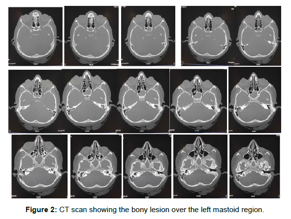
X-RAY of bilateral temporal bone was done which showed a well-defined, round to oval dense radio opacity in the left temporal region. Computed tomography was done which was reported as bony attenuation lesion along outer cortex of mastoid, part of temporal bone on the left side in the post auricular region (mastoid Osteoma). Surgical excision was done and the sample was sent for histopathological examination which confirmed the diagnosis of an Osteoma. Postoperatively, the patient was asymptomatic. On a regular follow up, that is 1 week, 1 month and 2 months there was no recurrence noted. Patient had a good cosmetic cover with no complications, recurrence or pain.
Keywords
Mastoid bone; Osteoma; Temporal bone; Post auricular incision; Benign lesions
Introduction
Osteoma is a benign tumour of Mesenchymal osteoplastic nature composed of well-differentiated osseous tissue with laminar structure. Temporal bone Osteomas in general constitute 0.1 to 1% of all benign tumours of the skull. In the temporal bone, the external auditory canal is the predominant location, rarely present in the mastoid, the squamous portion of the temporal bone, inner ear canal and middle ear. The Osteomas of the mastoids are slow growing benign tumours, made predominantly of mature bone. Clinically these tumours are asymptomatic, except for cosmetic deformities, and they are usually casual radiological findings. Surgical treatment is indicated for symptomatic Osteomas [1].
Clinically, it is difficult to classify the type of Osteoma because of its similar presentation. Mastoid Osteomas are of three types, namely Osteoma compactum, Osteoma canceller and Osteoma cartilaginous based on the histology. Non syndromic temporal bone Osteomas are not known and various possible causes have been reported which includes genetic origin, trauma, surgery, radiotherapy, chronic infections and pituitary dysfunctions [2].
Mastoid Osteomas are rare, benign tumours. If their growth significantly occludes the meatus, they may cause cosmetic deformities, conductive hearing loss, and recurrent external ear infections. Several other osseous lesions of the temporal bone should be considered in the differential diagnosis. Differential diagnosis includes osteosarcoma, osteoblastic metastasis, isolated eosinophilic granuloma, Paget’s disease, gaint cell tumour, osteoid Osteoma, calcified meningioma and monostotic fibrous dysplasia. The etiology of mastoid Osteomas is poorly understood. Non contrast CT scan is the imaging method of choice and surgical resection is the treatment of choice [3].
Case Report
Patient Information
A 29 year-old female reported to our OPD with a swelling behind left ear since 2 years. The patients consulted for the cosmetic deformity induced by the lesion. It was initially painless and she gradually developed pain over the swelling since the past 6 months. The swelling was initially small in size which gradually and progressively increased to the present size since the past 1 year with no aggravating or relieving factors. It was not associated with displacement of the left pinna. There was no history of trauma, headache, hearing impairment, otorrhea, dizziness, vomiting, facial weakness and neurological deficit [4].
Clinical findings
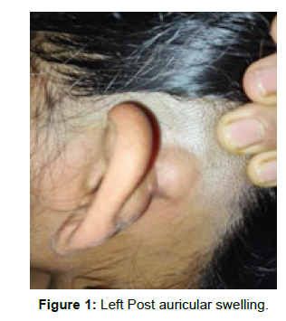
On examination, well defined smooth, bony hard, non-tender, non- pulsatile, non-reducible, non-compressible, swelling measuring 2x2 cm was present behind the left pinna not occluding the retro auricular groove and the swelling was fixed to underlying bone. Skin over swelling was free having normal local temperature (Figure 1). EAC showed no post-aural bulging. Tympanic membrane of left ear was intact. Right ear was normal. Audiometric evaluation showed normal hearing [5].
Timeline
The patient was followed up for a period of 3 months; i.e. 1 week, 1 month and 2 months post operatively.
Diagnostic Assessment
The diagnosis was based on the clinical presentation and noncontrast CT. X-RAY of bilateral temporal bone was done which showed a well-defined, round to oval dense radio-opacity in the left temporal region. Computed tomography was done which was reported as bony attenuation lesion along outer cortex of mastoid, part of temporal bone on the left side in the post auricular region (mastoid Osteoma) [6]. FNAC was not possible due to the hardness of the swelling (Figure 2).
Therapeutic Intervention
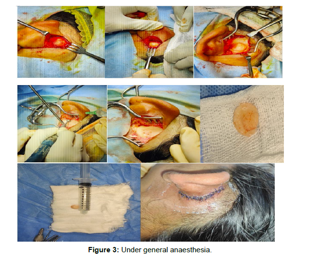
The indication for surgery in this case was the patient's sensation of a mass effect and pain over the swelling. The tumour was removed in its entirety via a post auricular approach. Patient posted for surgical excision of tumour (Figure 3) under general anaesthesia. Local infiltration was given using lignocaine and adrenaline over the left post auricular region. Post-auricular William Wilde’s incision was placed and dissection done in layers, exposing Osteoma.
Mastoid bone identified. Excision was done by drilling out a groove at base of Osteoma over the surface of mastoid cortex under continuous irrigation. Intact, pedunculated Osteoma was removed with chisel and hammer. After excision, the contour of mastoid cortex was found intact without any evidence of bony invasion. Excessive skin was removed and suturing was done in layers using 4-0 vicryl. Excised mass sent for histopathological examination [7].
The report mentioned the lesion showed dual population of mature lamellar and woven bone in trabecular pattern with haversian- like canals with intervening spaces filled with loose fibrous stroma. These features are consistent with the diagnosis of mastoid Osteoma. Patient was followed up after 7 days post operatively. Wound was healthy. Patient’s follow up was done for 3 months. There was no evidence of recurrence.
Discussion
Osteoma in head and neck region is a very rare entity. Incidence is generally 0.1-1% of all. Benign tumours of the skull, Temporal bone Osteomas are rare. They can be seen in all parts of the temporal bone like external auditory canal, mastoid and squamous portion, middle ear, Eustachian tube, petrous apex, internal auditory canal, zygomatic process, glenoid fossa and styloid process, External auditory canal being the most common site followed by mastoid region. Rarely the petrous part of the temporal bone may be involved along with the facial nerve.
The etiopathogenesis is controversial. Several factors have been proposed including chronic infection, trauma, hereditary, surgery, radiotherapy and glandular dysfunction (pituitary gland). Varboncoevr et al (8) mentioned that Osteomas origin from embryologic cartilaginous rest or persistent embryologic periosteum. Kaplan et al (10) proposed that the development of Osteoma might be due to trauma and muscle traction [8].
Osteoma of the temporal bone seems to arise from pre-osseous connective tissue from suture line which relatively has thick subcutaneous layers with rich blood supply. As the Osteomas are slow growing, they are asymptomatic and stable for many years. They present as a smooth surface with characteristic hard, bony consistency on palpation. Osteomas in the mastoid region are solitary and grow out from surface producing external swelling. Osteomas have been classified in many ways by their pattern of growth into outgrowing or ingrowing, unilateral or bilateral and on histopathological types; they are made up of discrete fibro vascular channels surrounded by lamellar bone.
a) OSTOMA COMPACTUM- It is the common type, hard and attached to cortex of mastoid process. Histologically it is composed of dense lamellate bone tissue and is traversed by few vessels. They have wide base and are very slow growing.
b) OSTEOMA CANCELLARE- It consists of fibrous cellular tissue and cancellous bone.
c) OSTEOMA CARTILAGENEUM- Rare, consisting of bone and cartilage.
d) OSTEOMA MIXTUM- It consists of mixture of types of bone found in Osteoma compactum and Osteoma canceller [9].
Extra canalicular Osteomas of temporal bone are primarily composed of mature bone and they have predominance in young females. Osteoma occurrence can be divided into syndromic and nonsyndromic. Etiology of non syndromic temporal bone Osteomas are not known and various possible causes have been reported. Osteomas of the external auditory canal has an estimated incidence of 0.5% of the total ear surgeries and they manifest as solitary, unilateral and pedunculated bony mass of unknown origin.
Osteomas of middle ear are extremely rare and when they do, they arise from promontory and results in progressive conductive hearing loss usually due to the involvement of the ossicular chain. Other sites involved are pyramidal eminence, hypo tympanum and lateral semicircular canal. Osteomas of internal auditory canal exhibit as a wide variety of symptoms including asymmetrical sensor neural hearing loss, vertigo, tinnitus, facial nerve palsy and vestibular dysfunction. Differential diagnosis may include osteosarcoma, osteoblastic metastasis, isolated eosinophilia granuloma, Paget’s disease, gaint cell tumour, osteoid Osteoma, calcified meningioma. Non contrast CT is the imaging method of choice. On imaging ivory Osteoma appears radio dense; similar to normal cortex, whereas mature stoma may demonstrate central marrow. It is seen as a high opacity, well demarcated and dense growth of sclerotic lesion.
In case of extra canalicular Osteoma, surgery is indicated if it is causing cosmetic disfigurement and reduced hearing. Surgery is the treatment of choice for external auditory canal Osteoma if it obstructs the canal or associated with external auditory canal cholesteatoma, for middle ear Osteoma if it is causing hearing loss and for symptomatic internal auditory canal Osteoma [10].
Conclusion
Mastoid Osteomas of temporal bone are rare benign slow growing asymptomatic tumours. They are usually asymptomatic unless it gradually increases in size to cause cosmetic disfigurement and sometimes pain. Radiological investigations are required to find out the extent with non-contrast CT as the modality of choice.
Complete surgical excision is the treatment of choice with drilling of the mastoid cortex if necessary to prevent recurrences.
References
- Carlos UP, de Carvalho RWF, de Almeida AMG, Rafaela ND (2009) Mastoid Osteoma. Int Arch Otorhinolaryngol 13: 350-353.
- El Fakiri M, El Bakkouri W, Halimi C, Aı¨t Mansour A, Ayache D et al (2011) Mastoid Osteoma. Eur Ann Otorhinolaryngol Head Neck Dis 128: 266-268.
- D’Ottavi LR, Piccirillo E, de Sanctis S, Cerqua N (1997) Mastoid Osteomas. Acta Otorhinolaryngol Ital 17: 136-139.
- Domı'nguez Pe'rez AD, Rodrı'guez Romero R, Domı´nguez Dura´n E, Riquelme Montan OP, Alca´ ntara Bernal R, et al. (2011) The mastoid Osteoma an incidental feature. Acta Otorrinolaringol Esp 62: 140-143.
- Denia A, Perez F, Canalis RR, Graham MD (1979) Extracanalicular Osteomas of the temporal bone. Arch Otolaryngol 105: 706-709.
- Ulku ÇH, Yucel A. (2013) Osteoma of the mastoid region. Kulak Burun Boğaz Uygulamaları (Turkey) 1: 135-138.
- Abdel Tawab HM, Kumar VR, Tabook SM (2015) Osteoma presenting as a painless solitary mastoid swelling. Case Rep Otolaryngol 201515: 590-783.
- Van Dellen JR A (1977) mastoid Osteoma causing intracranial complications. S Afr Med J. 51:597-598.
- Clerico DM, Jahn AF, Fontanella S (1994) Osteoma of the internal auditory canal. Ann Otol Rhinol Laryngol 103: 9-23.
- Ahmadi MS, Ahmadi M, Dehghan A (2014) Osteoid Osteoma presenting as a painful solitary skull lesion. Iran J Otorhinolaryngol 26: 115-118.
Google Scholar, Crossref, Indexed at
Google Scholar, Crossref, Indexed at
Google Scholar, Crossref, Indexed at
Google Scholar, Crossref, Indexed at
Google Scholar, Crossref, Indexed at
Google Scholar, Crossref, Indexed at
Google Scholar, Crossref, Indexed at
Google Scholar, Crossref, Indexed at
Citation: Gowda C, Chethana CS, Mahesh SG, Tanthry D (2022) A Unique Case of Mastoid Osteoma – A Case Report. Otolaryngol (Sunnyvale) 12: 481. DOI: 10.4172/2161-119X.1000483
Copyright: © 2022 Gowda C. This is an open-access article distributed under the terms of the Creative Commons Attribution License, which permits unrestricted use, distribution, and reproduction in any medium, provided the original author and source are credited.
Share This Article
Recommended Journals
Open Access Journals
Article Tools
Article Usage
- Total views: 2407
- [From(publication date): 0-2022 - Apr 06, 2025]
- Breakdown by view type
- HTML page views: 2079
- PDF downloads: 328


