Case Report Open Access
A Rare Location of Pseudoaneurysm in Chronic Pancreatitis: Left Sub-Capsular Renal Artery
Nitin J*, Reddy DN and Singh JRAsian Institute of Gastroenterology, Hyderabad, India
- *Corresponding Author:
- Nitin J
Asian Institute of Gastroenterology, Hyderabad, India
Tel: +91 4023378888
Fax: +91 4023324255
E-mail: docnits13@gmail.com
Received date: April 08, 2016; Accepted date: October 24, 2016; Published date: October 31, 2016
Citation: Nitin J, Reddy DN, Singh JR (2016) A Rare Location of Pseudoaneurysm in Chronic Pancreatitis: Left Sub-Capsular Renal Artery. J Gastrointest Dig Syst 6:474. doi:10.4172/2161-069X.1000474
Copyright: © 2016 Nitin J, et al. This is an open-access article distributed under the terms of the Creative Commons Attribution License, which permits unrestricted use, distribution, and reproduction in any medium, provided the original author and source are credited.
Visit for more related articles at Journal of Gastrointestinal & Digestive System
Case Report
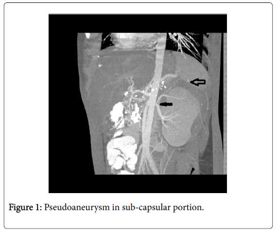
A 15 year old male with chronic pancreatitis was admitted with history of increasing abdominal pain, vomiting and fever since one month. His hemoglobin level was 8.9 (normal 13.0-17.0) g/l. Contrastenhanced computed tomography (CT) showed a large 10 × 6 cm subcapsular collection in left kidney with bleed within along with pseudo aneurysm in sub-capsular portion of left kidney from a proximal branch of left renal artery (Figure 1) with chronic calcific pancreatitis and intra ductal calculi.
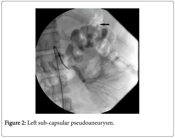
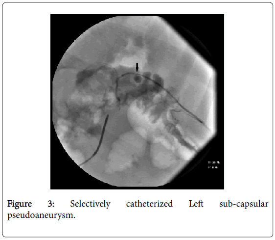
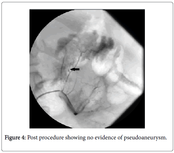
On conventional angiography, celiac angiogram was normal. Left renal artery arteriogram showed left sub-capsular pseudoaneurysm from a proximal branch of left renal artery (Figure 2), which was selectively catheterized (Figure 3) and coil embolization was done. Post procedure; check angiogram showed no evidence of pseudoaneurysm (Figure 4).
Conclusion
If abdominal pain and anemia develop in a patient with chronic pancreatitis, pseudoaneurysm should be considered. Pseudoaneurysmal bleeding may complicate 5% to 10% of all cases of chronic pancreatitis although pseudo aneurysms may be seen in up to 21% of patients with chronic pancreatitis undergoing angiography [1]. Severity and duration of pancreatitis, fluid collections and pancreatic surgery are the main risk factors. Splenic (60-65%), gastro-duodenal (20-25%) and pancreatico-duodenal (10-15%) arteries are most commonly affected.
Less frequently, involved are inferior phrenic, hepatic and superior mesenteric artery [2]. To best of our knowledge, this is first case reporting pseudoaneurysm of sub-capsular branch of left renal artery. Interventional radiological procedures have improved morbidity and mortality in ruptured pseudoaneurysms and reduced need of emergency surgery. Percutaneous angiographic embolization (coils or glue) is most commonly used [3].
References
- Balachandra S, Siriwardena AK (2005) Systematic appraisal of the management of the major vascular complications of pancreatitis. Am J Surg 190: 489-495.
- Nagar N, Dubale N, Jagadeesh R, Nag P, Reddy ND, et al. (2011) Unusual locations of pseudo aneurysms as a sequel of chronic pancreatitis. J Interv Gastroenterol 1: 28-32.
- Fankhauser GT, Stone WM, Naidu SG (2011) The minimally invasive management of visceral artery aneurysms and pseudoaneurysms. J Vasc Surg 53: 966-970.
Relevant Topics
- Constipation
- Digestive Enzymes
- Endoscopy
- Epigastric Pain
- Gall Bladder
- Gastric Cancer
- Gastrointestinal Bleeding
- Gastrointestinal Hormones
- Gastrointestinal Infections
- Gastrointestinal Inflammation
- Gastrointestinal Pathology
- Gastrointestinal Pharmacology
- Gastrointestinal Radiology
- Gastrointestinal Surgery
- Gastrointestinal Tuberculosis
- GIST Sarcoma
- Intestinal Blockage
- Pancreas
- Salivary Glands
- Stomach Bloating
- Stomach Cramps
- Stomach Disorders
- Stomach Ulcer
Recommended Journals
Article Tools
Article Usage
- Total views: 59502
- [From(publication date):
October-2016 - Apr 04, 2025] - Breakdown by view type
- HTML page views : 58679
- PDF downloads : 823


