A New Classification of Ovarian Granulosa Cell Tumor Based on Histopathology in Egyptian Cows and Buffaloes
Received: 15-Jan-2018 / Accepted Date: 23-Mar-2018 / Published Date: 27-Mar-2018
Abstract
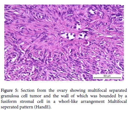
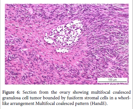
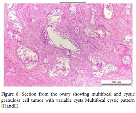
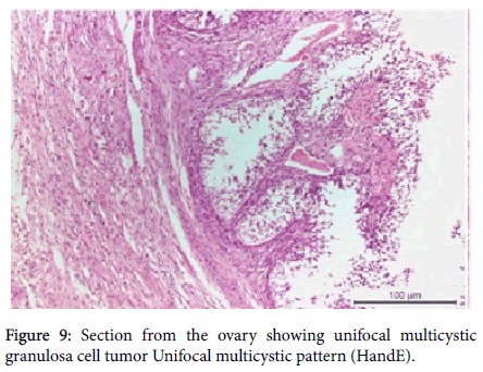
This study was carried out on 100 animals (79 cows and 21 buffaloes) to investigate the incidence and the growth patterns of granulosa cell tumors based on histopathological characteristics. Granulosa cell tumors were prevalent in 49 out of 73 cases having ovarian lesions. Unilateral localization was the most common. Different growth patterns evoked the idea of establishment of a new classification of bovine ovarian granulosa cell tumors. Ten patterns were recognized including 1- Single focal pattern, 2- Single focal pattern having sertoli-like cells, 3- Single focal pattern with theca-like cells around, 4- Multifocal separated pattern, 5- Multifocal coalesced pattern, 6- Multifocal and cystic pattern, 7- Unifocal multicystic pattern, 8- Multifocal with clusters pattern (rosettes), 9- Call Exner's cell formation, and 10- Adenomatous hyperplasia of rete ovary.
Keywords: Ovary; GCT; Cows; Buffaloes; Histopathology; Autoflourescence
Introduction
The ovary is a multi-function organ responsible for the differentiation, release of a mature oocyte, and for synthesizing and secreting hormones that essential for development of the fertilized ovum and maintenance of reproductive function [1]. Without the ovary; the animals will not reproduce. The ovarian pathologies include ovarian developmental anomalies which include: (ovarian hypoplasia and aplasia, freemartin, hermaphrodites, and white heifer disease), ovarian inflammatory conditions which include: (oophoritis and perioophoritis, ovarian abscess, ovaro-bursal adhesions and encapsulation, and ovarian sclerosis), ovarian functional disorders which include: (sub-estrum or silent heat, gestational estrus, persistent corpus luteum, ovulatory disturbances, ovarian cysts, and parovarian cysts), and finally ovarian neoplasms and associated conditions that include: (epithelial tumors, germ cell tumors, sex cord–stromal tumors «e.g. Granulosa Cell Tumors (GCTs), mesenchymal tumors, and hematoma) [2].
GCTs are considered the most common sex cord ovarian neoplasm in all domestic animals [3]. Its prevalence varies from 0.05% to 7.14% [2,4,5], as well as the incidence has been reported to be approximately 0.5%. Granulosa cell tumors affect all breeds of cattle but appear to occur more often in dairy cattle rather than beef cattle. These tumors have been diagnosed in every age of cow, ranging from virgin heifers to very old cows. Granulosa cell tumors have also been reported in pregnant cows, but the occurrence is rare [6,7]. This type of tumor was detected grossly and microscopically in native breeds of cows [8].
Implementation of a new classification of granulosa cell tumors (GCTs) is an important aid to understand the pathogenesis and different forms of these tumors, so the present study aimed to investigate the incidence and different growth patterns of ovarian granulosa cell tumors among native breeds of cows and buffaloes aged above 2 years based on histopathological characterization. As well as to detect autoflouresence ability of granulosa cells itself as a new record of some forms of GCTs in relation to normal granulosa cells of normal ovarian follicle.
Materials and Methods
100 slaughtered female cattle (79 cows and 21 buffaloes) were used in this study. The selected animals were vary in ages so they were allocated in two groups; the first group (11 animals) aged from 2-5 years while the second group (89 animals) aging above five years. Tissue specimens were taken from 100 paired ovaries of cows (Frisian and Balady) and buffaloes from different abattoirs (Beni-Suef, Beliffia, Nasr, and El-Basatin) of Beni-Suef and Cairo Provinces respectively (Table 1) during the period from September 2012 to March 2013.
| Species | Abattoir | Age | Total | ||
|---|---|---|---|---|---|
| 2-5 yrs | ˃5 yrs | ||||
| Cows | Frisian | Beni-Suef (1), Beliffia (12), Nasr (4) | 2 | 15 | 17 |
| Balady | Beliffia (49), Nasr (13) | 9 | 53 | 62 | |
| SellaffuB | El-Basatin (21) | 0 | 21 | 21 | |
| Total | 11 | 89 | 100 | ||
Table 1: Showing the animal species, breeds, abattoirs and the ages of selected animals in this study.
Both right and left ovaries were collected, examined grossly and photographed for recording the ovarian structures and /or any alterations, then cut longitudinally into two equal halves and fixed in 10% formalin solution immediately after collection for 48 hrs. The specimens then were processed by paraffin embedding method, sectioned 5-7 μ and stained with Hematoxylin and Eosin according to Bancroft and Gamble [9] for histopathological studies. Also use of Masson's trichrome and Periodic Acid Schiff's stains as specific stains.
Fluorescent microscopy was used to detect the autoflourescence ability of intimated GCTs in relation to granulosa cells of normal ovarian follicle. Stained sections routinely with hematoxylin and eosin (Haematoxylin: Fluka, AG, Switzerland, Buchs SG – Eosin Y: alcohol and water soluble, Winlap, UK), mounted in fluorescence- free D-P-X mountant (LOBA Chemie, India) [10]. The HandE stained ovarian tissue sections of both GCTs and normal graafian follicle were examined under fluorescence microscope (Euromex Oxion microscope Netherlands, 3 Watt LED for transmitted light, 85-240 V operation Reflected 100 W mercury-vapor light source for fluorescence, with power supply for 85-240 V operation). Fields were microphotographed optically using computer software.
Results
The Ovarian pathological alterations that were identified in this study after gross and histopathological examination of 100 right and left ovaries of both cows and buffaloes were seen in 73 cases from total 100 cases. While the rest 27 ovaries were cyclic normal ovaries not showing any pathological alterations.
The 73 cases exhibiting ovarian pathological alterations either present in single ovary or both ovaries were classified into four categories according to the type of this alteration as include 1- Granulosa cell tumor (by number of 49 cases from 73 cases) with 67.1% incidence 2- Ovarian cysts 21.9% (by number of 16 cases from 73 cases) 3- Single mixed case exhibiting both GCT and ovarian cyst, and final 4- Persistent corpus luteum 9.6% (by number of 9 cases from 73 cases). Table 2 showing the incidence of ovarian pathological alterations in cows (in Frisian and Balady cows) and buffaloes, also unilateral and bilateral localization of the lesion in the ovaries.
| Species Ovarian lesion (s) | Cows | Buffaloes | Total No. | ||
|---|---|---|---|---|---|
| Balady | Frisian | ||||
| Granulosa cell tumor (GCT) 67.1% | Unilateral | 33 (67.3%) | 4 (8.2%) | 9 (18.4%) | 49 |
| Bilateral | 3 (6.1%) | 0 | - | - | |
| Ovarian cysts 21.9% | Unilateral | 4 (25%) | 9 (56.25%) | 1 (6.25 %) | 16 |
| Bilateral | 2 (12.5 %) | 0 | - | - | |
| Mixed case of GCT and ovarian cyst 1.4% | 1 | 0 | 0 | 1 | |
| Persistent corpus luteum 9.6% | 7 | 0 | 0 | 7 | |
| Total No. | 50 | 13 | 10 | 73 | |
Table 2: Incidence of ovarian pathological alterations in cows (Frisian and Balady breeds) and buffaloes, also unilateral and bilateral localization of the lesion in the ovaries.
In this study, ovarian affections represented 73% from the collected cases from which granulosa cell tumors were seen in 67.1%.
The incidence of GCTs was 67.1% (49 out of 73). The affected ovaries were detected in 36 Balady cows, 4 Frisian cows, and 9 buffaloes. Thirty-seven out of forty-nine cases were unilaterally affected; the left ovary had GCTs in 31 cases whereas, six cases showed GCTs at the right ovary. Three cases were bilaterally affected. The nine cases of GCTs obtained from buffaloes were only unilateral.
Microscopical patterns of ovarian granulosa cells tumor
All 49 cases of granulosa cell tumor were demonstrated microscopically only without any gross morphological changes. These microscopical growth patterns of the tumor foci were categorized into different patterns as follow:
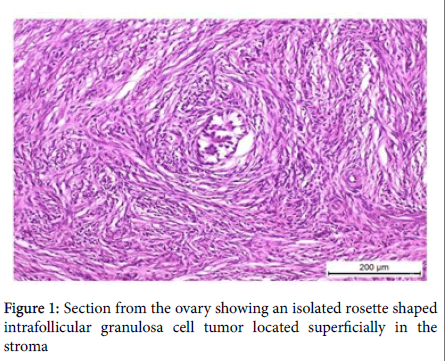
Single focal pattern: This pattern appeared as unifocal granulosa cell tumor with intrafollicular ('in situ' ) growth involving small area located on subscapular area of the ovary as an isolated rosette shaped structures located superficially in the stroma (Figure 1). The wall was bounded by irregular elliptical or angular cells that have circular arrangement, or the granulosa cells had pale unstained cytoplasm and a typical dark stained basal nuclei. The tumor had eosinophilic material as an early stage of Call Exner bodies formation. Mitotic activity was rare.
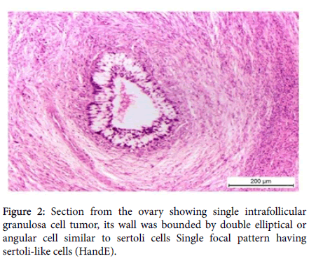
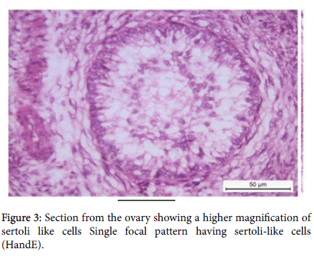
Single focal pattern having sertoli-like cells: Presence unifocal granulosa cell tumor and its wall was bounded by an elliptical or angular cell similar to sertoli cells (Figure 2). the contents were granulosa cells having pale unstained cytoplasm and atypical, bizarre nuclei (Figure 3). Mitotic activity was rare. No stromal invasion was found.
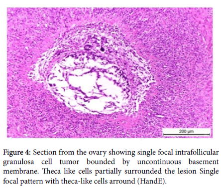
Single focal pattern with theca-like cells around: Microscopically, the tumor appeared oval or elliptical in cross section; its wall was bounded by uncontinuous basement membrane on which degenerating and necrotic cells lie. The granulosa cells had pale unstained cytoplasm and atypical, dark stained basal nuclei. The lumen of the tumor was trabeculated forming empty spaces having no contents. Mitotic activity was rare. Theca like cells partially surrounded the lesion (Figure 4). No stromal invasion was found.
Multifocal separted pattern: Multifocal granulosa cell tumor with intrafollicular ('in situ') growth involving small areas located subcapsular. Microscopically, showed a group of lobules appearing elliptical or oval; the wall of which was bounded by some fusiform stromal cells in a whorl-like arrangement (Figure 5). The contents were trabeculated, granulosa cells were large having pale unstained cytoplasm that may contain lipid, the nuclei were mostly polyhedral or rounded and dark. Mitotic activity was rare and there were empty areas in between the cells devoid of any content. No stromal invasion was found.
Multifocal coalesced pattern: Multifocal granulosa cell tumor appears microscopically, as a group of coalesced lobules of granulosa cells. The cells tend to form follicles. This group of lobulates was bounded by fusiform stromal cells in a whorl-like arrangement. The contents were trabeculated. The granulosa cells were large having pale unstained cytoplasm that may contain lipid. The nuclei were mostly rounded dark nuclei. Mitotic activity was rare. No common lumen is formed (Figure 6). No stromal invasion and no condensation of fibroblasts were detected.
Multifocal and cystic pattern: This pattern microscopically, showed a group of coalesced follicles of granulosa cells. The cells tend to form follicles. These groups of lobules were bounded by fusiform stromal cells in a whorl-like arrangement. The contents were trabeculated, granulosa cells were large having pale unstained cytoplasm that may contain lipid. The nuclei were mostly rounded dark nuclei. Mitotic activity was rare. No common lumen is formed; however, coalescence of some tubules occurred and there was empty areas in-between the cells devoid of any content. No stromal invasion and no condensation of fibroblasts were found (Figures 7 and 8).
Unifocal multicystic pattern: A group of dilated cystic structures lined by granulosa cells was arranged on trabeculae. Coalesced lobules of granulosa cells and separate focus un coalesced occurred to the side of the main collection. The cells tend to form follicles. This group of lobulates was bounded cells resembling theca cells and condensed fusiform stromal cells in a whorl-like arrangement. The contents were empty and unstained; granulosa cells were large having pale unstained cytoplasm that may contain lipid. The nuclei were mostly rounded dark nuclei. Mitotic activity was rare. The cysts were communicated and forming large cyst with incomplete septa (Figure 9). No stromal invasion and no condensation of fibroblasts around the lesion were found.
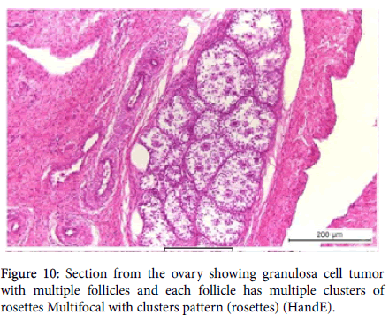
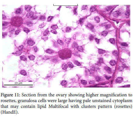
Multifocal with clusters pattern (rosettes): The ovary showed groups of eosinophilic materials as Call Exner's bodies each had a radiating granulosa cells and these cells tend to form rosettes like appearance. This rosette collected with each other’s to form clusters and their clusters were bounded by stromal cells. No common lumen for these groups of lesions is formed and there was empty areas in-between the cells. No stromal invasion and no condensation of fibroblasts around the lesion were found (Figure 10). Granulosa cells were large having pale unstained cytoplasm that may contain lipid (Figure 11).
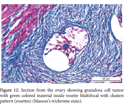
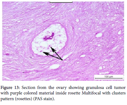
Specific stains (Masson's trichrome and Periodic Acid Schiff's) were used to determine what the structure of the eosinophilic material that present in the center of rosettes. By Masson's trichrome stain the eosinophilic material take the green color (Figure 12), while in other sections of GCTs; the eosinophilic material that present in the center of rosettes take the purple color by using of PAS stain (Figure 13).
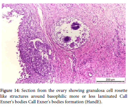
Call Exner's bodies formation: The microscopic picture of granulose cell tumor forming rosette like structures around basophilic blue colored material which was more or less laminated and/or globulated. These basophilic structures were similar to the microscopic picture of the wall of degenerating atretic follicles showing basophilia and lamination of Call Exner's bodies (Figure 14).
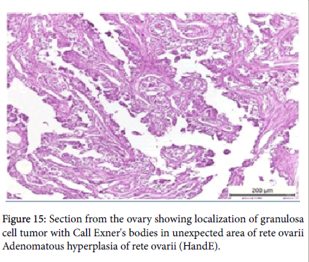
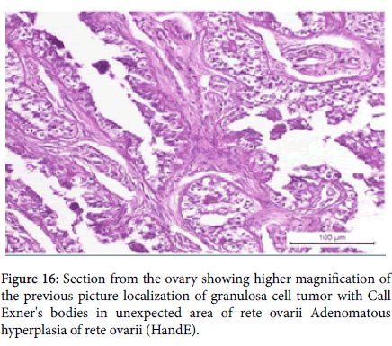
Adenomatous hyperplasia of rete ovarii: Papillary hyperplasia of the tubular structures of rete ovarii and localization of granulosa cell tumor with Call Exner's bodies in un expected area of rete ovarii were seen (Figures 15 and 16).
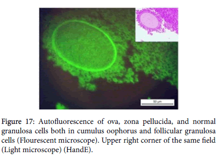
Fluorescent microscopy examination revealed that: Intrinsic autofluorescence was shown in ovarian tissue at various intensities such as ova, zona pellucida, and normal granulosa cells both in cumulus oophorus and follicular granulosa cells (Figure 17).
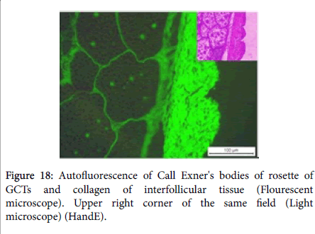
Call Exner's bodies formation was prominent structure to GCTs which appeared by HandE either eosinophilic or deeply basophilic in other cases (Figure 14), and when stained with Masson's trichrome stain, Call Exner's bodies has the same green color as collagen as seen in (Figure 12).
Neoplastic granulosa cells of ovarian tissue most commonly loses this autofluorescene property (Figure 18).
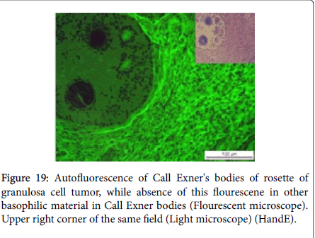
Call Exner's bodies when was eosinophilic in HandE stain emit autoflourescence and losses this ability of autoflouresence when become basophilic by HandE (Figure 19).
Discussion
Reproductive failure has been recognized today as one of the most serious problems affecting our economy through dairy cattle industry. Complete sterility is probably less important than sub-fertility or infertility, because sterile animals are few and easily identified in comparison with those having transient form of reproductive disorders. It is commonly observed that the individual females may be infertile due to a cause which does not involve other animals in a herd. As bovine; the most recognizable domestic animals in Egypt, any problems in their reproduction collaborate in great economic losses as most of the cows reach slaughterhouse for reasons of infertility.
Pathological conditions of ovary seriously interfere with normal functions of the entire reproductive tract, consequently, decreasing the reproductive potential of the animal [11].
In this study, the pathological findings of 73/100 cases of cows and buffaloes showing ovarian pathologies were A- Granulosa cell tumors (GCTs) which represent the highest incidence among other alterations by percentage of (67.1%) a total number of 49 cases from 73 total affected cases, B- Ovarian cysts were the second prevalent pathological condition affecting ovaries by total number of 16 cases that represent (21.9%), C- One mixed case having both cyst and granulosa cell tumor (1.4%), and finally, D- Persistent corpus luteum by percent of (9.6%) in which seven cases were affected.
As GCTs represent the highest incidence among the other ovarian abnormalities explains the importance to give more attention on this type of tumors.
In bovine, GCTs were reported first by Goldberg [12]. They arise from the granulosa and theca cell elements of the ovarian follicles, so they may be termed granulosa-theca cell tumors, granulosa cell tumors are usually benign with a very little tendency to be malignant [13].
Sex cord-stromal tumors are common in cows and mares, and less frequent in bitches and ewes [14]. Granulosa cell tumors were the most common ovarian tumor that affecting cattle [11,14], also Jubb et al. [15] confirmed that the tumors composed only of theca cells; thecoma or luteoma are much less common in all domestic animals than those arising from granulosa cells or containing a mixture of theca and granulosa cells, and this agreed with our results as the granulosa cells tumor was the most common lesion among other ovarian pathological alterations, as well as the only tumor type demonstrated also in all cases.
The clinical appearance and the behavior of the different types of ovarian tumors are extremely variable and histopathological examination is needed for treatment and to predict prognosis of the disease [12]. In our study the clinical findings could not be recorded as the specimens taken after slaughtering of the animals.
The incidence of granulosa cell tumor in buffaloes was 18.4% in this study while in the incidence in Indian buffaloes 0.2% according to Dwivedi [16], while McEntee [14] found 84 granulosa cell tumors out of 139 ovarian neoplasms in cattle by percent of 63.5%. In this study, GCTs not only the only tumor that found but also present with high incidence in both cows and buffaloes by 67.1% (as 49/73 cases) (81.6% and 18.4% respectively from 49 cases as 100%).
In this study, the incidence of ovarian granulosa cell tumor was highest among Balady cows more than Frisian cows, as GCTs were demonstrated in Balady cows 36/49 cases affected by percent of 75.4% from the total cases affected and in Frisian cows 4/49 by percent of 8.4% from the total cases affected, and this results disagreed with Moulton, (1978) who mentioned that there is no breed predilection has been found in any species.
Ages of bovine in the present study categorized into two groups; first group: two to five years, while the second group was above five years. The incidence of occurrence of granulosa cell tumors between the two groups was greater in the second one/or the elder ones as well as the younger animals also affected but much more lowest. Moulton (1978) mentioned that incidence is higher in old cows and also McEntee (1990) stated that bovine ovarian granulosa cell tumors were recorded at age ranging from new born to 19 years old with a medium age of 7 years but Nielsen and Kennedy [17] reported that GCTS tend to occur more often in younger animals.
In the present study the tumor foci did not interfere with pregnancy especially with presence of three pregnant cases having the tumor foci in their ovaries, and these results disagreed with Radostits et al. [18] who mentioned that the occurrence of granulosa cell tumors in pregnant cows is rare.
In the present study, GCTs founded to be unilateral affecting only one ovary by number of 42/49 cases (33 cases from cows and 9 cases from buffaloes), and three cases were bilaterally affected. The unilateral GCTs in the present study did not interfere with the function to the contralateral ovary (as the contralateral ovary was well functioning) and this results disagreed with Carlton and McGavin, [19] and Radostits et al. [18] who mentioned that granulosa cell tumors are most often unilateral and may suppress the function of the contralateral ovary, when this occurs; the contralateral ovary becomes atretic, while the ovary with the tumor may continue to grow.
Grossly, in this study, granulosa cell tumors were not detected in all 49 affected cases and these results disagreed with [8] who reported only 2 cases out of 140 non-pregnant cows had ovarian granulosa cell tumors which appeared grossly in a diameter more than of 1.5 cm. They were unilateral occupying nearly the whole ovarian stroma in the form of multicystic spaces that were separated by thin fibrous connective tissue septa, and these spaces contained clear yellowish fluid. Also Baumann [20] described ovarian granulosa cell tumor in bovine weighing 17.1 kg and measuring 48 × 38 × 19 cm, and Zinnebauer [21] reported 40 kg granulosa cell tumor in bovine ovary.
GCTs vary in size from relatively small, solid, yellow to white structures to large structures composed of multiple cysts, a single large cystic structure, or a combination of solid and cystic structures [20,21]. The surface of these tumors may be smooth or lobulated; they may be so highly vascularized that one can palpate fremitus in the middle uterine artery [18]. In contrast to our study as GCTs appeared only microscopically without gross changes on ovaries affected either in shape, size, color, consistency and/or cut sections.
GCTs microscopically, appeared in three growth patterns or forms; diffuse, trabecular, and follicular form, but the most common pattern was the follicular form according to [6]. Follicular and diffuse patterns of growth of the tumor were also reported by [14] as common GCTs growth pattern. Microscopically, granulosa cell tumors were found in one or both ovaries in 40 animals. The tumors were unilateral in 25 cases and bilateral in 13 cases distributed in the subcapsular area, cortex or in the ovarian hillus.
El-Nesr et al. [8] described the tumor foci of grossly detected as they had different patterns including, firstly, uniform populations of granulosa cells surrounded with fibrous connective tissue septa in which the granulosa cells appeared polyhedral with vesicular nuclei. The second pattern consisted of granulosa cells arranged in clusters around eosinophilic material (Call-Exner bodies) forming rosette pattern and surrounded by fibrovascular septa. In the third pattern, the tumor foci were appeared in the form multicystic spaces separated by fibrous t issues that were lined by granulosa cells. In some tumor masses, some cells were mitotically active with prominent nucleoli.
El-Nesr et al. [8] described four histopathological patterns of granulosa cell tumors in Balady cows; the first pattern had uniform populations of granulosa cells, that appeared as small polyhedral cells having an elliptical centrally located nuclei and their cytoplasm appeared lightly stained with poorly defined cell outlines. In the second pattern, granulosa cells were arranged in clusters around esinophilic secretions, Call-Exner's bodies, forming rosette pattern and were surrounded by fibrovascular septa. Granulose cell tumors in this pattern were found singular or in combination in the form of nests occupying nearly the whole ovarian stroma. In the third patterns, small to large cystic spaces containing eosinophilic material surrounded with one or multiple cell layers were seen in some cases. The fourth pattern showed groups of granulosa cell surrounded by a fibrous connective tissue membrane obliterating some cortical areas with the nearly absence of the Call-Exner's bodies.
While in our results, there was ten different histopathological patterns which are 1- single focal pattern, 2- single focal pattern having sertoli-like cells, 3- single focal pattern with theca- like cells around, 4- multifocal separated pattern, 5- multifocal coalesced pattern, 6- multifocal and cystic pattern,7- unifocal multicystic pattern, 8- multifocal with clusters pattern (rosettes multiple clusters pattern (rosettes), 9- Call Exner's cell formation, and 10- adenomatous hyperplasia of rete ovarii.
These forms of GCTs represent a new classification for these tumors as it is considered a prevalent ovarian tumor in our native animals. Also these 10 microscopical patterns of granulosa cell tumors might be a new type of pathogenesis that explains the different growing stages of the tumor.
Conclusion
In our results, Call Exner's bodies formation was prominent structure to GCTs which appeared by HandE either eosinophilic or deeply basophilic in other cases and when stained with Masson's trichrome stain, Call Exner's bodies has the same green color as collagen. Also, in further investigations with fluorescent microscope, the Call Exner's bodies when was eosinophilic in HandE stain emit autoflourescence and losses this ability of autoflouresence when become basophilic by HandE. As the different colors of Call Exner's bodies by HandE representing aging stages of Call Exner's bodies that is to say begin as secretions from granulosa cells which appeared; firstly, eosinophilic then after condensation and maybe calcification of this material was deeply basophilic, inspite of it was negative for Von kossa stain. By Masson's trichrome stain, the appearance of Call Exner's bodies take the same color of collagen in the trichrome stain together with emition of autoflouresence by the frourescent microscope revealed that this material belong to collagen, this collagenic materials produced through polymerization and esterification of secreted materials.
Stained sections routinely with hematoxylin and eosin (Haematoxylin: Fluka, AG, Switzerland, Buchs SG – Eosin Y: alcohol and water soluble, Winlap, UK), mounted in fluorescence- free D-P-X mountant (LOBA Chemie, India) [10].
Call Exner's bodies when was eosinophilic in HandE stain emit autoflourescence and losses this ability of autoflouresence when become basophilic by HandE as content of ovarian granulosa cell tumors, such as Call-Exner's bodies may or may not show fluorescence depending upon their chemical composition and cellular activity.
Kitano et al. [22] and El-Nesr et al. [8] mentioned that Call-Exner's bodies were numerous in the newly forming tumors in contrast to the large ones and may undergo hyalinosis in the form of multilayered basal laminae in some cases.
Finally, it is concluded that GCTs were the most common ovarian lesion in this which appeared only microscopically and based on histopathology, GCTs were classified into ten patterns.
References
- Barnett KR, Schilling C, Greenfeld CR, Tomic D, Flaws JA (2006) Ovarian follicle development and transgenic mouse models. Hum Reprod Update 12: 537-555.
- Purohit GN (2014) Ovarian and oviductal pathologies in the buffalo: Occurrence, diagnostic and therapeutic approaches. Asian Pac J Reprod 3: 156-168.
- Nielsen SW, Misdorp W, McEntee KE (1976) Tumours of the ovary. Bull World Health Organ 53: 203.
- Lagerlof N, Boyd H (1953) Ovarian hypoplasia and other abnormal conditions in the sexual organs of cattle of the Swedish Highland breed; results of postmortem examination of over 6,000 cows. Cornell Vet 43: 64-79.
- Izquierdo N, Zhelev V, Angelov AK (1983) Tumors of the ovaries in zebus. Vet Med Nauki 20: 71-78.
- Hunt JTC, Ronald D, Norval KW (1996) Veterinary Pathology. 6th Clinical Chemistry, Academic Press, New York, USA.
- El-Nesr KA, Kamel HH, Abd-El- Rahman AH (2006) Bovine ovarian granulose cell tumors: Pathological and clinicopathological studies. Egypt J Comp Path and Clinic Path 19: 228-245.
- Bancroft JD, Gamble M (2008) Theory and practice of histological techniques. (6th edn). Elsevier Health Sciences, North Hollywood, USA.
- Deeb S, Nesr KH, Mahdy E, Badawey M, Badei M (2008) Autofluorescence of routinely hematoxylin and eosinstained sections without exogenous markers. Afr J Biotechnol 7: 504-507.
- Ali RI, Raza MA, Jabbar AB, Rasool MH (2006) Pathological studies on reproductive organs of zebu cow. J Agri Social Sci 2: 91-95.
- Goldberg SA (1920) The occurrence of epithelial tumors in the domestic animals. J Am Vet Med Assoc 58:47-63.
- Moulton JE (1978) Tumors of the genital system. Tumors in domestic animals 1978: 309-345.
- McEntee K (1990) Ovarian neoplasms. In: Reproductive pathology of domestic mammals. San Diego, Academic Press, cap, New York, USA. pp. 69-93
- Jubb KVF, Kennedy PC, Palmer N (2007) Pathology of domestic animals. (5th edn). Saunders, Elsevier, Pheladelphia, USA.
- Diewedi JN, Singh CM (1975) Studies on the histopathology of uterus Indian buffalo. Indian J Anim Sci 45: 20-24.
- Nielsen SW, Kennedy PC (1990) Tumors of the genital systems. In: Atlas of tumor pathology (2nd series), fascicle. In: Moulton JE (ed.) Tumors in domestic animals (3rd edn). Armed Forces Institute of Pathology, Washington, University of California Press, Berkeley, Carlifornia, USA. pp. 153-173.
- Radostits OM, Gay CC (2001) Blood, DC and Hinchcliff, KW. Veterinary Medicine. (9th edn). pp. 1684-1688.
- Carlton W, McGavin MD, Thomson RG (1995) Thomson's special veterinary pathology/Special veterinary pathology.
- Baumann R (1935) Zur pathologischen anatomie der granulosazelltumoeren des eierstoches. Wien Tierarztl Monasschr 22: 193-202.
- Zinnebauer H (1961) Ein Besonders grosser ovarian tumor. Wein Tierarztl Monasschr 48: 944-947. Cited by McEntee (1990) Reproductive Pathology of domestic animals. (4th Edn). Academic Press, Inc., New York, USA.
- Kitano Y, Iwabuchi I, Keruma T, Ishikawa Y, Kadota K (1996) Ovarian Sex Cordâ€Stromal Tumours in Cattle, with Particular Reference to the Lamination of Basal Laminae. Transboundary and Emerging Diseases 43: 531-41.
Share This Article
Open Access Journals
Article Usage
- Total views: 9955
- [From(publication date): 0-2018 - Mar 28, 2025]
- Breakdown by view type
- HTML page views: 8599
- PDF downloads: 1356

