A Large Parathyroid Adenoma- A Textbook Case in Pathology
Received: 18-Jan-2016 / Accepted Date: 28-Jan-2016 / Published Date: 03-Feb-2016 DOI: 10.4172/2476-2024.1000106
6192Introduction
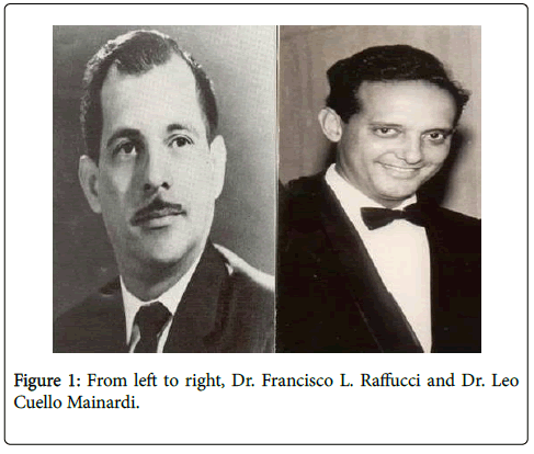
In 1963, I entered my residency in General Surgery at the University of Puerto Rico and Affiliated Hospitals in San Juan, Puerto Rico. At the time, Dr. Francisco L. Raffucci was Chairman of Surgery, and Dr. Leo Cuello Mainardi, Program Director in Surgery.
Dr. Francisco L. Raffucci, a pioneer in cardiovascular surgery in Puerto Rico, was also involved in treating patients with portal hypertension, liver failure, and pancreatic diseases. In 1961, he removed the first parathyroid adenoma in Puerto Rico.
My interest in parathyroid surgery was prompted by an assignment from Dr. Cuello Mainardi in 1964. He required that I conduct a seminar on the topic of hypercalcemia. For this purpose, I prepared a paper for the house staff on the differential diagnosis of hypercalcemia, which includes: sarcoidosis, milk-alkali syndrome, multiple myeloma, hypernephroma, metastatic bone disease, and hyperparathyroidism. The event was pivotal to what became a life-long career as a thyroid and parathyroid surgeon.
Upon completion of my training in 1968, and after serving a twoyear tour in the US Army, in 1970, I joined the faculty of the University of Puerto Rico School of Medicine’s Department of Surgery.
Case Presentation
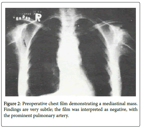
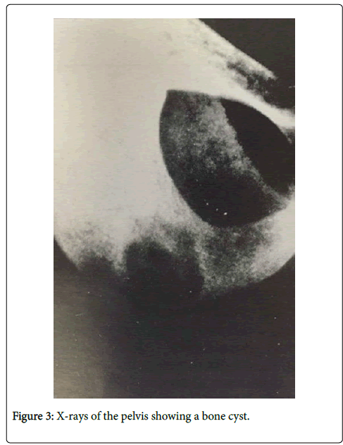
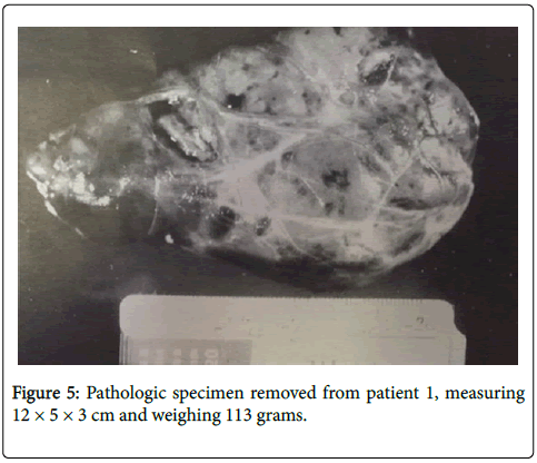
My first operation for hyperparathyroidism was performed in September 1970. The patient was a 41-year-old female who presented anorexia, weight loss, polydipsia, polyuria, and constipation, bone pain particularly in the pelvis, lacrimation, and burning sensation of the eyes. She also had repeated urinary infections. Her calcium levels ranged from 16.4 mg/dL to 18.1 mg/dL. The chest film was interpreted as normal, the intravenous pyelogram revealed nephrocalcinosis, and X-rays of the pelvis uncovered a pelvic bone cyst. A bone scan showed generalized demineralization. The neck was explored on September 21, 1970. No neck adenomas were detected. A large lower neck and mediastinal mass was found, requiring a median sternotomy for its removal. A large 12 cm×5 cm×3 cm mass, weighing 113.3 g, was removed. Although it was my first case as an attending surgeon, there was no way I could possibly miss such a large adenoma.
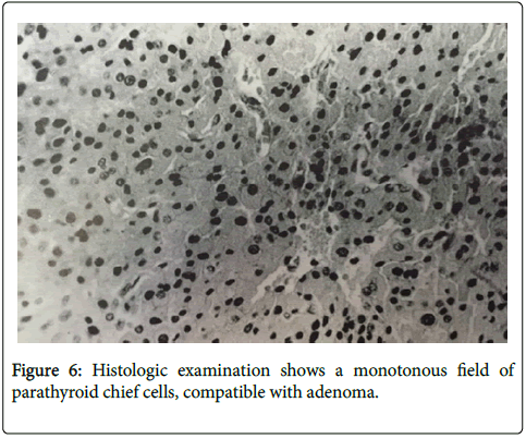
Histologic examination showed a monotonous field of parathyroid chief cells. A postoperative chest film confirmed absence of the previously undiagnosed mediastinal mass. The patient remained asymptomatic until 1990, when she was found to have mild hypercalcemia. Both the PTH-C terminal and the PTH intact tests were elevated as well.
To this day, this is the second largest parathyroid adenoma ever reported in the medical literature. The following Table 1 showing the parathyroid adenomas weighing over 50 g reported in the medical literature.
| Surgeon | Year | Location | Size (cm) | Weight (g) |
|---|---|---|---|---|
| Dresser | 1931 | R | 6.5×5.0×3.0 | 53.0 |
| Castleman | 1936 | R | 6.5×5.0×3.5 | 53.2 |
| Earll | 1969 | LL | 7×5×3 | 85.0 |
| Snell | 1936 | RL | 6×6×5 | 101.0 |
| Vazquez | 1970 | M | 12×6×3.5 | 113.3 |
| Sharpe | 1939 | LU | 7×6.5×4 | 120.0 |
Table 1: Parathyroid adenomas over 50 g in weight.
Discussion
This patient presented severe manifestations of hyperparathyroidism. At the time, in 1970, the diagnosis of hyperparathyroidism was made by excluding other causes of hypercalcemia, since PTH assays, thyroid sonograms, or CT scans were not available as diagnostic tools [1]. Nowadays, hyperparathyroidism can be diagnosed early either with mild symptoms or no symptoms at all.
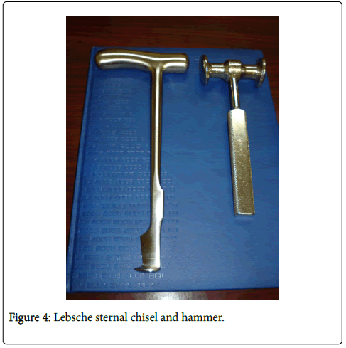
The above photo shows the instruments used to perform the sternotomy on this patient. At that time, the electric saw nowadays used for median sternotomies was not yet in use.
A review of the literature revealed that the largest parathyroid adenoma ever removed weighed 120 g; the procedure was done by Sharpe in 1939; however, it was smaller in size than the one in our case. Usually, adenomas weigh less than one or two grams. Our patient’s tumor weighed one hundred times the usual parathyroid lesion: 113 g, the second heaviest tumor reported so far in the literature.
Hyperparathyroidism is the most frequent cause of hypercalcemia in non-hospitalized patients, and the second most common cause of hypercalcemia after malignancy among those hospitalized patients [2]. In approximately 85% of cases the etiology of hyperparathyroidism is an adenoma; 10%-12% of cases are due to hyperplasia of all four parathyroid glands, and in approximately less than 3% of cases the cause is a parathyroid carcinoma. [3] Multiple adenomas are very rare [4,5]. The patient in question is alive 45 years after her operation. She is now 86 years old. Her most recent calcium level is 10 mg/dL and her PTH 72.8 pg/mL. Her serum creatinine is elevated, 1.8 mg/dL, and her creatinine clearance is low, 24.93 mL/min. She has no kidney stones. This woman still works as an assistant cook at a home for the elderly.
Summary
We have presented a patient with severe symptoms of hyperparathyroidism, markedly elevated calcium levels, bony cyst, nephrocalcinosis, and a large mediastinal adenoma. Forty-five years after the surgery, she is now 86 years old, with normal calcium and PTH intact levels. At the time of her operation, there were no localizing tests, nor the electric saw to open the sternum. This demonstrates that in the past, good results could be obtained with the tools that were available.
References
- Quintana VE, Quintana CS, Aguilo F Jr, Pagan-Saez H, Silva F (1989) Localization of parathyroid lesions: blind study for surgeons. Bol Asoc Med PR 81: 542-344.
- Clark O (1985) Endocrine Surgery of the thyroid and parathyroid. St. Louis: CV Mosby Co; 173.
- Clark OH, Quan-Yang D (1995) Parathyroid gland.In: Davis JH, Sheldon GF (eds.)Surgery, a problem solving approach.(2ndedn), St. Louis: CV Mosby Co., pp.2252.
- Verdonk CA, Edis AJ (1981) Parathyroid “double adenomasâ€: fact or fiction? Surgery 90: 523-526.
- Harness JK, Ramsburg SR, Nishiyama RH, Thompson NW (1979) Multiple adenomas of the parathyroid: do they exist? Arch Surg 114: 468-474.
Citation: Vazquez-Quintana E (2016) A Large Parathyroid Adenoma- A Textbook Case in Pathology. Diagn Pathol Open 1: 106. DOI: 10.4172/2476-2024.1000106
Copyright: ©2016 Vazquez-Quintana E. This is an open-access article distributed under the terms of the Creative Commons Attribution License, which permits unrestricted use, distribution, and reproduction in any medium, provided the original author and source are credited.
Share This Article
Open Access Journals
Article Tools
Article Usage
- Total views: 12496
- [From(publication date): 3-2016 - Apr 05, 2025]
- Breakdown by view type
- HTML page views: 11611
- PDF downloads: 885

