A Dangerous Method for Left Ventricular Lead Stabilization: Fixation with a Stylet
Received: 29-Mar-2018 / Accepted Date: 09-Apr-2018 / Published Date: 15-Apr-2018
Abstract
CRT is important in the treatment of heart failure. However, the physicians can encounter some serious difficulties in CRT implantations. Particularly, its difficulty is in the process of LV lead fixation. Although practical solutions such as permanent Stylet method have been developed to overcome this difficulty, they each can be problematic in separate ways. In our 58-year-old male patient adjacent organs perforation occurred because of permanent Stylet. In this report, we have showed that permanent Stylet method is a risky method.
Keywords: Stylet; CRT; LV lead; Heart failure
Introduction
The patients with symptomatic heart failure, despite medical therapy, are cured effectively with Cardiac Resynchronization Therapy (CRT) if there is left bundle branch block or wide QRS in electrocardiography (ECG). The electro physiologists have the greatest challenge in placing the left ventricular (LV) lead. The difficulty of the placement of the LV lead is depend on several factors, such as the unsuited coronary sinus anatomy, lead instability, the availability of inadequate pacing parameters, and the occurrence of extracardiac stimulation such as adjacent muscles or nerves stimulation [1]. They have developed a variety of methods such as retained guidewire [2] or Stylet [3], using new design leads or pericardial leads [4], stenting [5] etc. to overcome this problem. While some of these methods provide effective treatment in the short term, they cause serious complications in the long term.
In this case, we present the developing complications after retained Stylet within of the LV lead.
Case Presentation
A 58-year-old male was presented to our clinic with left sided chest pain and cough for three months. According to the New York Heart Association, effort capacity of patient was class II. In the physical exam of the patient, we heard 3/6 grade systolic murmur in mesocardiac area and with no other features. In his background and medical history, he has suffered a myocardial infarction six years ago and underwent triple vessel coronary artery bypass grafting surgery. In the followings, he was diagnosed with ischemic cardiomyopathy and Cardiac Resynchronization Therapy-Implantable Cardioverter Defibrillator (CRT-ICD) was placed due to incomplete left bundle branch block (LBBB).
Three years later, fracture was determined in patient’s LV lead, and new LV lead was replaced to the patient. His ECG revealed sinus rhythm with incomplete LBBB, QRS duration 0.10 second, pace rhythm for left and right ventricles of the heart. In his echocardiography, the left atrium and ventricular diameters were identified at the upper limit, LV ejection fraction of 45 percent and second-degree mitral regurgitation. Patient’s posteroanterior chest xray showed that cardiothoracic ratio was normal, CRT-ICD battery was in the left subclavian region, right atrial lead, right ventricular ICD lead, and LV lead were in the normal position, but previous LV lead was in the right atrium. It also was noticed a radiopaque thin line that originates from the previous LV lead, and it extended from the previous LV lead tip to the left lateral thoracic region (Figure 1)
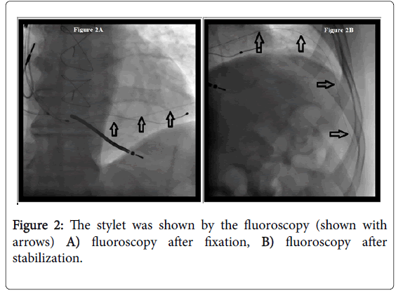
Also it confirmed under fluoroscopy, and was thought that it can be the Stylet which was left in the previous LV lead due to fixation and stabilization (Figures 2A and 2B).
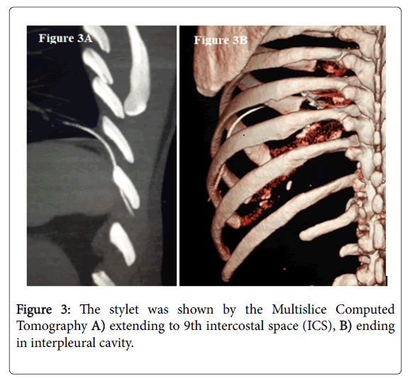
In the Multislice Computed Tomography, it was analyzed to be a Stylet, extending to 9th intercostal space (ICS) and ending in interpleural cavity (Figures 3A and 3B).
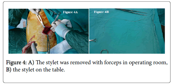
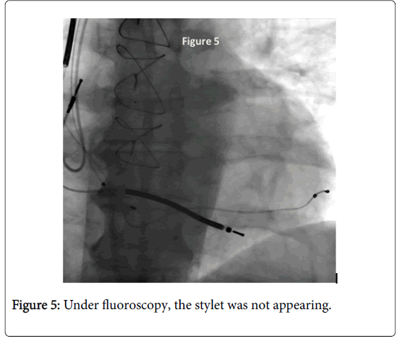
Patient’s condition was stable and consulted by cardiothoracic surgery and decided surgical removal of Stylet. Under general anesthesia, thoracotomy was performed from the 9th ICS and was entered to pleural cavity. Stylet was removed with forceps under the fluoroscopy in operating room (Figures 4 and 5).
The left sided chest tube was removed from the patient who does not develop any complications in post-operative first day. The patient was discharged in postoperative third day with antibiotics and wound care. Treatment of ischemic heart disease and heart failure in the patient was continued.
Discussion
The incidence of LV lead implantation failure is at a high level, in spite of the current progress in LV implantation methods. In intended appropriate LV lead implantation, failure rate is about 4% [6]. Appropriate LV lead implantation rate is improved by new leads and active fixation mechanisms [7-9]. At the same time, the scientists threw out an idea about the Stylet method within LV lead and it was a good method to stabilize appropriate LV lead location. Sharifkazemi et al. [3] study showed that after the use of permanent Stylet method, no LV lead dislocations were detected in CRT cases during the follow-up. In this way, they suggested using permanent Stylet method as the last resort when post-operative or intra-operative lead dislocation occurs, or if the electrode position is not stable enough and an alternative side branch is not available at the chosen location [3].
Osztheimer et al. [10] showed fracture of the LV lead in the right atrium and penetration of the Stylet into pulmonary lobe. They saw no effusion in the pericardial or pleural space and performed successful removal of all parts of the fractured LV lead through percutaneous lead extraction. This case was the first case reporting that permanent Stylet method poses danger because of penetration into an adjacent organ occurred [10]. However, the difference of our case is that it was too bad to be removed by percutaneous lead extraction method and it is need to be removed by surgical method.
Conclusion
In conclusion, the Stylet probe could be broken because of its stiff texture, and the broken tip can injure adjacent tissues. The permanent Stylet method to stabilize appropriate LV lead location is currently not suggested because of dangerous complications.
Disclosures
The authors have no conflicts of interest to disclose.
References
- Brignole M, Auricchio A, Baron-Esquivias G, Bordachar P, Boriani G, et al. (2013) 2013 ESC guidelines on cardiac pacing and cardiac resynchronization therapy: the task force on cardiac pacing and resynchronization therapy of the European Society of Cardiology (ESC).Developed in collaboration with the European Heart Rhythm Associate (EHRA). Eur Heart J 34: 228-329.
- De Cock CC, Jessurun ER, Allaart CA, Visser CA (2004) Repetitive intra-operative dislocation during transvenous lead implantation: usefulness of the retained guidewire technique. Pacing Clin Electrophysiol 27: 1589-1593.
- Sharifkazemi MB, Aslani A (2007) Stabilization of the coronary sinus lead position with permanent stylet to prevent and treat dislocation. Europace 9: 875-877.
- McALOON CJ, Anderson BM, Dimitri W, Panting J, Yusuf S, et al. (2016) Long-term follow-up of isolated epicardial left ventricular lead implant using a minithoracotomy approach for cardiac resynchronization therapy. Pacing Clin Electrophysiol 39: 1052-1060.
- Szilagyi S, Merkely B, Roka A, Zima E, Fulop G, et al. (2007) Stabilization of the Coronary Sinus Electrode Position with Coronary Stent Implantation to Prevent and Treat Dislocation. J Cardiovasc Electrophysiol 18: 303-307.
- Gras D, Boöcker D, Lunati M, Wellens HJ, Calvert M, et al. (2007) Implantation of cardiac resynchronization therapy systems in the CARE-HF Trial: procedural success rate and safety. Europace 9: 516-522.
- Nagele H, Azizi M, Hashagen S, Castel MA, Behrens S (2007) First experience with a new active fixation coronary sinus lead. Europace 9: 437-441.
- George H, Crossley MD, Mauro Biffi MD, Ben Johnson, Shelby Li, MS Keith Holloman, Derek BS, Exner V.
- Auricchio A, Delnoy PP, Regoli F, Seifert M, Markou T, et al. (2013) First-in-man implantation of leadless ultrasound-based cardiac stimulation pacing system: Novel endocardial left ventricular resynchronization therapy in heart failure patients. Europace 15: 1191-1197.
- Osztheimer I, Duray G, Huttl K, Merkely B (2016) Fracture and lung penetration of a left ventricular lead stabilized by retained stylet. Can J Cardiol 32: 1576.e19-1576.e20.
Citation: Islamoglu Y, Bucak E, Yildiz M, Demirtas S (2018) A Dangerous Method for Left Ventricular Lead Stabilization: Fixation with a Stylet. J Med Imp Surg 3: 119.
Copyright: © 2018 Islamoglu Y, et al. This is an open-access article distributed under the terms of the Creative Commons Attribution License, which permits unrestricted use, distribution, and reproduction in any medium, provided the original author and source are credited.
Share This Article
Recommended Conferences
42nd Global Conference on Nursing Care & Patient Safety
Toronto, CanadaRecommended Journals
Open Access Journals
Article Usage
- Total views: 3312
- [From(publication date): 0-2018 - Apr 09, 2025]
- Breakdown by view type
- HTML page views: 2469
- PDF downloads: 843


