A Case Report of Complication of Foreign Body in the Nose
Received: 20-Jan-2020 / Accepted Date: 24-Feb-2020 / Published Date: 02-Mar-2020 DOI: 10.4172/2161-119X.1000392
Abstract
Background: Foreign body in the nose, a common presentation in the ENT clinic and emergency room can have potentially fatal complications. Aim: To present a serious complication of foreign body in the nose.
Case report: A three years old female presented to our clinic with a history of foreign body in the nose for three days and left eye swelling of two days. Examination revealed a foreign body in the left nasal cavity complicated by acute left maxillary sinusitis and left periorbital cellulitis. She improved remarkably with foreign body removal and antibiotic administration.
Conclusion: Foreign body in the nose could have potentially fatal complications like sinusitis and orbital cellulitis, early presentation, foreign body removal and administration of antibiotics can effectively treat the complications.
Keywords: Foreign body in the nose; Acute sinusitis; Orbital cellulitis
Introduction
A foreign body in the nose is the presence of any material in the nasal cavity that was either inhaled or accidentally placed there [1].
This is more common in children as they tend to explore their body parts and cavities. Children aged two to five years are most likely to insert foreign bodies into their nose. Foreign bodies can also be seen in psychiatric patients or even healthy adults when insects fall into the nose. Children are not likely to inform their parents or care givers that a foreign body is in their nose so they are more at risk of complications. The immune system of children is also not as efficient as that of adults this also increases risk of complications.
Several complications have been noted from foreign bodies in those, they include; nasal septal perforation, meningitis, sinusitis, acute epiglottitis and respiratory arrest [2].
Case Report
A 3 year old female presented with a referral from a private hospital with complaints of foreign body in the left nasal cavity of three days duration and left eye swelling of two days duration.
Mother’s attention was drawn by patient to an object she inserted into her left nasal cavity; mother tried to remove foreign body but could not. There was associated foul smelling muco purulent left nasal discharge, however no pain and no epistaxis.
A day later, the mother noted left periorbital swelling which progressively increased in size associated with redness of the left eye. The swelling progressively increased to involve left maxillary area. There was associated fever, no visual loss, no epiphora. There was no dysphagia, odynophagia or refusal of meals, she also had no ear complains.
Patient thus presented at a private hospital, the private hospital had no ear nose and throat specialist so patient was counselled and referred to the Ear, Nose, Throat, Head and Neck Clinic of the University of Benin Teaching Hospital, the closest teaching hospital from referal centre also located in Benin city, Nigeria.
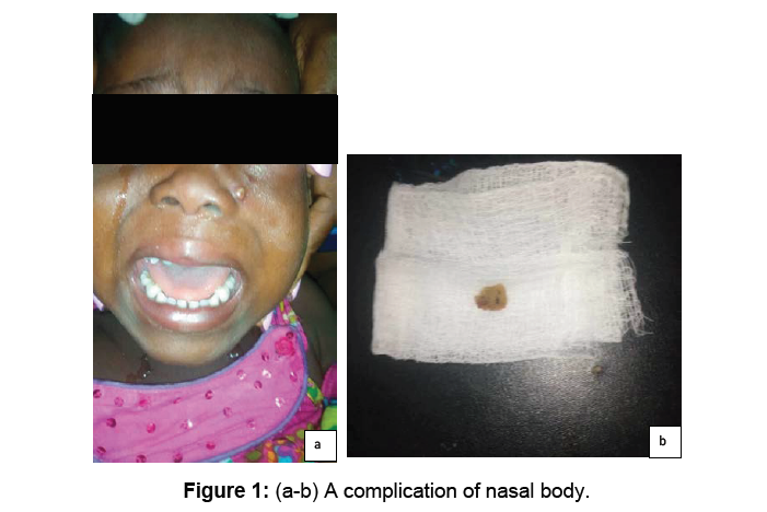
On examination at the Ear, Nose, Throat, Head and Neck Clinic UBTH, she was febrile to touch, not pale, anicteric, there was no peripheral lymph node enlargement. She had periorbital swelling with differential warmth. The maxillary area was erythematous and swollen. It was also tender to touch with differential warmth. Left nasal cavity was covered by mucopurulent discharge. On nasal toileting, a foreign body was noted. Right nasal cavity appeared normal. Mucopurulent post nasal discharge was noted on the posterior pharyngeal wall.
Examination of the neck yielded no abnormal findings. The pinna were well formed, both external auditory canals were obscured by wax so tympanic membrane could not be visualised.
An assessment of foreign body in the left nasal cavity was made with acute left maxillary sinusitis and left periorbital cellulitis.
Patient had foreign body removed with a eusthachian tube catheter, patient was properly held between mother’s legs and there was no need for local or general anaesthesia, foreign body was non negative in nature, it was a piece of foam. The procedure was fast and well tolerated by the child. She was commenced on syrup co-amoxiclav, Ibuprofen, pseudoephedrine hydrochloride and menthol crystal steam inhalation.
She was also placed on cerumol ear drops and subsequently had bilateral ear syringing with Normal saline irrigation via an 18 G cannular and a 20 mls syringe. She was subsequently referred to the eye clinic, computed tomography scan of the paranasal sinuses and brain were requested, parents declined the CT scan for financial reasons and she was told to come for follow up in a week.
Patient did not present at clinic for one week review and a call was placed to the father who said she has improved remarkably and will not need follow-up.
Discussion
Nasal foreign body is a relatively common finding in the ENT practice with foam as found in this patient being one of the common incriminated foreign body [3].
This highlights the need for materials like foam or other small household items that can be put in the nose to be kept far from children.
The patient had a unilateral foul smelling mucopurulent discharge which is the most consistent finding in patients with nasal foreign body [4]. This underscored the need for appropriate history-taking by primary care physicians and paediatricians who are a lot of times the first point of contact. Parents and caregivers are also to take this finding seriously and it should form part of health education lectures.
This patient presented with left eye swelling and further examination revealed she had acute maxillary sinusitis and left periorbital cellulitis. These are recognized and documented complications of sinusitis [4]. It could not be ascertained clinically if other paranasal sinuses especially the ethmoidal sinuses were affected, patient however declined a Computed Tomography (CT) scan which would have assessed all the sinuses for financial reasons (Figure 1).
Acute sinusitis is a disease entity with severe extra cranial and intracranial complications [5].
Orbital complications of sinusitis like preseptal cellulitis as found in this patient are commoner in children [6]. However, the prognosis is favourable and the index patient also had a favourable prognosis
The index patient was not registered on any of the health insurance schemes this may explain why the parents declined follow up visits when the child’s symptoms resolved.
This shows the need for Universal Health Coverage as the parents would have been highly encouraged to come for follow up if the hospital fees were not out of pocket.
Conclusion
Index case was a child who had a foreign body in her nose complicated by acute sinusitis and periorbital celluilitis. The child responded favourably and promptly to foreign body removal and medication administration. This may however not have been the case if presentation was further delayed and complications of acute rhino sinusitis allowed to set in.
We need to highlight the need for appropriate evaluation of unilateral mucopurulent nasal discharge and prompt referral of foreign body in the nose as fatal complications could arise.
The need for Universal Health Coverage cannot be overemphasized and our government and her agencies should be made to discourage out of pocket health expenditure.
Conflict of Interest
There is no conflict of interest.
References
- Virteeka S, Baranowski K (2019) Foreign body, nose. Katherine Baranowski, Treasure Island (FL): Stat Pearls Publishing.
- Okoye BC, Onotai LO (2006) Foreign body in the nose. Niger J med 15: 301-304.
- Kalan A, Taariq M (2000) Foreign bodies in the nasal cavities: A comprehensive review of the aetiology, diagnostic pointers and therapeutic measures. Postgrad Med J 76: 484–487.
- Hazarika P, Nayak R, Krishnan B (2013) Textbook of Ear, Nose, Throat and Head & Neck Surgery. Clinical and Practical. (3rdedn), CBS Publishers and Distributors Put Ltd., New Delhi, India.
- Al-Madani MV, Khatatbeh AE, Rawashdeh RZ, Al-Khtoum NF, Shawagfeh NR (2013) The prevalence of orbital complications among children and adults with acute rhinosinusitis. Braz J Otorhinolaryngol 7: 716-719.
Citation: Eustace OE, John A (2020) A Case Report of Complication of Foreign Body in the Nose. Otolaryngol (Sunnyvale) 10: 392. DOI: 10.4172/2161-119X.1000392
Copyright: © 2020 Eustace OE, et al. This is an open-access article distributed under the terms of the Creative Commons Attribution License, which permits unrestricted use, distribution, and reproduction in any medium, provided the original author and source are credited.
Share This Article
Recommended Journals
Open Access Journals
Article Tools
Article Usage
- Total views: 2870
- [From(publication date): 0-2020 - Apr 07, 2025]
- Breakdown by view type
- HTML page views: 2096
- PDF downloads: 774

