A Case Report of Calcified Submitral Aneurysm Associated with Takayasus Arteritis
Received: 17-Feb-2018 / Accepted Date: 27-Feb-2018 / Published Date: 05-Mar-2018 DOI: 10.4172/2167-7964.1000293
Abstract
Submitral aneurysm is a rare entity, possibly arising due to the disjunction between the left ventricular musculature and the left atrium-mitral valve region. We describe a rare case of a calcified submitral aneurysm arising from the inferomedial posterior mitral valve annulus associated with right subclavian artery aneurysm and left subclavian artery occlusion.
Keywords: Submitral aneurysm; Subclavian aneurysm; Takayasu’s arteritis
Introduction
Submitral Aneurysm (SMA) was first described by Corvistart in 1812 and the first case was reported in 1962 [1]. SMA invariably occurs adjacent to the posterior annulus of the mitral valve. We report a rare case of a calcified submitral aneurysm arising from the inferomedial portion of the posterior mitral valve annulus extending adjacent to the non-coronary cusp of the aortic valve. It was associated with the right subclavian Artery (SCA) aneurysm and left SCA occlusion.
Case Report
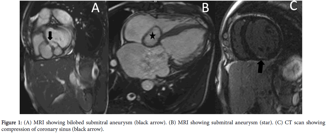
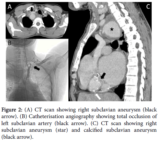
A 32-year-old Indian male presented with dyspnoea on exertion associated with paroxysmal nocturnal dyspnoea (NYHA IV) and palpitations (NYHA II) for 8 months. Clinical examination revealed absence of left radial pulse and severe mitral regurgitation with no signs of heart failure. Blood investigations showed raised erythrocyte sedimentation rate. His electrocardiogram showed atrial fibrillation and left ventricular hypertrophy. Two-dimensional echocardiography with color doppler showed a submitral aneurysm posteromedial to left atrium (LA) and severe mitral regurgitation. Magnetic resonance imaging (MRI) revealed bilobed aneurysm (Figure 1A-1C) arising from the inferomedial part of the posterior mitral valve annulus and extending to adjacent part of the non-coronary cusp of the aortic valve and posteromedial part of the tricuspid annulus. Mitral regurgitation and communication of the aneurysm into the left atrial cavity was present. On late gadolinium enhanced images, patchy areas of fibrosis was noted along the basal inferior wall in the noncoronary distribution indicating myocarditis. Contrast-enhanced computed tomography revealed the presence of calcification within the aneurysmal sac (Figure 2A-2C) and compression of the coronary sinus (Figure 1C). Additionally, a large saccular aneurysm with mural thrombus was noted arising from the proximal part of the SCA (Figure 2A) causing mass effect on the superior vena cava and occlusion of the left SCA just distal to the origin of left vertebral artery. On imaging, there were no signs of tuberculosis. Coronary angiogram revealed normal coronaries and a right SCA angiogram revealed a large aneurysm in the right SCA (Figure 2B) and occlusion of the left SCA. A covered stent was deployed just distal to the origin of the right common carotid artery and the aneurysm was occluded. The patient was put on medical follow up as he denied surgical repair of the submitral aneurysm.
Discussion
SMA has been described mainly from African countries [1] and case reports from the Indian subcontinent are increasing in the recent past. Most likely etiology has been described as a disjunction between the left ventricular musculature and the left atrium-mitral valve region due to the disturbance of complex embryogenesis, which ties up the left atrium, LV and the mitral valve ensuring electrical isolation [2,3]. Takayasu’s arteritis, rheumatic endocarditis, and infections including tuberculosis may predispose to SMA in addition to congenital etiology [2-4]. In our case, the SMA was associated with the right SCA aneurysm and left SCA occlusion which indicates that the SMA is due to Takayasu’s arteritis more than a congenital disjunction between left ventricle and left atrial musculature. MRI is a useful tool in diagnosing and characterizing the SMA. Aneurysm extension and its neck can be accurately defined. Cine images are useful in the grading of MR and communication of SMA into LA. Cardiac CT adds on additional information regarding the calcification of SMA and compression of adjacent structures such as coronary arteries. Surgical closure of the neck of SMA with mitral valve repair is the preferred treatment.
In conclusion, calcified submitral aneurysm is a rare entity. Possible association with Takayasu’s arteritis should be looked for in treating these patients.
Compliance with Ethical Standards
Funding: No funding obtained for this study.
Conflict of Interest: The authors declare that they have no conflict of interest
Ethical approval: All procedures performed in studies involving human participants were in accordance with the ethical standards of the institutional and/or national research committee and with the 1964 Helsinki declaration and its later amendments or comparable ethical standards.
Informed consent: Informed consent was obtained from all individual participants included in the study.
References
- Abrahams DG, Barton CJ, Cockshott WP, Edington GM, Weaver EJM (1962) Annular subvalvular left ventricular aneurysms. Q J Med 31: 345-360.
- Du Toit HJ, Von Oppell UO, Hewitson J, Lawrenson J, Davies J (2003) Left ventricular sub-valvar mitral aneurysms. Interact Cardiovasc Thorac Surg 2: 547-551.
- Nayak VM, Victor S (2006) Sub-mitral membranous curtain: a potential anatomical basis for congenital sub-mitral aneurysms. Ind J Thorac Cardiovasc Surg 22: 205-211.
- Deshpande J, Vaideeswar P, Sivaraman A (2000) Subvalvular left ventricular aneurysms. Cardiovasc Pathol 9: 267-271.
Citation: Hamsini BC, Gowda GD, Viswamitra S, Sola S (2018) A Case Report of Calcified Submitral Aneurysm Associated with Takayasu’s Arteritis. OMICS J Radiol 7: 293. DOI: 10.4172/2167-7964.1000293
Copyright: © 2018 Hamsini BC, et al. This is an open-access article distributed under the terms of the Creative Commons Attribution License, which permits unrestricted use, distribution, and reproduction in any medium, provided the original author and source are credited.
Share This Article
Open Access Journals
Article Tools
Article Usage
- Total views: 4309
- [From(publication date): 0-2018 - Apr 19, 2025]
- Breakdown by view type
- HTML page views: 3536
- PDF downloads: 773


