Case Report Open Access
A Case Report of Acute Lobar Nephronia Caused by Enterobacter cloacae
Tuck Yean Yong1* and Kareeann Sok Fun Khow2,31Internal Medicine, Flinders Private Hospital, Australia
2Aged and Extended Care Services, The Queen Elizabeth Hospital, Central Adelaide Local Health Network, Australia
3School of Medicine, University of Adelaide, Adelaide, South Australia, Australia
- *Corresponding Author:
- Tuck Yean Yong
c/-156A Grand Junction Road
Rosewater, South Australia
Tel: +61 8 8241 2121
Fax: +61 8 8240 0879
E-mail: tyyong@hotmail.com
Received date: January 02, 2016 Accepted date: January 11, 2016 Published date: January 24, 2016
Citation: Yong TY, Khow KSF (2016) A Case Report of Acute Lobar Nephronia Caused by Enterobacter cloacae. J Emerg Infect Dis 1:104. doi: 10.4172/2472-4998.1000104
Copyright: © 2016 Yong TY, et al. This is an open-access article distributed under the terms of the Creative Commons Attribution License, which permits unrestricted use, distribution, and reproduction in any medium, provided the original author and source are credited.
Visit for more related articles at Journal of Infectious Disease and Pathology
Abstract
Acute lobar nephronia is an uncommon type of urinary tract infection affecting the kidneys and more commonly reported in the pediatrics population. It can be a precursor to renal abscess formation if undertreated. Escherichia coli is the commonest pathogen observed in adults with this condition but other organisms have also been reported. This case report describes a 65-year-old woman whose work-up for fever, nausea and vomiting led to the diagnosis of acute lobar nephronia caused by Enterobacter cloacae. The acute lobar nephronia was identified on ultrasound of the kidneys. She was successfully treated with intravenous gentamicin followed by oral ciprofloxacin. Repeat ultrasound showed resolution of the nephronia. This case highlights the need for clinicians to be aware of this condition and ensure appropriate antibiotic treatment to prevent progression into a renal abscess.
Keywords
Acute lobar nephronia; Enterobacter cloacae; Urinary tract infection
Introduction
Acute lobar nephronia is an uncommon type of urinary tract infection which was first described by Rosenfield in 1979 [1]. Pathologically, it is a focal area of infected kidney without tissue liquefaction [2]. It is thought to represent a disease state midway between tissue inflammation and abscess formation [3]. On radiological imaging, acute lobar nephronia can be mistaken for a neoplastic condition if acute symptoms are not taken into account [4]. Most of the published case reports or series have focused mainly on the pediatric population [3,5]. Only a few cases have been reported in adults [4,6,7]. Most cases of acute lobar nephronia are associated with Escherichia coli infection [4,7]. In this present case report will describe a patient with acute lobar nephronia caused by Enterobacter cloacae.
Case report
A 65-year-old woman first presented with a three-day history of fever (38.4°C), nausea and vomiting. She had no acute urinary symptoms or prior urological history. Her medical history included Type 2 diabetes with no known end-organ complications and hypertension. Her regular medications included Metformin 1000 mg twice a day, Gliclazide modified release 60 mg once a day and Irbesartan 300 mg once a day. Eight weeks prior to admission, her glycated haemoglobin was recorded as 69 mmol/mol (or 8.5%).
On physical examination, she was febrile and tachycardic (pulse of 100 beats/min). Her blood pressure was 125/75 mmHg, respiratory rate was 16 breaths/min and oxygen saturation was 96% on room air. Examination of her chest revealed normal findings. Examination of her abdomen and pelvis did not reveal any focus of tenderness. However urinalysis was positive for nitrites and leucocyte esterase.
Her laboratory investigation results initially showed haemoglobin was 123 g/L (reference interval, RI: 115-155), white cell count was 15.0 × 10⁹ cells/L (RI: 4.0-11.0 × 10⁹), neutrophil count was 12.6 × 10⁹ cells/L (RI: 1.8-7.5 × 10⁹) and platelet count was 242 × 109 cells/L (RI: 150-450 × 109). Electrolytes were all within reference range including serum creatinine of 86 µmol/L (RI: 50-100) and estimated glomerular filtration rate of 61 ml/min/1.73 m2. Her C-reactive protein was 260 mg/L (RI: <8.0) Blood and urine culture revealed growth of Enterobacter cloacae. This organism was only susceptible to gentamicin and ciprofloxacin.
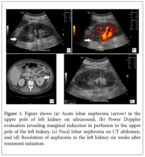
Ultrasound of the patient’s renal tract revealed a loss of corticomedullary differentiation of the upper pole of the left kidney (Figure 1a). On power Doppler evaluation, a marginal reduction in the amount of perfusion to the upper pole of the left kidney was observed (Figure 1b). These findings were indicative of an evolving focal lobar nephronia. CT abdomen also revealed a mass in the upper pole of the left kidney which consistent with acute lobar nephronia (Figure 1c).
Figure 1: Figure shows (a) Acute lobar nephronia (arrow) in the upper pole of left kidney on ultrasound, (b) Power Doppler evaluation revealing marginal reduction in perfusion to the upper pole of the left kidney, (c) Focal lobar nephronia on CT abdomen, and (d) Resolution of nephronia in the left kidney six weeks after treatment initiation.
The patient was treated empirically with intravenous Ampicillin 1 g four times a day and Gentamicin 320 mg once a day (for three days) prior to the availability of microbiology results. When the results of antibiotic susceptibility became available, the patient was treated with oral Ciprofloxacin. Clinically her fever and vomiting resolved after a few days of antibiotics. She was treated with antibiotics for a total of two weeks. During her admission, her glycaemia control was also optimised.
She was reviewed in outpatient clinic six weeks after discharge and she had remained well. Her repeat ultrasound scan showed resolution of the nephronia (Figure 1d).
Discussion
Acute lobar nephronia is an uncommon type of renal tract infection. It is an acute localized non-liquefactive infection of the kidney caused by bacterial infection [2]. If undertreated, it has been observed to result in renal abscess formation. Many of the published cases of this condition have been in the pediatric population and relatively scarce in adults [3,5]. Most of the reported cases of acute lobar nephronia in adults have involved E. coli as the causative organism [4,7]. To our knowledge, this is the first reported case of acute lobar nephronia involving E. cloacae.
The main manifestations of acute lobar nephronia have previously been described as fever, chills, abdominal pain, flank pain and tenderness of the costovertebral angles [2]. However localizing features such as abdominal or flank pain may not always be present [7]. The present case of acute lobar nephronia had non-specific symptoms and diagnosis was directed to the urinary tract with the positive microbiological findings in the urine and blood culture. Diagnosis of acute lobar nephronia relies on findings on ultrasound or computed tomography (CT) imaging [7,8]. It is important to interpret the imaging findings and clinical features because acute lobar nephronia may sometimes be mistaken for a renal mass [4]. Repeat scan, usually with ultrasound, is often recommended four to six weeks after initial presentation, to ensure resolution of the nephronia.
E. coli is one of the commonest organism identified in patients with acute lobar nephronia but other organisms such as Proteus mirabilis, Klebsiella species, Pseudomonas aeruginosa and enterococci [9]. However Enterobacter has not been reported in any of the previous cases of acute lobar nephronia in adults. E. cloacae is a well-known nosocomial and multiresistant bacterial pathogen contributing to bacteremia and urinary tract infection [10]. E. cloacae has an intrinsic resistance to Ampicillin, Amoxycillin, first-generation and second-generation Cephalosporins because of the production of constitutive AmpC β-lactamase [11]. In the present case, the E. cloacae was susceptible to Ciprofloxacin and her clinical symptoms improved after a course of this antibiotic.
Previous reports of acute lobar nephronia has involved patients who were immunocompromised including one with human immunodeficiency virus and hepatitis C while a few others were kidney transplant recipients [9,12-14]. The current case had Type 2 diabetes that was sub optimally controlled at the time of presentation, which may have affected patient’s immune system and predisposed to this type of infection. However any association between Type 2 diabetes and acute lobar nephronia has not been reported previously. This association may warrant additional investigation. In conclusion, acute lobar nephronia is usually considered the midpoint in the spectrum of upper urinary tract infections between acute pyelonephritis and renal abscess. Its presentation may not involve abdominal or urinary symptoms. Urine culture, ultrasound and CT imaging are useful to identify the presence of acute lobar nephronia and the causative pathogen. Appropriate treatment should be administered in patients with this condition to avoid progression to a renal abscess.
Acknowledgement
The author has no funding.
Conflict of Interest
The author has no conflict of interest.
References
- Rosenfield AT, Glickman MG, Taylor KJ, Crade M, Hodson J (1979) Acute focal bacterial nephritis (acute lobar nephronia). Radiology 132:553-561.
- Li Y, Zhang Y (1996) Diagnosis and treatment of acute focal bacterial nephritis. Chinese medical journal 109: 168-172.
- Cheng CH, Tsau YK, Lin TY (2010) Is acute lobar nephronia the midpoint in the spectrum of upper urinary tract infections between acute pyelonephritis and renal abscess? J Pediatr 156:82-86.
- Kwong T, Coker C, Simpson E (2012) A rare and unusual presentation of urological infection on a patient on steroids. BMJ case reports.
- Cheng CH, Tsau YK, Chang CJ, Chang YC, Kuo CY, et al. (2010) Acute lobar nephronia is associated with a high incidence of renal scarring in childhood urinary tract infections. Pediatr Infect Dis J 29:624-628.
- Harris EE, Sweat M, Katsanis WA, Aronoff GR (1992) Case report: acute focal bacterial pyelonephritis (lobar nephronia)-presentation as a palpable abdominal mass. Am J Med Sci 304:303-305.
- Conley SP, Frumkin K (2014) Acute lobar nephronia: a case report and literature review. J Emerg Med 46:624-626.
- McCoy RI, Kurtz AB, Rifkin MD, Kodroff MB, Bidula MM (1985) Ultrasound detection of focal bacterial nephritis (lobar nephronia) and its evolution into a renal abscess. Urol Radiol7:109-111.
- Dave SS, Noursadeghi M, Rickards D, Cartledge JD, Miller RF (2005) Atypical presentation of lobar nephronia in an adult co-infected with HIV and hepatitis C. Sex Transm Infect 81:183.
- Davin-Regli A, Pages JM (2015) Enterobacter aerogenes and Enterobacter cloacae; versatile bacterial pathogens confronting antibiotic treatment. Front Microbiol 6:392.
- Mezzatesta ML, Gona F, Stefani S (2012)Enterobacter cloacae complex: clinical impact and emerging antibiotic resistance. Future microbiology 7:887-902.
- Thomalla JV, Gleason P, Leapman SB, Filo RS (1993) Acute lobar nephronia of renal transplant allograft. Urology 41:283-286.
- Joss N, Baxter G, Young B, Buist L, Rodger RS(2005) Lobar nephronia in a transplanted kidney. Clin Nephrol 64:311-314.
- Tai HC, Yang PJ, Lee PH, Chung SD, Chueh SC, et al. (2008) Acute lobar nephronia in a renal allograft: a case report and literature review. Transplant Proc 40:1737-1740.
Relevant Topics
Recommended Journals
Article Tools
Article Usage
- Total views: 17178
- [From(publication date):
March-2016 - Jul 02, 2025] - Breakdown by view type
- HTML page views : 16209
- PDF downloads : 969

