A Case of Recurrent Basal Cell Carcinoma Diagnosed on Fine Needle Aspiration Cytology
Received: 04-Apr-2014 / Accepted Date: 03-Jun-2014 / Published Date: 05-Jun-2014 DOI: 10.4172/2161-0681.1000177
Abstract
Basal Cell Carcinoma (BCC) is the most common malignant skin tumor. Its diagnosis is made on clinical grounds followed by confirmation on histopathology. Fine Needle Aspiration Cytology (FNAC) can be reliably used to evaluate skin tumors especially BCC. Here a case of recurrent BCC of skin successfully diagnosed through FNAC is reported.
Keywords: Skin tumors; BCC; FNAC
314306Introduction
Basal Cell Carcinoma (BCC) is the most common malignant skin tumor and is defined by the World Health Organization Committee on the skin tumors as “a locally invasive, slowly spreading rarely metastasizing tumor, arising in the epidermis” [1-5].
It is usually observed in older patients, especially in those frequently and excessively exposed to ultraviolet radiation during their lives [6].
The diagnosis of BCC is usually clinical, followed by microscopic confirmation of the lesion either through biopsy or surgical excision [7]. Although histology is the gold standard for diagnosing BCC, cutaneous cytology especially Fine-Needle Aspiration Cytology (FNAC) can also be successfully employed for diagnosing skin tumors, including BCC, due to simplicity of the procedure as well as easy staining techniques [8]. FNAC is a simple, quick, non-invasive procedure that can accurately confirm or exclude the diagnosis of malignancy. Most of the studies in literature have shown high sensitivity and specificity of FNAC for diagnosing BCC both during the initial workup as well as in cases of recurrence [9,10]. We report here a case of recurrent BCC of skin diagnosed on FNAC.
Case Report
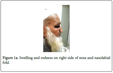
A 70 years old man presented in ENT OPD with pain and swelling on right side of nose and nasolabial fold for the past 3 months (Figure 1a). He was a diagnosed case of basal cell carcinoma involving right cheek and ala of nose and had undergone surgical resection followed by reconstruction and photodynamic therapy three years back. Histopathology of the resected lesion revealed basal cell carcinoma involving right cheek and right ala of nose with clearance of surgical resection margins.
Currently, the swelling, which is progressively increasing in size is involving right side of nose and nasolabial fold and is associated with pain. Clinically, the lesion was suspicious for recurrent BCC. The patient was sent to histopathology department on OPD basis for FNAC of the lesion to rule out recurrence. FNAC was performed and smears were obtained from swelling involving right side of nose and nasolabial fold. Air dried and wet, alcohol fixed, slides were prepared.
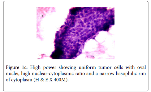
The alcohol fixed slides were later stained with H & E and PAP stains. Microscopic examination of the smears revealed cohesive clusters and sheets of atypical cells having round to ovoid, hyperchromatic nuclei displaying mild to moderate pleomorphism. Cytoplasm was scant. Focally, peripheral palisading and eosinophilic nucleoli seen. Background showed blood and few acute and chronic inflammatory cells (Figure 1b,1c).
Keeping in view the clinical history, the morphological features were consistent with previously diagnosed basal cell carcinoma. Patient was later referred for CT scan of face and a reconstructive surgery was planned.
Discussion
Basal cell carcinoma is a locally invasive, rarely metastasizing, common skin tumor [1-5]. Exposure to excessive sunlight is a major predisposing factor. Biopsy or surgical resection of the lesion with histopathological evaluation remains the mainstay for the diagnosis. However, FNAC can also be successfully employed for the diagnosis of BCC provided the yield is adequate and cytopathologist has expertise in interpreting skin lesions. The advantages of FNAC are that it is a quick and easy procedure to perform requiring minimum special equipment and little training. Limitations include inadequate yield, lack of experience by cytopathologist in interpreting skin tumors and failure to exactly assess variants of BCC.
The cytological features of BCC include 1) tightly cohesive cellular fragments, 2) small size of the tumor cells, 3) morphologically uniform tumor cells, 4) oval or fusiform and sometimes round nuclei with blurred chromatin structures, 5) high nuclear-cytoplasmic ratio with a narrow basophilic rim of cytoplasm, 6) nucleoli usually not evident and 7) some fragments with distinct sharp borders [11].
The differential diagnosis of basal cell carcinoma on FNAC include tumors containing basaloid cells i.e., basaloid squamous cell carcinoma, adenoid cystic carcinoma, small cell neuroendocrine carcinoma, pilomatricoma, seborrheic keratosis and eccrine gland carcinoma of the skin (11). Hence, one should be careful in interpreting tumors with basaloid morphology.
The high diagnostic accuracy of the cytology for BCC was first reported by Ruocco, in a study comprising of 500 cases [12]. Powell et al. performed a study on 82 skin tumors, and reported that the sensitivity and specificity of cytological examination in diagnosing BCC were 91% and 87%, respectively [13]. In a study done by Naraghi et al., the sensitivity and specificity of the cytology in identifying BCC variants were 87.3% and 95.3%, respectively [11]. Another study from south of Xinjiang on 22 cases of BCC skin revealed that there were no false-positive cases, but one false-negative due to inadequate sampling, giving a diagnostic accuracy of 95.65% [14]. In a recent study conducted by Kassi et al the sensitivity and specificity of fine-needle aspiration cytology for basal cell carcinoma were 94.3% and 100%, respectively [10].Thus, studies have confirmed that cytological examination of the smears obtained from the suspected BCC lesions can be a useful alternative method of diagnosis and can guide the dermatologist for subsequent confirmation by histopathology, thus expediting the treatment of the patient. Alternatively, in an already diagnosed case of BCC with suspected recurrence, FNAC can be used as a sole modality for confirming the diagnosis and initiating further appropriate management as was in this case.
Conclusion
In conclusion, FNAC is highly accurate, reliable, simple and very useful for diagnosing BCC. It can be performed in outpatient's clinics, thus saving both the time and cost in clinic and laboratory. This case report highlights the usefulness of FNAC in diagnosing BCC as an initial diagnostic tool as well as for confirming recurrence which can eventually be further confirmed by histopathology.
References
- Nakayama M, Tabuchi K, Nakamura Y, Hara A (2011) Basal cell carcinoma of the head and neck. J Skin Cancer 2011: 496910.
- Oram Y, Orengo I, Alford E, Green LK, Rosen T, et al. (1994) Basal cell carcinoma of the scalp resulting in spine metastasis in a black patient. J Am AcadDermatol 31: 916-920.
- Spates ST, Mellette JR Jr, Fitzpatrick J (2003) Metastatic basal cell carcinoma. DermatolSurg 29: 650-652.
- Lo JS, Snow SN, Reizner GT, Mohs FE, Larson PO, et al. (1991) Metastatic basal cell carcinoma: report of twelve cases with a review of the literature. J Am AcadDermatol 24: 715-719.
- Akinci M, Aslan S, Markoç F, Cetin B, Cetin A (2008) Metastatic basal cell carcinoma. ActaChirBelg 108: 269-272.
- Situm M, Buljan M, Bulat V, Lugović Mihić L, Bolanca Z, et al. (2008) The role of UV radiation in the development of basal cell carcinoma. CollAntropol 32 Suppl 2: 167-170.
- Kate MS, Jain P, Patne SS (2013) Basal cell carcinoma diagnosed on Fine-Needle Aspiration Cytology – A Pathological Case Report. SEAJCRR 2: 275-80.
- Jain M, Madan NK, Agarwal S, Singh S (2012) Pigmented basal cell carcinoma: Cytological diagnosis and differential diagnoses. J Cytol 29: 273-275.
- Vega-Memije E, De Larios NM, Waxtein LM, Dominguez-Soto L (2000) Cytodiagnosis of cutaneous basal and squamous cell carcinoma. Int J Dermatol 39: 116-120.
- Kassi M, Kasi PM, Afghan AK, Marri SM, Kassi M, et al. (2012) The role of fine-needle aspiration cytology in the diagnosis of Basal cell carcinoma. ISRN Dermatol 2012: 132196.
- Naraghi Z, Ghaninejad H, Akhyani M,Akbari D (2005) Cytological diagnosis of cutaneous Basal Cell Carcinoma, ActaMedicaIranica43:50-54.
- V Ruocco (1992)Attendibilitadelladiagnosicitologicadiobasalioma. G ItalDermatoVenereol 127:23-29.
- Powell CR, Menon G, Harris AI (2000) Cytological examination of basal cell carcinoma-a useful tool for diagnosis? Br J Dermatol 143:71.
- Fang X, Ma B (1999) Fine needle aspiration cytology of basal cell carcinoma of the skin: a clinical and cytopathological appraisal. J Dermatol 26: 640-646.
Citation: Moatasim A (2014) A Case of Recurrent Basal Cell Carcinoma Diagnosed on Fine Needle Aspiration Cytology. J Clin Exp Pathol 4:177. DOI: 10.4172/2161-0681.1000177
Copyright: © 2014 Moatasim A. This is an open-access article distributed under the terms of the Creative Commons Attribution License, which permits unrestricted use, distribution, and reproduction in any medium, provided the original author and source are credited.
Share This Article
Recommended Journals
Open Access Journals
Article Tools
Article Usage
- Total views: 16049
- [From(publication date): 9-2014 - Dec 19, 2024]
- Breakdown by view type
- HTML page views: 11637
- PDF downloads: 4412


