Case Report Open Access
A Case of Genital Erosion Associated with Cutaneous Pseudomonas putida Infection and Review of the Literature
| Y├?┬▒ld├?┬▒ray Yeniay1 and Gürol Aç├?┬▒kgöz2* | ||
| 1Gölcük Military Hospital, Department of Dermatology, Kocaeli, Turkey | ||
| 2Gülhane School of Medicine, Department of Dermatology, Ankara, Turkey | ||
| Corresponding Author : | Gürol Açikgöz Gülhane School of Medicine Department of Dermatology, 06018, Etlik, Ankara, Turkey Tel: +903123044459 Fax: +903123044450 E-mail: gacikgoz@gata.edu.tr |
|
| Received June 30, 2014; Accepted October 16, 2014; Published October 31, 2014 | ||
| Citation: Yeniay Y, Açikgöz G (2014) A Case of Genital Erosion Associated with Cutaneous Pseudomonas putida Infection and Review of the Literature. J Infect Dis Ther 2:178. doi: 10.4172/2332-0877.1000178 | ||
| Copyright: © 2014 Yeniay Y, et al. This is an open-access article distributed under the terms of the Creative Commons Attribution License, which permits unrestricted use, distribution, and reproduction in any medium, provided the original author and source are credited. | ||
Related article at Pubmed Pubmed  Scholar Google Scholar Google |
||
Visit for more related articles at Journal of Infectious Diseases & Therapy
Abstract
Erosive genital lesions are mainly associated with infectious factors, which are subdivided into two groups, including sexually transmitted infections and non-sexually transmitted infections. Although sexually transmitted infections well investigated in the literature, non-sexually transmitted infections associated with genital erosive lesions have not been identified clearly. Here we presented a case of genital erosion associated with cutaneous Pseudomonas putida infection. In literature cutaneous manifestations and clinical spectrum of cutaneous P. putida infection has not been well discussed. In addition to this, all previous case reports were complicated skin infections, which result in soft tissue infection. Here we summarized cutaneous manifestation of P. putida infection and represent a rare etiological factor for erosive genital lesions.
| Introduction |
| Genital erosions or ulcers are associated with various kinds of conditions including infections, drug reactions, immunobullous disorders, Behçet’s disease, inflammatory bowel disease, erosive lichen planus, lichen sclerosis et atrophicus, premalignant/malignant conditions, dermatitis artefacta, hidradenitis suppurativa and pyoderma gangrenosum [1]. Although a broad spectrum of etiological factors responsible for genital erosive lesions, sexually transmitted infections are usually considered in the first place [2]. In this case report, we described a rare infectious cause of genital erosion associated with cutaneous Pseudomonas putida infection and performed a brief literature review regarding the clinical characteristics of P. putida infections. We aimed to enlighten the clinical spectrum of P. putida infection and inform physicians about this rare etiological factor for erosive genital lesions. |
| Case |
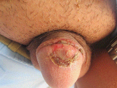
| A 21-year-old previously healthy male patient with two months history of genital erosive lesion on glans penis was attended to our outpatient clinic. He had never had a similar lesion previously and his past medical history was unremarkable. The lesion was painful and pruritic. He had no history of sexually transmitted infections or suspicious sexual contact. He informed us that he received topical mupirocin cream for this condition prescribed by his family doctor without showing any sign of healing. The treatment resistance caused doubt about the previous diagnosis and patient was advised to attend our clinic. Physical examination revealed well-defined, erythematous, round shaped erosive lesion on the glans penis (Figure 1). The lesions had a smooth surface, which was partially covered by yellow exudate, without any induration. There were no sign of vesicles or bullae. No penile discharge or enlarged inguinal lymph nodes were observed. |
| In laboratory examination; complete blood count, sedimentation rate, renal and hepatic function tests were within normal limits. Swabs were taken from the lesion for microbiological examinations and meanwhile topical rifampicin therapy was initiated as empiric therapy. We performed serological studies for sexually transmitted infections including herpes simplex virus (HSV), cytomegalovirus (CMV), treponema pallidum, chlamydia trachomatis and human immunodeficiency virus (HIV), but no positive result was observed. Microscopic examination of the specimens did not identify any fungal or bacterial infectious agent. Swap cultures revealed a pure growth of P. putida. P. putida strains were also identified by an automatized system (BD Phoenix System, Beckton Dickinson, USA) and antimicrobial susceptibility tests were performed by Kirby-Bauer disc diffusion method according to the guidelines of the Clinical and Laboratory Standards Institute (CLSI). In Kirby-Bauer disc diffusion method, turbidity of bacterial suspension was adjusted to 0.5 McFarland's standard and then it was spread on the surface of Mueller-Hinton agar (MHA) plates using sterile cotton swab. Ciprofloxacin, gentamycin, ceftazidime, piperacillin/tazobactam, ticarcillin/clavulanic acid and aztreonam disks were placed on the agars. The zone of inhibition was measured and interpreted according to the CLSI guidelines. In our case report, the organism was sensitive to ciprofloxacin, gentamycin, ceftazidime, piperacillin/tazobactam but resistant to ticarcillin/clavulanic acid and aztreonam. According to culture results, intravenous cefotaxime treatment (2 g/day) was initiated. After 5 days of treatment patient’s lesion was resolved without any complication. |
| Discussion |
| Genital erosion was usually considered as a sign of sexually transmitted infections including herpes simplex virus, treponema pallidum, haemophilus ducreyi, or chlamydia trachomatis. In addition to this, Ebstein Barr virus, tuberculosis, leishmaniasis, HIV/AIDS related ulcer and amoebiasis were described as infectious causes of erosive genital lesions other than sexually transmitted infections [3]. Instead of all these infectious etiologic factors of erosive genital lesions, here we presented a rare case of cutaneous P. putida infection associated with genital erosion. |
| P. putida, a member of the fluorescent group of pseudomonas, is a gram-negative, aerobic, non-lactose fermenting, flagellated bacillus. This bacillus survives in moist environment and usually colonized on inanimate surfaces, patients’ mucous membranes and indwelling devices in health care units [4]. Although pathogenicity of P. aeruginosa, another member of pseudomonas group, is well-defined, pathogenic potential of P. putida just recognized in last decades [5]. According to the literature various conditions associated with P. putida infection identified, but no skin infection caused by P. putida colonization reported previously [6]. In addition to this, a case series report presented that only 5% of P. putida infections were associated with soft tissue infections [7]. |
| We performed literature searches with PubMed from 1990 to 2014 using the search term “pseudomonas putida” and investigated articles and case reports associated with soft tissue or skin infections caused by P. putida (Table 1). Including our case report, a total of 10 cases of skin or soft tissue infections associated with P. putida were reported. Of these 10 cases; three had traumatic wound infection, two had wound infection without any detailed information, two had soft tissue infection associated with morbidities including malignancy and peripheral vascular disease, one had blast injury infection, one had history of contaminated water exposure, and our case had uncomplicated skin infection [4,6-10]. According to the reported cases, disturbance of the skin barrier upon injury is main predisposing factor that facilitates P. putida infections. Although we could not clearly identify a trauma history in our patient, we considered that friction in genital area during sexual intercourse might be a traumatizing factor for glans penis. Repeated friction of the skin cause loss of skin barrier function. Impaired skin barrier function and natural moisture of genital region can easily enhance the colonization of P. putida, which result in skin infection. |
| In dermatology, topical antibiotics are widely used in the treatment of skin infections. Polymyxin B, bacitracin, silver sulfadiazine, ciprofloxacin gentamycin and tobramycin are effective topical antibiotics that are used in the treatment of cutaneous pseudomonas infections [11,12]. Our patient previously received topical mupirocin which is a monoxycarbolic acid class antibiotic that was originally isolated from P. fluorescens. Although, mupirocin is active primarily against to gram-positive organisms including S. aureus, S. epidermidis, S. pyogenes, S. agalactiae and S. viridans, it has no effect on pseudomonas infections. In addition to this treatment, we initiated topical rifampicin therapy as empiric therapy. Although naturel strains of P. putida are sensitive to the rifampicin, pathogenic strains of P. putida are intrinsically resistant to the rifampicin [13]. This situation explains why we did not observe any response to rifampicin therapy until initiation of intravenous cefotaxime treatment. We could not suggest any treatment regime for cutaneous P. putida infections, due to limited literature information about P. putida infections. It was emphasized that, empiric therapy similar to other P. species especially P. aeruginosa could be taken into account regarding to the severity of the infection [6]. |
| Conclusion |
| In conclusion, cutaneous P. putida infection was underestimated in dermatology practice and published studies were inadequate to enlightened clinical spectrum of this infection. In our case, genital erosion was the main clinical manifestation of our patient, which could be easily misdiagnosed as sexual transmitted disease or non-pseudomonal bacterial infection. It should be considered that genital erosive lesions could be associated with P. putida infection. In addition to this, P. species should usually kept in mind especially in treatment refractory cutaneous infection. |
References
- Sehgal VN, Pandhi D, Khurana A (2014) Nonspecific genital ulcers.ClinDermatol 32: 259-274.
- Gomes Naveca F, Sabidó M, AmaralPires de Almeida T, AraújoVeras E, Contreras MejíaMdel C, et al. (2013) Etiology of genital ulcer disease in a sexually transmitted infection reference center in Manaus, Brazilian Amazon. PloS one 8, e63953.
- Bohl TG (2004) Vulvar ulcers and erosions--a dermatologist's viewpoint.DermatolTher 17: 55-67.
- Thomas BS, Okamoto K, Bankowski MJ, Seto TB (2013) A Lethal Case of Pseudomonas putidaBacteremia Due to Soft Tissue Infection.Infect Dis ClinPract (BaltimMd) 21: 147-213.
- Yoshino Y, Kitazawa T,Kamimura M, Tatsuno K, Ota Y, et al. (2011) Pseudomonas putidabacteremia in adult patients: five case reports and a review of the literature. Journal of infection and chemotherapy: official journal of the Japan Society of Chemotherapy 17: 278-282.
- Carpenter RJ, Hartzell JD, Forsberg JA, Babel BS, Ganesan A (2008) Pseudomonas putidawar wound infection in a US Marine: a case report and review of the literature.J Infect 56: 234-240.
- Yang CH, Young T, Peng MY, Weng MC (1996) Clinical spectrum of Pseudomonas putidainfection.J Formos Med Assoc 95: 754-761.
- Chen CH, Hsiu RH, Liu CE, Young TG (2005) Pseudomonas putidabacteremia due to soft tissue infection contracted in a flooded area of central Taiwan: a case report. J MicrobiolImmunol Infect 38: 293-295.
- Kim SE, Park SH, Park HB, Park KH, Kim SH, et al. (2012) Nosocomial Pseudomonas putidaBacteremia: High Rates of Carbapenem Resistance and Mortality.Chonnam Med J 48: 91-95.
- Lombardi G, Luzzaro F, Docquier J, Riccio ML, Perilli M, et al. (2002) Nosocomial infections caused by multidrug-resistant isolates of Pseudomonas putidaproducing VIM-1 metallo-beta-lactamase. Journal of clinical microbiology 40: 4051-4055.
- Barankin B, Levy J (2012) Dermacase. Can you identify this condition? Pseudomonas aeruginosa infection.Can Fam Physician 58: 1103-1104.
- Zhu CY, Zhang GX, Yu ZZ, Li ZJ, Fan YM (2014) Pseudomonas aeruginosaecthymagangrenosum in a woman with recurrent Graves' disease. International journal of infectious diseases: IJID: official publication of the International Society for Infectious Diseases 21: 19-20
- Molina L, Udaondo Z, Duque E, Fernández M, Molina-Santiago C, et al. (2014) Antibiotic resistance determinants in a Pseudomonas putidastrain isolated from a hospital.PLoS One 9: e81604.
Tables and Figures at a glance
| Table 1 |
Figures at a glance
 |
| Figure 1 |
Relevant Topics
- Advanced Therapies
- Chicken Pox
- Ciprofloxacin
- Colon Infection
- Conjunctivitis
- Herpes Virus
- HIV and AIDS Research
- Human Papilloma Virus
- Infection
- Infection in Blood
- Infections Prevention
- Infectious Diseases in Children
- Influenza
- Liver Diseases
- Respiratory Tract Infections
- T Cell Lymphomatic Virus
- Treatment for Infectious Diseases
- Viral Encephalitis
- Yeast Infection
Recommended Journals
Article Tools
Article Usage
- Total views: 15715
- [From(publication date):
December-2014 - Jul 02, 2025] - Breakdown by view type
- HTML page views : 11165
- PDF downloads : 4550
